

The combination of chemical fixation procedures with high pressure freezing and freeze substitution preserves highly labile tissue ultrastructure for electron tomography applications
- PMID: 17962040
- PMCID: PMC2459253
- DOI: 10.1016/j.jsb.2007.09.002
The combination of chemical fixation procedures with high pressure freezing and freeze substitution preserves highly labile tissue ultrastructure for electron tomography applications
Abstract
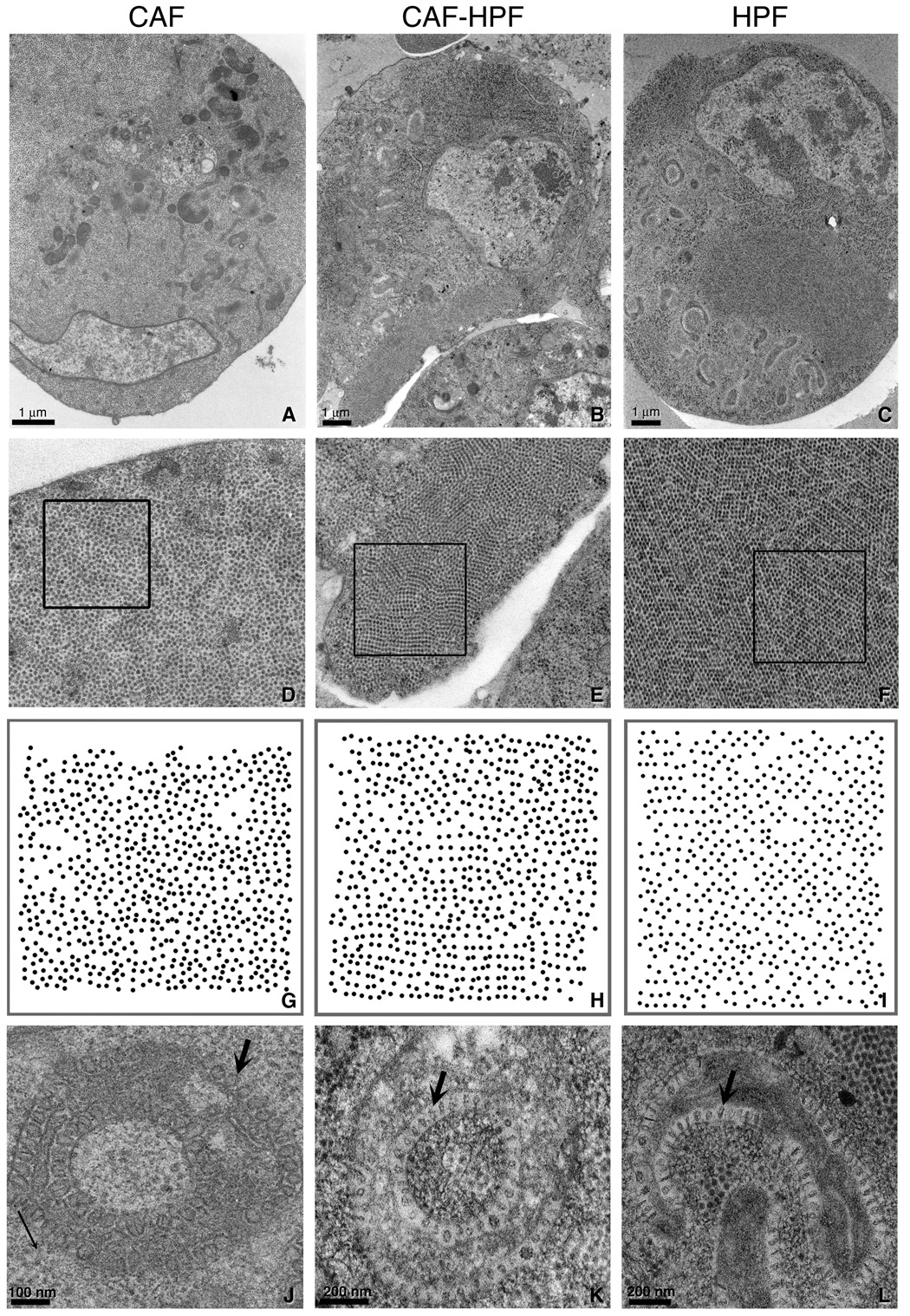
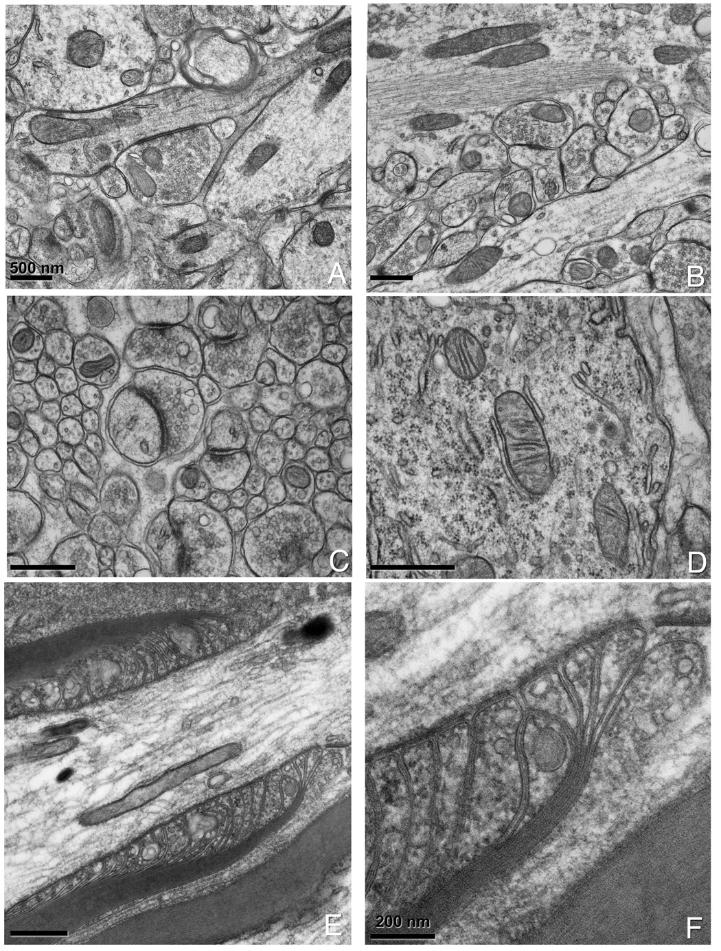
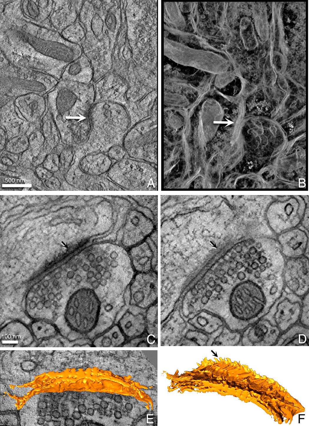
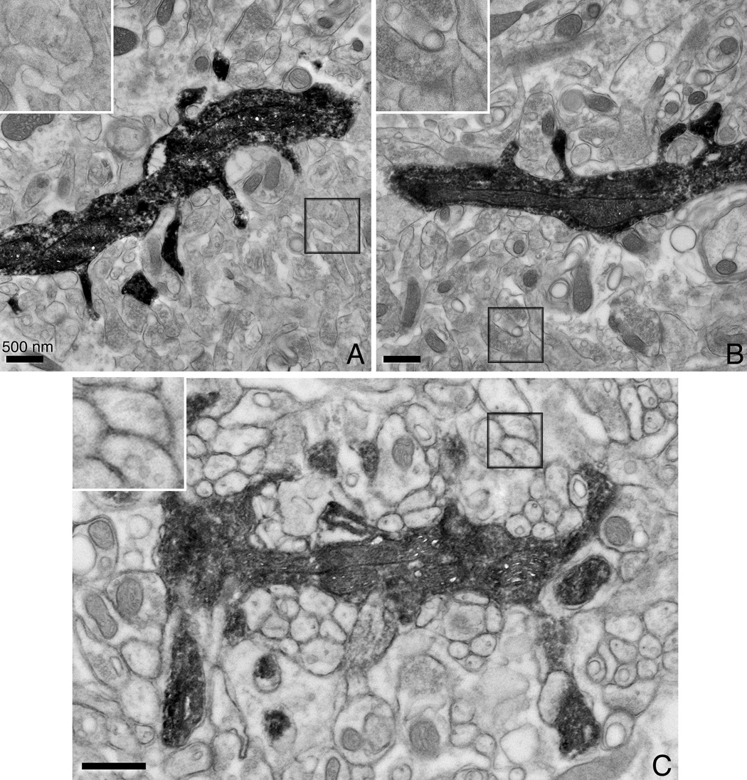
The emergence of electron tomography as a tool for three dimensional structure determination of cells and tissues has brought its own challenges for the preparation of thick sections. High pressure freezing in combination with freeze substitution provides the best method for obtaining the largest volume of well-preserved tissue. However, for deeply embedded, heterogeneous, labile tissues needing careful dissection, such as brain, the damage due to anoxia and excision before cryofixation is significant. We previously demonstrated that chemical fixation prior to high pressure freezing preserves fragile tissues and produces superior tomographic reconstructions compared to equivalent tissue preserved by chemical fixation alone. Here, we provide further characterization of the technique, comparing the ultrastructure of Flock House Virus infected DL1 insect cells that were (1) high pressure frozen without fixation, (2) high pressure frozen following fixation, and (3) conventionally prepared with aldehyde fixatives. Aldehyde fixation prior to freezing produces ultrastructural preservation superior to that obtained through chemical fixation alone that is close to that obtained when cells are fast frozen without fixation. We demonstrate using a variety of nervous system tissues, including neurons that were injected with a fluorescent dye and then photooxidized, that this technique provides excellent preservation compared to chemical fixation alone and can be extended to selectively stained material where cryofixation is impractical.
Figures

References
-
- Al-Amoudi A, Dubochet J, Norlen L. Nanostructure of the epidermal extracellular space as observed by cryo-electron microscopy of vitreous sections of human skin. J Invest Dermatol. 2005;124:764–777. - PubMed
-
- Belichenko PV, Dahlstrom A. Studies on the 3-dimensional architecture of dendritic spines and varicosities in human cortex by confocal laser scanning microscopy and Lucifer yellow microinjections. J Neurosci Methods. 1995;57:55–61. - PubMed
-
- Buhl EH. Intracellular injection in fixed slices in combination with neuroanatomical tracing techniques and electron microscopy to determine multisynaptic pathways in the brain. Microsc Res Tech. 1993;24:15–30. - PubMed
-
- Calle M, Corstens GJ, Wang L, Kozicz T, Denver RJ, Barendregt HP, Roubos EW. Evidence that urocortin I acts as a neurohormone to stimulate alpha MSH release in the toad Xenopus laevis. Brain Res. 2005;1040:14–28. - PubMed
Publication types
MeSH terms
Grants and funding
- GM34220/GM/NIGMS NIH HHS/United States
- NS14718/NS/NINDS NIH HHS/United States
- R01 DA016602/DA/NIDA NIH HHS/United States
- GM065937/GM/NIGMS NIH HHS/United States
- RR004050/RR/NCRR NIH HHS/United States
- DA016602/DA/NIDA NIH HHS/United States
- R01 GM034220/GM/NIGMS NIH HHS/United States
- R37 GM034220/GM/NIGMS NIH HHS/United States
- P41 RR004050/RR/NCRR NIH HHS/United States
- R01 NS014718/NS/NINDS NIH HHS/United States
- R01 GM072881/GM/NIGMS NIH HHS/United States
- R01 GM065937/GM/NIGMS NIH HHS/United States
- GM072881/GM/NIGMS NIH HHS/United States
- S10 RR016699/RR/NCRR NIH HHS/United States
LinkOut - more resources
Full Text Sources
Other Literature Sources
