Review
. 2008 Jan;27(1):89-110.
doi: 10.1016/j.preteyeres.2007.08.002. Epub 2007 Sep 7.

Zebrafish: a model system for the study of eye genetics

Affiliations
Review

Zebrafish: a model system for the study of eye genetics

James M Fadool et al. Prog Retin Eye Res. 2008 Jan.

Abstract

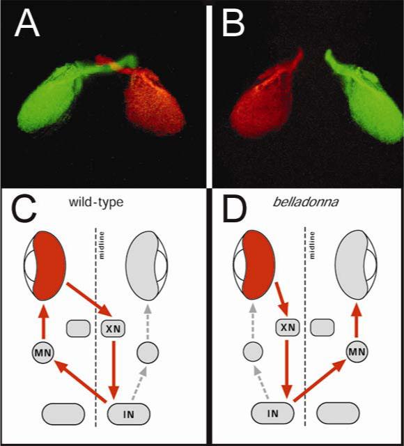
Over the last decade, the use of the zebrafish as a genetic model has moved beyond the proof-of-concept for the analysis of vertebrate embryonic development to demonstrated utility as a mainstream model organism for the understanding of human disease. The initial identification of a variety of zebrafish mutations affecting the eye and retina, and the subsequent cloning of mutated genes have revealed cellular, molecular and physiological processes fundamental to visual system development. With the increasing development of genetic manipulations, sophisticated techniques for phenotypic characterization, behavioral approaches and screening strategies, the identification of novel genes or novel gene functions will have important implications for our understanding of human eye diseases, pathogenesis, and treatment.


Figures

Figure 1
Figure 1
Histology of the zebrafish retina. (A–C) Fluorescent double immunolabelling of specific cell types in transverse section of the eye of a zebrafish larvae and DAPI (4’, 6-diamidino-2-phenylindole) counterstaining reveal the archetypical laminar arrangement of the retina. (A) Labeling with the Zn-8 monoclonal antibody specific for ganglion cells (gc) and the red-cone opsin. (B) Labeling for the rod photoreceptors with the 1D1 antibody and Müller glia with anti-CAZ. (C) Labeling of amacrine cells (amc) with the 5E11 antibody and co-labeling for UV opsin. (D) Mosaic organization of the larval retina revealed by labeling red and green cones with the zrp1 antibody and DAPI counterstaining of nuclei. The red and green cones can be distinguished by the greater fluorescent labeling of the red cones, and the positions of the blue and ultraviolet cones can be discerned by labeling with DAPI. The identity of the cone subtypes are diagrammatically represented and color-coded. (E) Transverse section through the adult retina demonstrating EGFP fluorescence in rod photoreceptors (left panel in green) and counterstaining with DAPI (blue) to reveal the tiering of the cone nuclei (relative positions of the cone subtypes are diagrammatically represented). The rod cell bodies appear white due to double labeling for EGFP and DAPI. The rod outer segments are also immunolabeled for opsin. The plane of section in (F) is indicated by the arrow. (F) EGFP fluorescence of the rods, immunolabeling of the UV sensitive opsin, DAPI labeling of the cone nuclei in a tangential section of the adult retina and the merged image. Note the regular arrangement of the rod myoids around the outer segment of the UV-cone outer segments. (E & F from Fadool, 2003; reprinted with the permission of the publisher).
Figure 2
Figure 2
Micrographs showing development of the zebrafish eye, cornea and lens. Histological sections at 24 hpf (A), 36 hpf (B), and 48 hpf (C). Neurogenesis and lamination of the retina progresses rapidly between 36 and 38 hpf. The lens vesicle detatches from the overlying ectoderm by 24 hpf and differentiation of the fiber cells also progresses rapidly between 36 and 48 hpf. The periocular mesenchyme is present at 24 hpf, but more prominent at 36 and 48 hpf (arrows). Hyaloid vasculature is indicated with asterisk. (D) Diagram of the embryonic zebrafish eye. Insets in (D) indicate corresponding histological sections through the 3 dpf lens transition zone (E), lens epithelium (F) and posterior lens (G). TEM micrographs (H,I) show developing lens epithelium (H), interdigitating lens fiber cells and establishment of rudimentary lens sutures (arrow, I). (Adapted from Soules and Link, 2005; reprinted with permission of the original author as defined by the publisher).
Figure 3
Figure 3
Electroretinograms (ERGs) from 6-day-old OKR+ (a) and pob mutant larvae (b). The responses were elicited with a short 0.01 s flashes of green (520 nm) light at the same intensity. In (a), only the b-wave is evident but both the a- and b-waves are present in (b). The vertical bars represent 50 μV (a) or 100 μV (b). Time markers = 0.1 ms. (c), Comparison of the spectral sensitivities of pob and normal sibling larvae. Spectral sensitivities were determined by ERG analysis. The inverse of the number of photons required to generate a threshold (20 μV) b-wave response was calculated at each wavelength and normalized to the sensitivity of the normal larvae at 430 nm. Note the pob mutant larvae are about 2 log units less sensitive to red light. (From Brockerhoff et al., 1997; reprinted with permission of the publisher).
Figure 4
Figure 4
Electron micrographs of cone terminals in wild-type (A and B) and nrc mutant larva (C). (A) In the wild-type retina, bipolar and horizontal cell processes invaginate the pedicle in a tight bundle (arrow). Horizontal cell processes (H) are easily recognized by their large size, electron lucent cytoplasm and characteristic densities (small arrowheads). Synaptic ribbons (R) are associated with the presynaptic membrane via an arciform density (curved arrow). (B) Basal contacts (B) are found in wild-type cones between the ribbon synapses. Inset, Under high power, the basal contacts show dense cytoplasmic material on both sides of the junction. Synaptic vesicles (V) surround the synaptic ribbons (R). (C) In the nrc retina, synaptic ribbons (R) in most of the pedicles appear to be floating in the cytoplasm, unassociated with an arciform density and the presynaptic membrane. Few postsynaptic processes invaginate the pedicles. Many of these processes have small densities (arrowheads) suggesting they are horizontal cell processes. Basal contacts are made onto bipolar cells at the base of the pedicle (B). Synaptic vesicles (V) often clump and fail to distribute evenly in the pedicle. However, they surround synaptic ribbons as they do in wild-type pedicles (small arrows). Scale bar, 0.5 μm. (From Allwardt et al., 2001; reprinted with permission of the publisher)
Figure 5
Figure 5
Projection defect of retinal ganglion cells in bel mutant larvae revealed by injection of DiI (left eye, red) and DiO (right eye, green) into either eye. (A) Wild-type larvae have a complete contralateral projection with the optic nerves crossing at the chiasm. (B) bel mutant larva demonstrating complete ipsilateral projection with no formation of the optic chiasm. (C and D) Model of reversal in bel mutant. (C) In wild-type larvae, the visual stimulus is perceived in the stimulated eye (red) and transferred across the midline into the OKR-mediating nucleus (XN). This nucleus connects to an integrator nucleus (IN), which in turn controls the motor nucleus (MN) after crossing the midline. The IN also controls the movement of the unstimulated eye, albeit less robustly. (D) In bel mutant larvae the only defect is that the initial connections do not cross the midline but instead innervate the ipsilateral OKR-mediating nucleus. The result is the stimulated eye (red) drives the movement of the unstimulated eye. (Modified from Rick et al., 2000; reprinted with permission of the publisher)
Figure 6
Figure 6
(a) Dark adaptation curves for wild-type (circles) and two nba fish (triangles) determined by behavioral testing. The biphasic curve for the wild-type larva reflects cone dark adaptation (dashed line) and the second phase reflects rod adaptation (solid line). (b) Full-field ERGs of wild-type (left) and nba (right) fish to white light stimuli. a, a-wave; b, b-wave. Calibration bars (right lower) signify 0.2 s horizontally and 50 μV vertically. (c) Histological sections showing the photoreceptor layer of 13-month-old wild-type (wt) and nba retina. Note the thinning of the rod outer segments (r) in the nba retina and the accumulation of lipid droplets in the PE (arrow). (c, cones; in, inner nuclear layer). (From Li and Dowling, 1997; reprinted with permission of the publisher, copyright 1993−2005 by The National Academy of Sciences of the United States of America, all rights reserved.)

Similar articles

Cited by

References

    1. Ahmad I, Dooley CM, Polk DL. Delta-1 is a regulator of neurogenesis in the vertebrate retina. Dev. Biol. 1997;185:92–103. - PubMed
    1. Ahmad I, Tang L, Pham H. Identification of neural progenitors in the adult mammalian eye. Biochem. Biophys. Res. Commun. 2000;270:517–521. - PubMed
    1. Allende ML, Amsterdam A, Becker T, et al. Insertional mutagenesis in zebrafish Identifies two novel genes, pescadillo and dead eye, essential for embryonic development. Genes. Dev. 1996;10:3141–55. - PubMed
    1. Allwardt BA, Lall AB, Brockerhoff SE, Dowling JE. Synapse formation is Arrested in retinal photoreceptors of the zebrafish nrc mutant. J. Neurosci. 2001;21:2330–42. - PMC - PubMed
    1. Amsterdam A, Hopkins N. Mutagenesis strategies in zebrafish for identifying genes involved in development and disease. Trends Genet. 2006;22:473–478. - PubMed

Publication types