. 2007;35(21):7336-47.
doi: 10.1093/nar/gkm863. Epub 2007 Oct 25.

Expression of a novel non-coding mitochondrial RNA in human proliferating cells

Affiliations

Expression of a novel non-coding mitochondrial RNA in human proliferating cells

Jaime Villegas et al. Nucleic Acids Res. 2007.

Abstract

Previously, we reported the presence in mouse cells of a mitochondrial RNA which contains an inverted repeat (IR) of 121 nucleotides (nt) covalently linked to the 5' end of the mitochondrial 16S RNA (16S mtrRNA). Here, we report the structure of an equivalent transcript of 2374 nt which is over-expressed in human proliferating cells but not in resting cells. The transcript contains a hairpin structure comprising an IR of 815 nt linked to the 5' end of the 16S mtrRNA and forming a long double-stranded structure or stem and a loop of 40 nt. The stem is resistant to RNase A and can be detected and isolated after digestion with the enzyme. This novel transcript is a non-coding RNA (ncRNA) and several evidences suggest that the transcript is synthesized in mitochondria. The expression of this transcript can be induced in resting lymphocytes stimulated with phytohaemagglutinin (PHA). Moreover, aphidicolin treatment of DU145 cells reversibly blocks proliferation and expression of the transcript. If the drug is removed, the cells re-assume proliferation and over-express the ncmtRNA. These results suggest that the expression of the ncmtRNA correlates with the replicative state of the cell and it may play a role in cell proliferation.


Figures
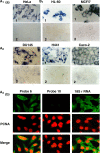
Figure 1.
Figure 1.
Expression of the hypothetical human ncmtRNA. (a) ISH of the indicated cells with probe 6 (panels 1) or the control sense probe 10 (panels 2). Note cytoplasmic and perinuclear hybridization signals (×100). (b) Melanoma cell line expressing the transcript are also expressing PCNA. The cells were subjected to ISH with probe 6 or probe 10 together with immunocytochemistry of PCNA. The merge figure confirmed the co-expression of both molecules. As a positive control, the same cells were subjected to ISH for the 18S rRNA and immunocytochemistry of PCNA (×63).
Figure 2.
Figure 2.
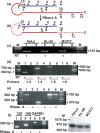
The human ncmtRNA. (a) Theoretical structure of the human ncmtRNA. The 5′ end of the sense 16S mtrRNA (red line) is linked to a fragment of the antisense 16S mtrRNA or IR (blue line) forming a double-stranded structure and a loop of unknown length. The position of the reverse primers (under the lines) and the forward primers (over the lines) are indicated (see Materials and Methods section). (b) Digestion of the loop and the single-stranded region of the ncmtRNA by RNase A is also illustrated. (c) Amplification of the cDNA obtained from three tumor cell lines using primers 1 and 2. An amplicon of ∼210 bp was obtained only when the reaction was carried out with reverse transcriptase (c, odd lanes). (d) Amplification of the cDNA of HeLa cells by PCR using primers 1 in combination with primers 3 (lanes 1 and 2), 4 (lanes 3 and 4), 5 (lanes 5 and 6) and 6 (lanes 7 and 8), respectively. Amplicons of ∼500, 700 and 800 bp were obtained. No amplification products were generated with primer 1 and 6 (lanes 7 and 8) or without reverse transcriptase (d, even lanes). M, 100 bp ladder. (e) RNA from HeLa cells in 2× SSC was incubated without (odd lanes) or with 50 μg/ml of RNase A for 15 min at 25°C (even lanes). The RNA was recovered and amplified by RT-PCR using primers 1 and 5 (lanes 1 and 2), primers 10 and 11 (lanes 3 and 4) or with primers 7 and 5 (lanes 5 and 6). Amplicons of 800 and 350 bp were obtained with primer 1 and 5, and 10 and 11, respectively, only with untreated RNA (lanes 1 and 3, respectively). Amplification of the fragment of 750 bp obtained with primers 7 and 5 was not affected by the nuclease treatment (lanes 5 and 6). f) Amplification of the 12S mtrRNA (lanes 1 and 2), 18S rRNA (lanes 3 and 4) and GAPDH mRNA (lanes 5 and 6) after digestion with RNase A (mock experiment, odd lanes). (g) About 1 μg of RNA from the indicated cell lines was digested with RNase A and the digestion products were resolved by electrophoresis on a 1.5% agarose gel. After blotting, the membrane was probed with a 32P-labeled PCR fragment targeted to the double-stranded region of the ncmtRNA (see Materials Methods section). A single hybridization band corresponding to a transcript of ∼800 nt was detected.
Figure 3.
Figure 3.
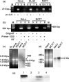
The IR is contiguous with the 16S mtrRNA. (a) Total RNA from HeLa (lanes 1 to 4), HL-60 (lanes 5 to 8) and MCF/7 (lanes 9 to 12) cells were separated into polyA+ and polyA− fractions. A total of 100 ng of each fraction was used to synthesize cDNA, which was then amplified by PCR using primers 1 and 2, to generate the 215 bp amplicon as indicated. Odd lanes correspond to reactions carried out in the absence of RT. (b) cDNA was synthesized from the polyA+ fraction of HeLa or MCF/7 cells, using oligo dT (lanes 1, 2, 5 and 6) or primer 12 (lanes 3, 4, 7 and 8). A single amplicon of 500 bp was obtained after PCR amplification of the cDNAs with primers 1 and 3 only when reverse transcriptase was included in the reaction mixture (even lanes). (c) About 3 μg of HeLa and MCF/7 cells RNA was resolved by electrophoresis on a 1% native agarose gel and subjected to northern blot. The membrane was probed with 32P-labeled primer 13 (Figure 2a). The probe hybridized with a single transcript, which migrated below the 1353 bp DNA marker (M = λDNA/HindIII and φDNA/HaeIII). The size of this transcript, deduced from the dsDNA ladder, corresponds to 2280 nt. (d) Northern blot was carried out with RNA from the indicated cells, under denaturing electrophoretic conditions. In this case, probe 13 hybridized with a single band that migrated on top of the 18S rRNA and corresponding to a transcript of 2200 nt. (e) For S1 protection assay, an asymmetric PCR fragment 215 nts containing the 31 nt of the sense 16S mtrRNA plus 184 nt of the IR was synthesized and labeled with digoxigenin (see Materials and Methods section). After denaturation at 100°C for 5 min, the probe was incubated overnight at 50°C either alone (lane 2), with 20 μg of HeLa RNA (lane 3) or with 20 μg of yeast RNA (lane 4). After hybridization, the products were digested with S1 nuclease and the products resolved by 2.5% native agarose gel electrophoresis and blotted to a nylon membrane. The products of digestions were reveled with anti-digoxigenin antibody conjugated to alkaline phosphatase (see Materials and Methods section). Lane 1 represents the probe alone without treatment.
Figure 4.
Figure 4.
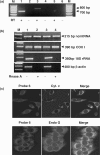
Mitochondrial localization of the ncmtRNA. (a) The mitochondrial fraction from HeLa cells was treated with RNase A, previously to RNA extraction. Amplification of the mitochondrial RNA with primer 1 in combination with primer 4 (lanes 1 and 2) or primer 5 (lanes 3 and 4) yielded the expected amplicons of 700 and 800 bp, respectively. No amplification was obtained with primers 1 and 6 (lanes 5 and 6) or when the reaction was carried out without reverse transcriptase (RT−, lanes 2, 4 and 6). (b) Total RNA extracted from two different preparations of HeLa mitochondria (lanes 1and 2, and 3 and 4) treated without (lanes 1 and 3) or with RNase A (lanes 2 and 4) was used to amplify the 215 bp fragment of the ncmtRNA, and the indicated amplicons of COX I mRNA, 18S rRNA and β-actin mRNA. Note that RNase A treatment abolished contamination with cytoplasmic transcripts. (c) Co-localization of the ncmtRNA with the mitochondrial markers cytochrome c and endonuclease G. HeLa cells were subjected to FISH to detect the ncmtRNA and immunocytochemistry to detect cytochrome c or endonuclease G (see Methods section) and analyzed by confocal microscopy.
Figure 5.
Figure 5.
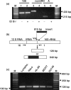
Synthesis of the ncmtRNA requires mitochondrial transcription. (a) Total RNA was extracted from HeLa cells incubated without (odd lanes) or with (even lanes) 1 μg/ml of ethidium bromide for 28 days. The RNA was amplified by RT-PCR using specific primers for 18S rRNA (lanes 1 and 2) (see Methods section), primers 9 and 11 (lanes 3 and 4) and primers 1 and 2 for the ncmtRNA (lanes 5 and 6). (b) Theoretical structure of an anomalous mtDNA containing an insert of 815 bp between the tRNAval and the 16S mtrRNA genes. (c) mtDNA from the indicated cell lines was amplified between primer 16 positioned close to the 3′ end of the 12S gene and primer 1 positioned on the 16S mtrRNA gene as shown in (b). All samples yielded a single amplicon of 128 nt. (a) M, 100 bp ladder. (c) 50 bp ladder.
Figure 6.
Figure 6.
Expression of the ncmtRNA in proliferating cells. (a) The indicated cells were subjected to ISH with probe 13 (panels 1) or with corresponding control sense probe (panels 2). Strong cytoplasmic and perinuclear hybridization signals were found (×100). No hybridization was found with the control sense probe. (b) ISH and immunocytochemistry to determine the expression of the ncmtRNA and PCNA, respectively, in brain, smooth muscle and liver cells. Each panel of tissues was also stained with hematoxylin-eosin (×40).
Figure 7.
Figure 7.
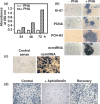
Reversibility of the ncmtRNA expression. (a) Human lymphocytes were incubated without (white bars) or with PHA (hatched bars) for 24, 48 and 72 h. At each time period, the cells were incubated with BrdU for 16 h and the incorporation of the nucleoside was determined by ELISA (see Materials and Methods section). The incorporation of BrdU is expressed as the absorbance at 450 nm. (b) About 100 000 lymphocytes incubated without (−PHA) or with PHA (+PHA) for 72 h were subjected to immunocytochemistry to determine the expression of Ki-67, PCNA and phospho-histone H3 (PO4-H3), and to ISH with probe 13 to determine the expression of the ncmtRNA (×40). (c) PHA-stimulated lymphocytes for 72 h were hybridized with probe 13 (ncmtRNA) or with the corresponding sense control probe (Control sense) (×20). (d) DU145 cells were incubated twice with aphidicolin (see Methods section) and subjected to ISH together with a control culture. The blocked cells were changed to normal medium for 48 h and then subjected to ISH (Recovery) (×40).

Similar articles

Cited by

References

    1. Nass MM, Nass S, Afzelius BA. The general occurrence of mitochondrial DNA. Exp. Cell Res. 1965;37:516–539. - PubMed
    1. Anderson S, Bankier AT, Barrell BG, de Brujin MH, Coulson AR, Drouin J, Eperon IC, Nierlich DP, Roe BA, et al. Sequence and organization of the human mitochondrial genome. Nature. 1981;290:457–465. - PubMed
    1. Ojala D, Montoya J, Attardi G. tRNA punctuation model of RNA processing in human mitochondria. Nature. 1981;290:470–474. - PubMed
    1. Chomyn A, Attardi G. Recent advances on mitochondrial biogenesis. In: Ernster L, editor. Molecular Mechanisms in Biogenesis. Amsterdam: Elsevier Science Publishers B. V.; 1992. pp. 483–509.
    1. Shadel GS, Clayton DA. Mitochondrial DNA maintenance in vertebrates. Ann. Rev. Biochem. 1997;66:409–435. - PubMed

Publication types