

Insight into the neuroendocrine site and cellular mechanism by which cortisol suppresses pituitary responsiveness to gonadotropin-releasing hormone
- PMID: 17962347
- PMCID: PMC2219297
- DOI: 10.1210/en.2007-0773
Insight into the neuroendocrine site and cellular mechanism by which cortisol suppresses pituitary responsiveness to gonadotropin-releasing hormone
Abstract
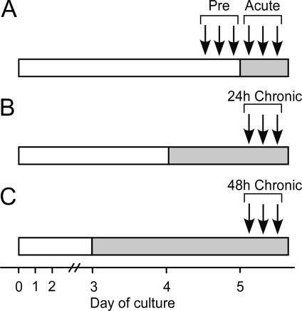
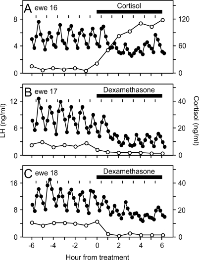
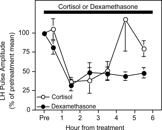
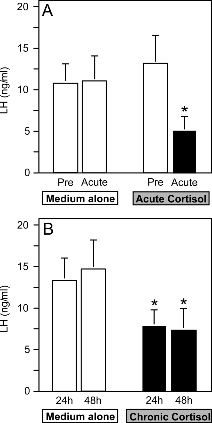
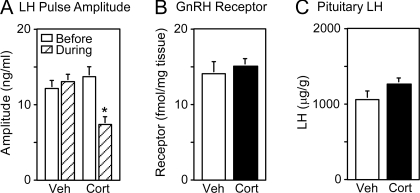
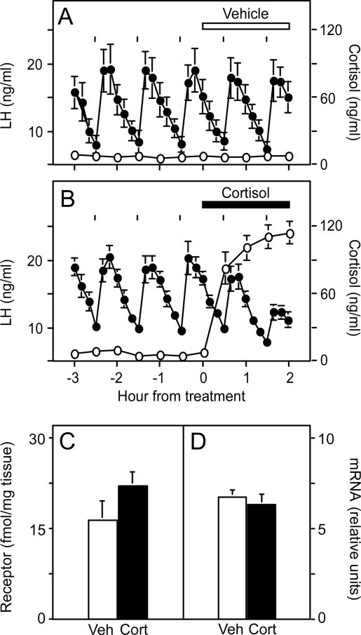
Stress-like elevations in plasma glucocorticoids rapidly inhibit pulsatile LH secretion in ovariectomized sheep by reducing pituitary responsiveness to GnRH. This effect can be blocked by a nonspecific antagonist of the type II glucocorticoid receptor (GR) RU486. A series of experiments was conducted to strengthen the evidence for a mediatory role of the type II GR and to investigate the neuroendocrine site and cellular mechanism underlying this inhibitory effect of cortisol. First, we demonstrated that a specific agonist of the type II GR, dexamethasone, mimics the suppressive action of cortisol on pituitary responsiveness to GnRH pulses in ovariectomized ewes. This effect, which became evident within 30 min, documents mediation via the type II GR. We next determined that exposure of cultured ovine pituitary cells to cortisol reduced the LH response to pulse-like delivery of GnRH by 50% within 30 min, indicating a pituitary site of action. Finally, we tested the hypothesis that suppression of pituitary responsiveness to GnRH in ovariectomized ewes is due to reduced tissue concentrations of GnRH receptor. Although cortisol blunted the amplitude of GnRH-induced LH pulses within 1-2 h, the amount of GnRH receptor mRNA or protein was not affected over this time frame. Collectively, these observations provide evidence that cortisol acts via the type II GR within the pituitary gland to elicit a rapid decrease in responsiveness to GnRH, independent of changes in expression of the GnRH receptor.
Figures
Similar articles
-
Does the type II glucocorticoid receptor mediate cortisol-induced suppression in pituitary responsiveness to gonadotropin-releasing hormone?Endocrinology. 2004 Jun;145(6):2739-46. doi: 10.1210/en.2004-0123. Epub 2004 Mar 19. Endocrinology. 2004. PMID: 15033919
-
Psychosocial stress inhibits amplitude of gonadotropin-releasing hormone pulses independent of cortisol action on the type II glucocorticoid receptor.Endocrinology. 2009 Feb;150(2):762-9. doi: 10.1210/en.2008-0757. Epub 2008 Oct 1. Endocrinology. 2009. PMID: 18832098 Free PMC article.
-
Does cortisol inhibit pulsatile luteinizing hormone secretion at the hypothalamic or pituitary level?Endocrinology. 2004 Feb;145(2):692-8. doi: 10.1210/en.2003-1114. Epub 2003 Oct 23. Endocrinology. 2004. PMID: 14576178
-
New insights regarding glucocorticoids, stress and gonadotropin suppression.Front Neuroendocrinol. 2006 Jul;27(2):233-45. doi: 10.1016/j.yfrne.2006.03.335. Epub 2006 May 19. Front Neuroendocrinol. 2006. PMID: 16712908 Review.
-
Multifarious effects of estrogen on the pituitary gonadotrope with special emphasis on studies in the ovine species.Arch Physiol Biochem. 2002 Apr;110(1-2):62-73. doi: 10.1076/apab.110.1.62.898. Arch Physiol Biochem. 2002. PMID: 11935402 Review.
Cited by
-
Social subordination and polymorphisms in the gene encoding the serotonin transporter enhance estradiol inhibition of luteinizing hormone secretion in female rhesus monkeys.Biol Reprod. 2009 Dec;81(6):1154-63. doi: 10.1095/biolreprod.109.079038. Epub 2009 Jul 15. Biol Reprod. 2009. PMID: 19605783 Free PMC article.
-
Neurochemically distinct circuitry regulates locus coeruleus activity during female social stress depending on coping style.Brain Struct Funct. 2019 May;224(4):1429-1446. doi: 10.1007/s00429-019-01837-5. Epub 2019 Feb 14. Brain Struct Funct. 2019. PMID: 30767070 Free PMC article.
-
The Roles of Androgens in Humans: Biology, Metabolic Regulation and Health.Int J Mol Sci. 2022 Oct 8;23(19):11952. doi: 10.3390/ijms231911952. Int J Mol Sci. 2022. PMID: 36233256 Free PMC article. Review.
-
Neural and endocrine mechanisms underlying stress-induced suppression of pulsatile LH secretion.Mol Cell Endocrinol. 2019 Dec 1;498:110579. doi: 10.1016/j.mce.2019.110579. Epub 2019 Sep 12. Mol Cell Endocrinol. 2019. PMID: 31521706 Free PMC article. Review.
-
Role of estradiol in cortisol-induced reduction of luteinizing hormone pulse frequency.Endocrinology. 2009 Jun;150(6):2775-82. doi: 10.1210/en.2008-1754. Epub 2009 Jan 29. Endocrinology. 2009. PMID: 19179435 Free PMC article.
References
-
- Ferin M 1999 Stress and the reproductive cycle. J Clin Endocrinol Metab 84:1768–1774 - PubMed
-
- Dobson H, Smith RF 2000 What is stress, and how does it affect reproduction? Anim Reprod Sci 60–61:743–752 - PubMed
-
- Tilbrook AJ, Turner AI, Clarke IJ 2002 Stress and reproduction: central mechanisms and sex differences in non-rodent species. Stress 5:83–100 - PubMed
-
- Rivier C, Rivest S 1991 Effect of stress on the activity of the hypothalamic-pituitary-gonadal axis: peripheral and central mechanisms. Biol Reprod 45:523–532 - PubMed
-
- Tilbrook AJ, Turner AI, Clarke IJ 2000 Effects of stress on reproduction in non-rodent mammals: the role of glucocorticoids and sex differences. Rev Reprod 5:105–113 - PubMed
