

Food restriction alters neuronal morphology in the hypothalamic ventromedial nucleus of male rats
- PMID: 17962353
- PMCID: PMC2194610
- DOI: 10.1210/en.2007-0008
Food restriction alters neuronal morphology in the hypothalamic ventromedial nucleus of male rats
Abstract
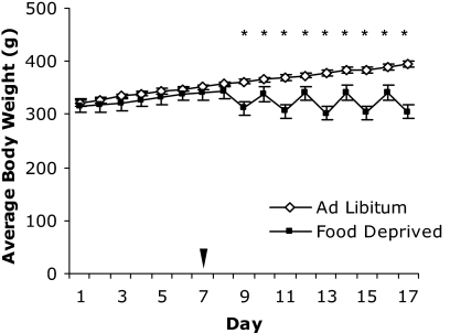
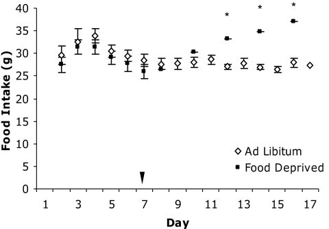
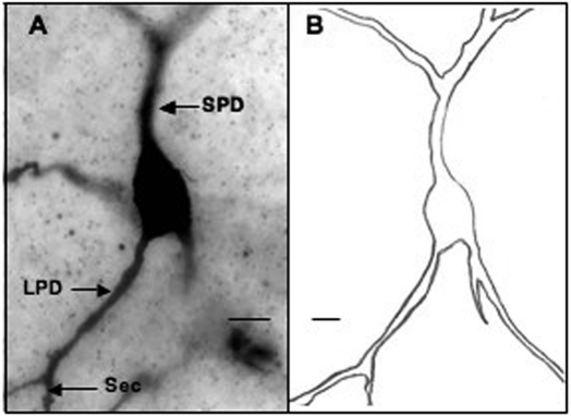
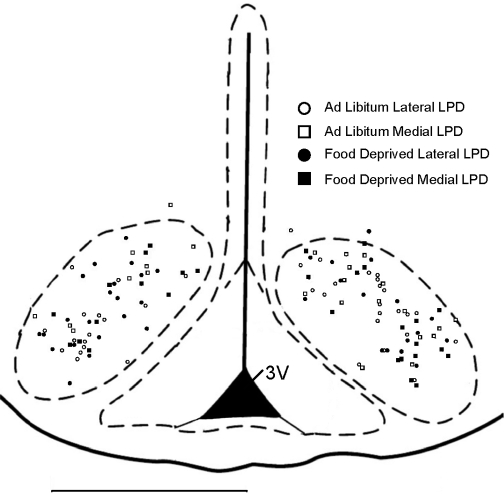
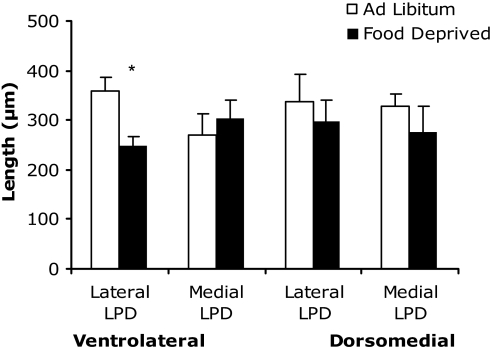
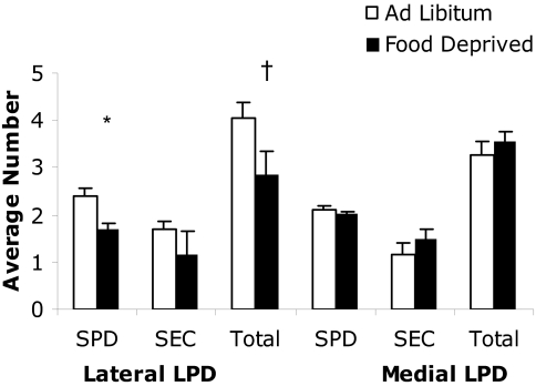
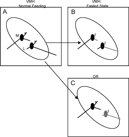
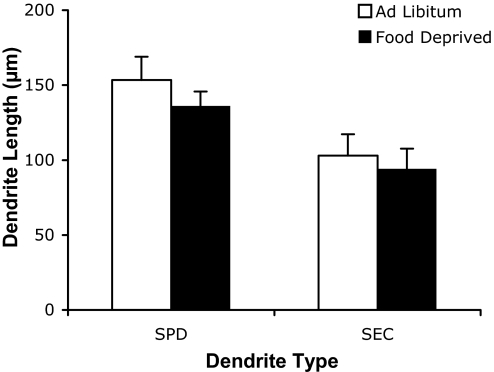
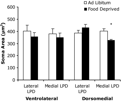
Several lines of evidence have implicated the hypothalamic ventromedial nucleus (VMH) in the control of caloric homeostasis. For example, the activity of VMH neurons depends on energy availability. We tested the hypothesis that energy balance may involve the remodeling of the dendritic arbor of VMH neurons. We compared two groups of animals: one group had ad libitum access to food, and the other experienced 10-d restricted access to food. As expected, the food-deprived group lost body weight and had reduced levels of glucose, insulin, and leptin. VMH neurons were visualized after Golgi impregnation, and dendrite length was measured. Food deprivation had differential effects on VMH neurons. In particular, within the ventrolateral VMH, for neurons with long primary dendrites (LPDs) that extended in the lateral, but not medial, direction, the LPDs were 31% shorter. These same neurons exhibited a 32% reduction in the number of other dendrites without a change in soma size. In contrast, within the dorsomedial VMH, for neurons with medially, but not laterally, extended LPDs, the soma area was reduced by 28%. However, neurons in the dorsomedial VMH did not display a change in the length or number of dendrites, regardless of LPD direction. Thus, although structural changes during calorie depletion occur in both the dorsomedial and ventrolateral VMH, only the latter exhibits a remodeled dendritic arbor. These results also suggest that the direction of the LPD may be an important marker of neuronal function in the VMH.
Figures
Similar articles
-
Sex differences in the dendritic arbor of hypothalamic ventromedial nucleus neurons.Physiol Behav. 2009 May 25;97(2):151-6. doi: 10.1016/j.physbeh.2009.02.019. Epub 2009 Feb 28. Physiol Behav. 2009. PMID: 19254731 Free PMC article.
-
Genetic and dietary effects on dendrites in the rat hypothalamic ventromedial nucleus.Physiol Behav. 2009 Oct 19;98(4):511-6. doi: 10.1016/j.physbeh.2009.08.005. Epub 2009 Aug 19. Physiol Behav. 2009. PMID: 19698729 Free PMC article.
-
Dendritic arbor of neurons in the hypothalamic ventromedial nucleus in female prairie voles (Microtus ochrogaster).Horm Behav. 2013 Jan;63(1):173-9. doi: 10.1016/j.yhbeh.2012.10.001. Epub 2012 Oct 8. Horm Behav. 2013. PMID: 23058474
-
The rise, fall, and resurrection of the ventromedial hypothalamus in the regulation of feeding behavior and body weight.Physiol Behav. 2006 Feb 28;87(2):221-44. doi: 10.1016/j.physbeh.2005.10.007. Epub 2006 Jan 18. Physiol Behav. 2006. PMID: 16412483 Review.
-
Ovarian hormone action in the hypothalamic ventromedial nucleus: remodelling to regulate reproduction.J Neuroendocrinol. 2011 Jun;23(6):465-71. doi: 10.1111/j.1365-2826.2011.02143.x. J Neuroendocrinol. 2011. PMID: 21518031 Free PMC article. Review.
Cited by
-
Estradiol and progesterone differentially regulate the dendritic arbor of neurons in the hypothalamic ventromedial nucleus of the female rat (Rattus norvegicus).J Comp Neurol. 2008 Oct 20;510(6):631-40. doi: 10.1002/cne.21816. J Comp Neurol. 2008. PMID: 18698598 Free PMC article.
-
Role of synaptic plasticity and EphA5-ephrinA5 interaction within the ventromedial hypothalamus in response to recurrent hypoglycemia.Diabetes. 2014 Mar;63(3):1140-7. doi: 10.2337/db13-1259. Epub 2013 Nov 12. Diabetes. 2014. PMID: 24222347 Free PMC article.
-
Ovarian hormone-induced reorganization of oxytocin-labeled dendrites and synapses lateral to the hypothalamic ventromedial nucleus in female rats.J Comp Neurol. 2010 Nov 15;518(22):4531-45. doi: 10.1002/cne.22470. J Comp Neurol. 2010. PMID: 20886620 Free PMC article.
-
Metabolic context regulates distinct hypothalamic transcriptional responses to antiaging interventions.Int J Endocrinol. 2012;2012:732975. doi: 10.1155/2012/732975. Epub 2012 Aug 27. Int J Endocrinol. 2012. PMID: 22934110 Free PMC article.
-
Controlling feeding behavior by chemical or gene-directed targeting in the brain: what's so spatial about our methods?Front Neurosci. 2013 Dec 18;7:182. doi: 10.3389/fnins.2013.00182. Front Neurosci. 2013. PMID: 24385950 Free PMC article. Review.
References
-
- Powley TL 1977 The ventromedial hypothalamic syndrome, satiety, and a cephalic phase hypothesis. Psychol Rev 84:89–126 - PubMed
-
- Reeves AG, Plum F 1969 Hyperphagia, rage, and dementia accompanying a ventromedial hypothalamic neoplasm. Arch Neurol 20:616–624 - PubMed
-
- Muller D, Roeder F, Orthner H 1973 Further results of stereotaxis in the human hypothalamus in sexual deviations. First use of this operation in addiction to drugs. Neurochirurgia (Stuttg) 16:113–126 - PubMed
-
- Miki T, Liss B, Minami K, Shiuchi T, Saraya A, Kashima Y, Horiuchi M, Ashcroft F, Minokoshi Y, Roeper J, Seino S 2001 ATP-sensitive K+ channels in the hypothalamus are essential for the maintenance of glucose homeostasis. Nat Neurosci 4:507–512 - PubMed
Publication types
MeSH terms
Substances
Grants and funding
LinkOut - more resources
Full Text Sources
Research Materials
