. 2007 Oct 30;104(44):17424-9.
doi: 10.1073/pnas.0708561104. Epub 2007 Oct 25.

Human G(salpha) mutant causes pseudohypoparathyroidism type Ia/neonatal diarrhea, a potential cell-specific role of the palmitoylation cycle

Affiliations

Human G(salpha) mutant causes pseudohypoparathyroidism type Ia/neonatal diarrhea, a potential cell-specific role of the palmitoylation cycle

Noriko Makita et al. Proc Natl Acad Sci U S A. .

Abstract

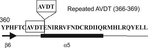
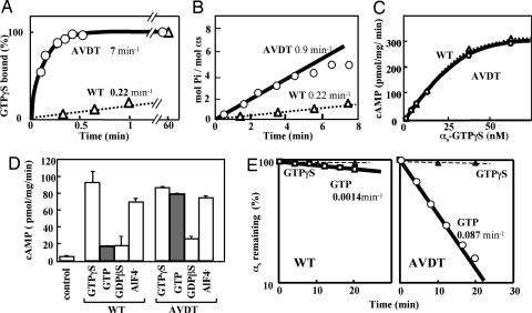
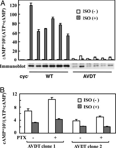
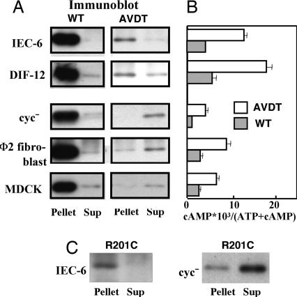
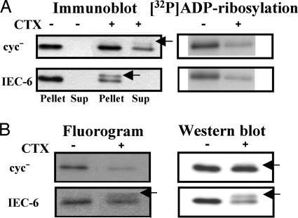
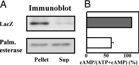
Pseudohypoparathyroidism type Ia (PHP-Ia) results from the loss of one allele of G(salpha), causing resistance to parathyroid hormone and other hormones that transduce signals via G(s). Most G(salpha)mutations cause the complete loss of protein expression, but some cause loss of function only, and these have provided valuable insights into the normal function of G proteins. Here we have analyzed a mutant G(salpha) (alphas-AVDT) harboring AVDT amino acid repeats within its GDP/GTP binding site, which was identified in unique patients with PHP-Ia accompanied by neonatal diarrhea. Biochemical and intact cell analyses showed that alphas-AVDT is unstable but constitutively active as a result of rapid GDP release and reduced GTP hydrolysis. This instability underlies the PHP-Ia phenotype. alphas-AVDT is predominantly localized in the cytosol, but in rat and mouse small intestine epithelial cells (IEC-6 and DIF-12 cells) alphas-AVDT was found to be localized predominantly in the membrane where adenylyl cyclase is present and constitutive increases in cAMP accumulation occur in parallel. The likely cause of this membrane localization is the inhibition of an activation-dependent decrease in alphas palmitoylation. Upon the overexpression of acyl-protein thioesterase 1, however, alphas-AVDT translocates from the membrane to the cytosol, and the constitutive accumulation of cAMP becomes attenuated. These results suggest that PHP-Ia results from the instability of alphas-AVDT and that the accompanying neonatal diarrhea may result from its enhanced constitutive activity in the intestine. Hence, palmitoylation may control the activity and localization of G(salpha) in a cell-specific manner.


Conflict of interest statement

The authors declare no conflict of interest.

Figures

Fig. 1.
Fig. 1.
Carboxyl-terminal sequence of αs-AVDT. The AVDT (366–369) amino acid moiety that is present as a repeat sequence in the αs-AVDT mutant is boxed.
Fig. 2.
Fig. 2.
Biochemical properties of recombinant WT (αs-WT) and mutant (αs-AVDT) αs proteins. (A) Rates of GTP[γS] binding. Recombinant αs-WT (triangles) and αs-AVDT (circles) proteins (each at 40 nM) were incubated at 22°C with 1 μM [35S]GTP[γS] (2.2 × 10⁵ cpm/pmol) in buffer A [20 mM Na-Hepes, pH 7.4/10 mM MgSO4/0.1 mM EDTA/3 mM 2-mercaptoethanol/0.025% polyoxyethylene (10) lauryl ether (C12E10)]. At the times indicated, the reaction was terminated, and GTP[γS] binding was quantitated by filtration on nitrocellulose membranes. The apparent on rates of GTP[γS] binding (kapp) were then calculated by fitting the data to the equation B = Beq (1 − e−kt) as described in Materials and Methods, where B is the concentration of bound GTP[γS], Beq is the equilibreated concentration of binding sites, k is the constant describing the rate of approach of the reaction to Beq, and t is time. (B) Steady-state GTPase. Forty nanomolar of recombinant αs-WT (triangles) or αs-AVDT (circles) was incubated for the indicated times at 22°C with 2 μM [γ-32P]GTP (2 × 10⁵ cpm/pmol−1) in buffer A. At the times indicated, the reactions were terminated, and phosphate release was quantified by charcoal adsorption. The steady-state GTPase rates (kss) were then calculated. Pi, inorganic phosphate. (C) cAMP synthesis stimulated by different concentrations of αs-WT or αs-AVDT in the presence of GTP[γS]. Reactions were performed at 20°C for 10 min in 75-μl volumes containing 22.5 μg of cyc cell membranes, as described previously (12, 47) apart from minor changes to the buffer conditions. These included 50 mM Na-Hepes (pH 7.4), 6 mM MgCl2, 0.01% C12E10, and 50 μg ml−1 BSA. Before the assay, the αs proteins were incubated with 100 μM GTP[γS] for 30 min (αs-WT) or 1 min (αs-AVDT). (D) The effects of the indicated different guanine nucleotides on cAMP synthesis in the presence of 15 nM αs-WT or αs-AVDT. Reactions were conducted at 20°C for 10 min as described in C. Before the assay commenced, the αs proteins were incubated with GTP[γS], GTP, or guanosine 5′-[β-thio]diphosphate (GDP[βS]), each at 100 μM, or with 20 μM AlCl3 and 10 mM NaF for 30 min (αs-WT) or 1 min (αs-AVDT). (E) The protein stability of αs-WT or αs-AVDT assessed in vitro by [35S]GTP[γS] binding. Recombinant αs-WT or αs-AVDT (each at 30 nM) was incubated at 22°C with the indicated concentrations of GTP or GTP[γS]. At the times indicated, 5-μl aliquots were withdrawn and assayed for GTP[γS] binding in 20-μl reaction mixtures. The values represent the means ± SD of triplicate determinations, and each set of results is representative of at least two additional experiments.
Fig. 3.
Fig. 3.
cAMP accumulation and the expression of recombinant αs proteins in S49 cyc stable clones expressing HA-tagged αs-WT or HA-tagged αs-AVDT. (A) For each transfected cyc cell clone, the open bars and filled bars indicate the cAMP accumulation levels after 30 min in cells without or with 10 μM isoproterenol, respectively (see Materials and Methods). (Lower) For immunoblotting analysis, proteins from these clones were detected by using the monoclonal antibody 12CA5 directed against the HA tag, after immunoprecipitation with the same antibody. (B) Two stable cyc clones expressing αs-AVDT were incubated without or with 200 ng/ml pertussis toxin (PTX) for 12 h. cAMP accumulation without or with 10 μM isoproterenol was then assayed as described in the Materials and Methods. The values shown in these analyses represent the means ± SD of triplicate determinations. Each set of results is representative of at least two additional experiments.
Fig. 4.
Fig. 4.
Localization of recombinant αs proteins and measurement of cAMP accumulation. (A) The localization of recombinant αs proteins in cells stably expressing αs-WT or αs-AVDT. IEC-6 (2.0 × 10⁷), DIF-12 (2.0 × 10⁷), cyc (4.0 × 10⁷), Φ2 (2.0 × 10⁷), or MDCK cells (2.0 × 10⁷) stably expressing αs-WT or αs-AVDT, derived from 40-ml culture volumes, were fractionated and HA-tagged αs proteins were then detected by immunoblotting with the monoclonal antibody 12CA5 after immunoprecipitation with the same antibody, as described in Materials and Methods. (B) cAMP accumulation in cells stably expressing αs-WT or αs-AVDT. cAMP accumulation of the stable cells expressing αs-WT or αs-AVDT described in A was assayed as described in Materials and Methods. Briefly, cells were seeded in 24-well plates at 1.5 × 10⁵ (cyc cells) or at 0.75 × 10⁵ (other cells) cells per well and labeled with [3H]adenine (4 μCi/ml, Amersham Pharmacia) for an additional 16 h. Cells were then washed once with DMEM and incubated in the presence of 3-isobutyl-1-methylxanthine (IBMX) for 30 min. cAMP and ATP fractions were resolved, and cAMP accumulation was estimated by the radioactivity of cAMP and ATP. Values represent means ± SD of triplicate determinations. (C) Localization of recombinant αs proteins in IEC-6 cells and cyc cells stably expressing αs-R201C. The cells were fractionated, and HA-tagged αs proteins were detected by immunoblotting with 12CA5 after immunoprecipitation with that same antibody. Each set of results is representative of at least two additional experiments. Sup, supernatant.
Fig. 5.
Fig. 5.
Effects of activation by cholera toxin (CTX) on the localization (A) and palmitoylation (B) of recombinant αs proteins in cyc or IEC-6 cells expressing HA-tagged αs-WT. (A) cyc (1.0 × 10⁷) or IEC-6 (0.5 × 10⁷) cells derived from 10-ml culture volumes were fractionated, and HA-tagged αs proteins were detected by immunoblotting with monoclonal antibody 12CA5 after immunoprecipitation with the same antibody. Membrane fractions of these cells were also prepared in which the αs was [32P]ADP-ribosylated with a 50 μg/ml concentration of activated cholera toxin. [32P]ADP-ribosylated HA-tagged αs proteins were then visualized by autoradiography after immunoprecipitation as described in Materials and Methods. Before fractionation, the cells were incubated without or with 1 μg/ml cholera toxin for 4 h. (B) cyc (5.0 × 10⁷) or IEC-6 (2.5 × 10⁷) cells derived from 50-ml culture volumes were incubated for 2 h in DMEM containing 10% dialyzed FBS, 5 mM sodium pyruvate, and 0.5 mCi/ml [9,10-3H]palmitic acid. After labeling, the cells were incubated without or with cholera toxin for 4 h and fractionated. Palmitoylated HA-tagged αs proteins were visualized by fluorography (30-day exposure), and HA-tagged αs proteins were detected by immunoblotting after immunoprecipitation as described in Materials and Methods. Arrows indicate αs proteins that have been ADP-ribosylated by cholera toxin. Each set of results is representative of at least two additional experiments.
Fig. 6.
Fig. 6.
Effects of APT1 or LacZ adenovirus infection on the localization of HA-tagged αs-AVDT (A) and on the accumulation of cAMP (B) in IEC-6 cells expressing HA-tagged αs-AVDT. (A) IEC-6 cells stably expressing αs-AVDT derived from a 40-ml culture volume were infected by recombinant APT1 adenovirus or LacZ adenovirus (at a multiplicity of infection of 50). After 48 h, the cells were fractionated, and HA-tagged αs-AVDT was detected by immunoblotting with the 12CA5 antibody. Each set of results is representative of at least two additional experiments. Palm. esterase, palmitoyl esterase; Sup, supernatant. (B) cAMP accumulation in stable cells in A was assayed as described in Materials and Methods. Values represent the means ± SE of three independent experiments with triplicate determinations.

Similar articles

Cited by

References

    1. Farfel Z, Bourne HR, Iiri T. N Engl J Med. 1999;340:1012–1020. - PubMed
    1. Spiegel AM, Weinstein LS. Annu Rev Med. 2004;55:27–39. - PubMed
    1. Aldred MA. J Pediatr Endocrinol Metab. 2006;19:S635–S640. - PubMed
    1. Lania AG, Mantovani G, Spada A. Nat Clin Pract Endocrinol Metab. 2006;2:681–693. - PubMed
    1. Iiri T, Farfel Z, Bourne HR. Nature. 1998;394:35–38. - PubMed

Publication types

MeSH terms

Substances