. 2007 Nov 6;104(45):17813-8.
doi: 10.1073/pnas.0704975104. Epub 2007 Oct 25.

A common mechanism of PLP/DM20 misfolding causes cysteine-mediated endoplasmic reticulum retention in oligodendrocytes and Pelizaeus-Merzbacher disease

Affiliations

A common mechanism of PLP/DM20 misfolding causes cysteine-mediated endoplasmic reticulum retention in oligodendrocytes and Pelizaeus-Merzbacher disease

Ajit-Singh Dhaunchak et al. Proc Natl Acad Sci U S A. .

Abstract

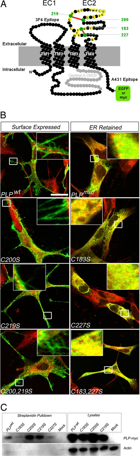
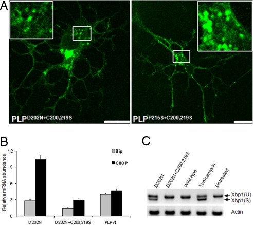
A large number of mutations in the human PLP1 gene lead to abnormal myelination and oligodendrocyte death in Pelizaeus-Merzbacher disease (PMD). Here we show that a major subgroup of PMD mutations that map into the extracellular loop region of PLP/DM20 leads to the failure of oligodendrocytes to form the correct intramolecular disulfide bridges. This leads to abnormal protein cross-links and endoplasmic reticulum retention and activates the unfolded protein response. Importantly, surface expression of mutant PLP/DM20 can be restored and the unfolded protein response can be reverted by the removal of two cysteines. Thus, covalent protein cross-links emerge as a cause, rather than as a consequence, of endoplasmic reticulum retention.


Conflict of interest statement

The authors declare no conflict of interest.

Figures

Fig. 1.
Fig. 1.
Topology of proteolipid protein PLP/DM20 and the role of cysteine residues in subcellular trafficking. (A) Two-dimensional model of PLP (276 residues shown as black beads) and its splice isoform, DM20, lacking 35 residues (marked in gray) from an intracellular loop. The orientation of four transmembrane domains (TM1–TM4) positions both N and C termini in the cytoplasm. Within the second extracellular domain (EC2), the position of four cysteine residues (marked in green) forming two disulfide bridges (marked in red) is indicated. Also indicated are C-terminal epitope tags used in this study (EGFP or myc) and the approximate positions of extracellular (3F4) and intracellular (A431) antibody binding sites common to PLP and DM20. Positions of amino acids in EC2 that are substituted in patients with PMD are marked in yellow. Those that have been studied in detail here carry the single-letter code of the wild-type sequence, labeled in red. (B) In transfected, fixed, and permeabilized oli-neu cells, mutant PLP–myc (shown in green) colocalized with the endogenous chaperone and thiol-disulfide oxidoreductase PDI (shown in red). Only merged images are shown. (Left) Wild-type protein, PLPWT, reached the cell surface, as demonstrated by green fluorescent microspikes at the tip of processes (magnified in Insets). (Right) In contrast, PLPMSD (derived from jimpy-msd mice) failed to reach the cell surface. There are no labeled microspikes (magnified in Insets), but there is substantial overlap between PLPMSD and PDI (shown in yellow). Note also the paucity of cellular processes. From two disulfide bridges in EC2, the “outer” one (Cys200–Cys219) is dispensable for folding and cell-surface expression. To test the function of each disulfide bridge (see A) for PLP folding and colocalization with PDI (shown in red), single and double cysteine-to-serine substitutions were engineered for each disulfide bridge. Replacing one or both cysteines of the outer bridge did not interfere with cell-surface labeling of PLP (shown in green), as indicated by fluorescent microspikes (Left and magnified in Insets). In contrast, replacing one or both cysteines of the membrane-proximal bridge (Cys183–Cys227) led to severe misfolding, as visualized by ER retention and colocalization of PLP with PDI (shown as yellow overlay in Insets), similar to PLPMSD (topmost Right). Note also the paucity of cellular processes. (Scale bar: 20 μm.) (C) To obtain independent biochemical evidence that PLPC200S and PLPC219S (lacking the outer bridge) are cell-surface expressed in COS7 cells, all membrane proteins were biotinylated with membrane-impermeable sulfo-NHS biotin before cell lysis, and proteins were pulled down with streptavidin-conjugated agarose beads. Only myc-epitope labeled PLPWT, PLPC200S, and PLPC219S could be detected on Western blots (the first, third, and fourth lanes). (Left) The absence of actin in these six lanes confirms that only live cells were biotinylated. Total lysates served as positive controls for transfection and loading. Mock, transfected with plasmids lacking a cDNA insert.
Fig. 2.
Fig. 2.
Critical cysteine residue in PLP/DM20 for ER retention and protein dimerization. (A–D) oli-neu cells were transfected to express a myc-epitope-labeled natural PMD mutant, PLPC219Y (A and B), or a double mutant, PLPC200S,C219Y (C and D). Only merged images are shown. The PMD mutant was retained as visualized 24 h after transfection by lack of processes and colocalization of PLP (shown in green) with the ER marker PDI (shown in red in A). Mutant PLP (shown in green) also showed segregation from the late endosomal/lysosomal marker LAMP1 (shown in red in B and magnified in Inset). In contrast, the double mutant PLP behaved like a wild-type protein (see Fig. 1B and SI Fig. 5A), with reduced colocalization with PDI and the branched morphology of oli-neu cells (shown in C) and the emergence of green fluorescent microspikes on processes (shown in D and magnified in Inset). PLPC200S,C219Y also overlapped with the endosomal marker (shown as the yellow area in D). Thus, it is not Y219 but an unpaired C200 that emerges as the cause of PMD. (Scale bar: 10 μm.) (E) When oli-neu cell extracts were analyzed by semiquantitative Western blotting, mutant PLPC219Y revealed predominantly a dimer band, whereas PLPWT and the “rescued” PLPC200S,C219Y were monomeric. In the presence of ME, all forms were monomeric, demonstrating that dimerization of PLPC219Y can be attributed to cysteine oxidation. (F) SDS/PAGE of cellular extracts from primary oligodendrocytes (OL) and oli-neu cells with immunodetection of PLP/DM20. When analyzed under nonreducing conditions, endogenous PLP/DM20 expression in cultured oligodendrocytes (lane 1) led to a small percentage of dimerized PLP and DM20, detected by antibody 3F4. Absence of dimers in the presence of ME suggests that these are cysteine-mediated cross-links. Transfected oli-neu cells, expressing the slightly larger epitope-tagged PLP–myc (lane 2), also exhibited a small percentage of ME-sensitive PLP dimers, detected with antibody 3F4. Note that antibody 3F4 detects endogenous and induced PLP/DM20 expression. (G) Detection of mutant PLP as dimers in oli-neu glial cells, transfected to express the natural PMD mutant PLPD202N. By performing Western blot analysis of nonreducing gels (using antibody 3F4), >50% of total PLP was detectable in the 52-kDa putative dimeric form (lane 1). When PLP was further modified by the substitution of C200 and C219 for serine (see also Fig. 3), such dimer formation was largely prevented (lane 2) with most PLP being monomeric, similar to wild-type PLP (compare with F). All dimers were completely absent in the presence of 150 mM ME in the gels (lanes 4 and 5), demonstrating the involvement of cysteine cross-links. The same results were found for PLPR204G (data not shown). Note that endogenous DM20 (in mock-transfected cells) migrated exclusively as a monomer, even under nonreducing conditions (lanes 3 and 6).
Fig. 3.
Fig. 3.
PMD-causing PLP mutations can be rescued by the replacement of cysteines. (A) To distinguish PLP at the cell surface from PLP in intracellular compartments, oli-neu cells that express various EGFP-tagged mutant PLP isoforms (shown in green) were additionally live-stained for PLP by using monoclonal antibody 3F4 (shown in red; see Fig. 1A). Only merged images are shown. The same results were obtained with COS7 cells (data not shown). We analyzed various natural PMD-causing mutations that map into EC2 (PLPD202N, PLPR204G, PLPV208D, PLPL209H, and PLPP215S) for cell-surface expression of PLP in the absence (Left) or the presence (Right) of additional point mutations that substituted C200 and C219 for serine. Remarkably, in the absence of C200 and C219, the PMD-causing mutants were fully rescued from ER retention, and PLP was localized at the cell surface (labeled in red) and in the late endosomal/lysosomal compartment (shown in green). In the presence of EC2 cysteines, all PMD mutants were negative for 3F4 immunoreactivity and confined to the ER. Note also the lack of glial processes. This reveals that the ER retention of many natural PMD-causing PLP mutants, which do not alter cysteine residues, instead require cysteines for ER retention. Grayscale images, shown on the right, better reveal the distribution of double-labeled PLP at the cell surface (3F4) and in intracellular compartments (GFP). (Scale bar: 10 μm.) (B) PLPD202N trafficking to the cell surface in the presence or absence of C200 and C219. The percentage of cells live-stained by antibody 3F4 for surface-expressed PLP (in a population of cells expressing EGFP-tagged mutant PLP) were calculated from transfection experiments (n = 3). The PMD mutation PLPD202N was fully retained in the ER. Note that, in the absence of C200 and C219, PLPD202N was rescued from ER retention, because 95% of GFP-positive cells were stained by antibody 3F4.
Fig. 4.
Fig. 4.
Rescue of PLP trafficking in primary oligodendrocytes and the attenuation of the UPR. (A) Confocal analysis of primary oligodendrocytes expressing either EGFP-tagged PLPD202N+C200,219S (Left) or EGFP-tagged PLPP215S+C200,219S (Right). Note that both proteins traffic to the cell surface and accumulate in an endosomal compartment (shown at higher magnification in Insets), similar to EGFP-tagged wild-type PLP (data not shown) and similar to their behavior in oli-neu cells (see Fig. 3). (B) Relative mRNA levels of the UPR markers BIP and CHOP in PLP-expressing oli-neu cells. For quantitative real-time PCR, cDNA was obtained 24 h after transfection. Expression of PLPD202N (an ER-retained mutant) resulted in the up-regulation of BIP and CHOP mRNA when compared with cells expressing wild-type PLP. Note that triple mutant PLP is comparable with wild-type PLP. BIP and CHOP mRNA levels were normalized to PLP expression. (C) Xbp1 mRNA was spliced in response to ER stress. oli-neu cells expressing ER-retained PLPD202N induced splicing of a Xbp1 mRNA, whereas cells expressing triple mutant PLP only exhibited unspliced Xbp1. Xbp1(U) and Xbp1(S) denote unspliced and spliced mRNA, respectively. Treatment of cells with tunicamycin served as a positive control to trigger the UPR.

References

    1. Hurtley SM, Helenius A. Annu Rev Cell Biol. 1989;5:277–307. - PubMed
    1. Ellgaard L, Helenius A. Nat Rev Mol Cell Biol. 2003;4:181–191. - PubMed
    1. Ellgaard L, Molinari M, Helenius A. Science. 1999;286:1882–1888. - PubMed
    1. Rutkowski DT, Kaufman RJ. Trends Cell Biol. 2004;14:20–28. - PubMed
    1. Patil C, Walter P. Curr Opin Cell Biol. 2001;13:349–355. - PubMed

Publication types

MeSH terms