

Neutrophil interactions with keratocytes during corneal epithelial wound healing: a role for CD18 integrins
- PMID: 17962453
- PMCID: PMC2228250
- DOI: 10.1167/iovs.07-0562
Neutrophil interactions with keratocytes during corneal epithelial wound healing: a role for CD18 integrins
Abstract
Purpose: To determine the role of keratocytes and leukocyte beta(2) (CD18) integrins in neutrophil (PMN) migration through the corneal stroma after epithelial scrape injury.
Methods: Using C57BL/6 wild-type and CD18(-/-) mice, corneas were excised at 6 hours (wild-type) or 24 hours (CD18(-/-)) after central corneal epithelial abrasion, time points determined previously to have similar levels of emigrated PMNs. Corneas were prepared for ultrastructural morphometric analysis of PMNs, keratocyte networks, and collagen.
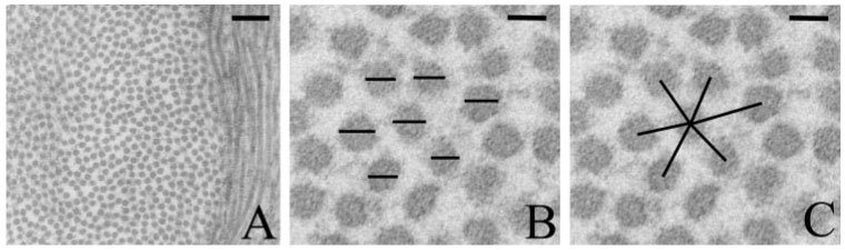
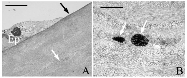
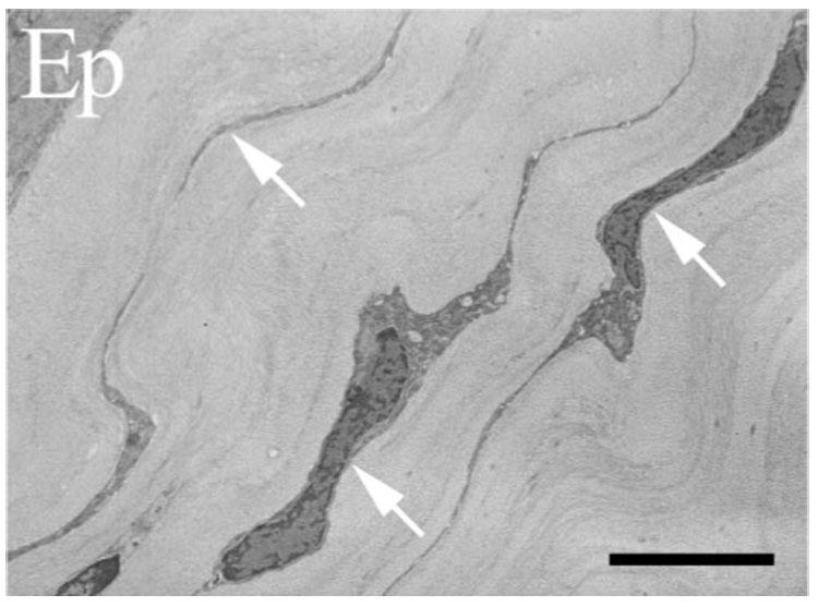
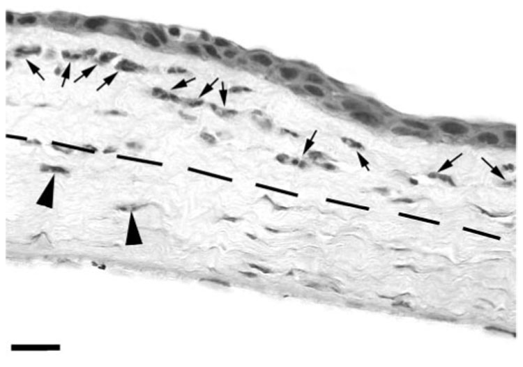
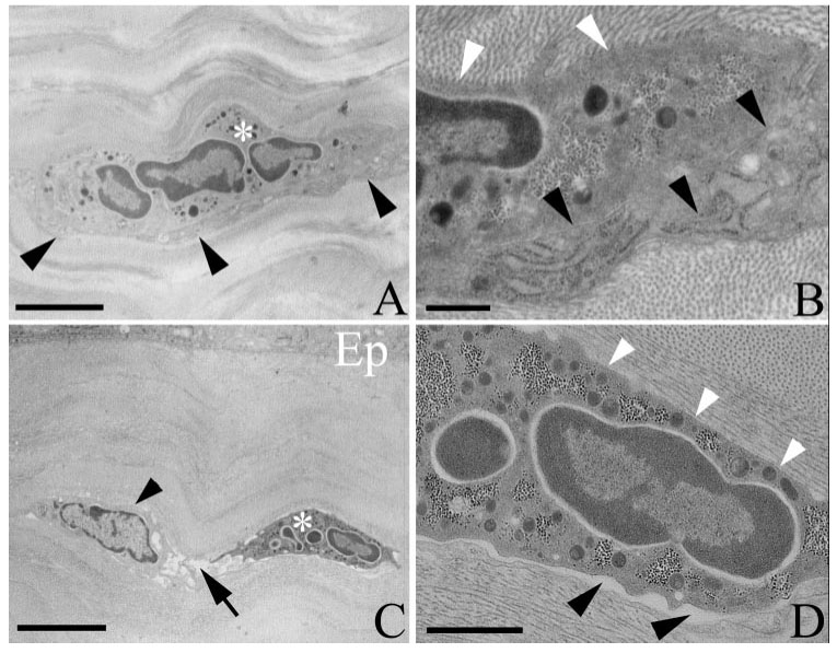
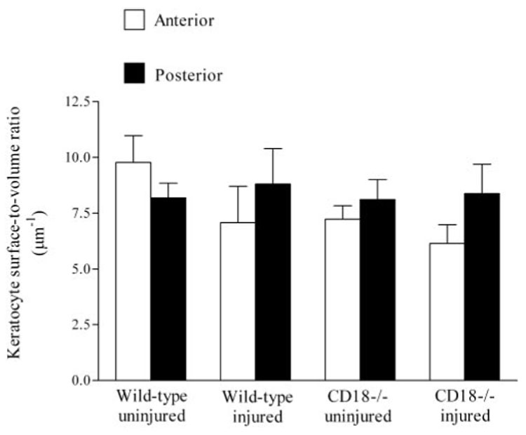
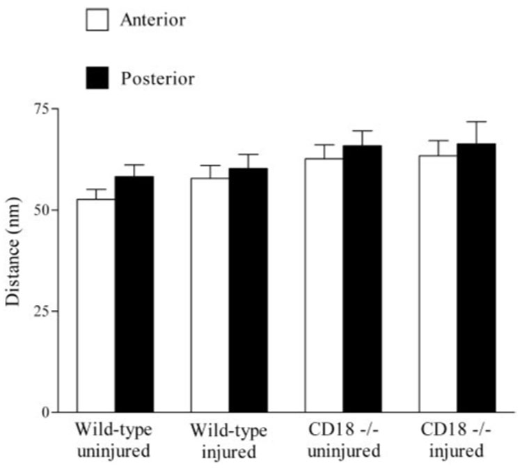
Results: Transmission electron microscopy revealed intact keratocyte networks within the paralimbus that were morphometrically similar, regardless of epithelial injury or mouse genotype. Secondary to epithelial abrasion, extravasated PMNs within the paralimbus developed close contacts with keratocytes and collagen. In wild-type mice, 40% of the PMN surface was in contact with the keratocyte surface, and this value decreased to 10% in CD18(-/-) mice. PMN contact with collagen was similar in wild-type and CD18(-/-) mice, with approximately 50% of the PMN surface contacting the collagen fibrils. Since corneal edema resulting from scrape injury was similar, regardless of genotype and did not involve structural changes in collagen fibrils, these data favor a direct role for CD18 in mediating PMN contact with keratocytes.
Conclusions: The data show that in response to epithelial scrape injury, PMN migration in the corneal stroma involves close contact between keratocytes and collagen. Although PMN-keratocyte contacts require CD18 integrins, contact with collagen is CD18 independent. Fundamentally, PMN migration along keratocyte networks constitutes the beginning of a new experimental concept for understanding leukocyte migration within the wounded cornea.
Figures

Similar articles
-
ICAM-1 mediates surface contact between neutrophils and keratocytes following corneal epithelial abrasion in the mouse.Exp Eye Res. 2010 Nov;91(5):676-84. doi: 10.1016/j.exer.2010.08.007. Epub 2010 Aug 14. Exp Eye Res. 2010. PMID: 20713042 Free PMC article.
-
Integrin-dependent neutrophil migration in the injured mouse cornea.Exp Eye Res. 2014 Mar;120:61-70. doi: 10.1016/j.exer.2014.01.004. Epub 2014 Jan 24. Exp Eye Res. 2014. PMID: 24462632 Free PMC article.
-
Two waves of neutrophil emigration in response to corneal epithelial abrasion: distinct adhesion molecule requirements.Invest Ophthalmol Vis Sci. 2006 May;47(5):1947-55. doi: 10.1167/iovs.05-1193. Invest Ophthalmol Vis Sci. 2006. PMID: 16639002
-
The corneal fibrosis response to epithelial-stromal injury.Exp Eye Res. 2016 Jan;142:110-8. doi: 10.1016/j.exer.2014.09.012. Exp Eye Res. 2016. PMID: 26675407 Free PMC article. Review.
-
Regulatory Mechanism of Collagen Degradation by Keratocytes and Corneal Inflammation: The Role of Urokinase-Type Plasminogen Activator.Cornea. 2016 Nov;35 Suppl 1:S59-S64. doi: 10.1097/ICO.0000000000000995. Cornea. 2016. PMID: 27661072 Review.
Cited by
-
CCL20, γδ T cells, and IL-22 in corneal epithelial healing.FASEB J. 2011 Aug;25(8):2659-68. doi: 10.1096/fj.11-184804. Epub 2011 Apr 25. FASEB J. 2011. PMID: 21518851 Free PMC article.
-
Integrins: An Important Link between Angiogenesis, Inflammation and Eye Diseases.Cells. 2021 Jul 6;10(7):1703. doi: 10.3390/cells10071703. Cells. 2021. PMID: 34359873 Free PMC article. Review.
-
Ly6G+ Neutrophils and Interleukin-17 Are Essential in Protection against Rodent Malaria Caused by Plasmodium berghei ANKA.Research (Wash D C). 2024 Dec 19;7:0559. doi: 10.34133/research.0559. eCollection 2024. Research (Wash D C). 2024. PMID: 39703777 Free PMC article.
-
Dendritic cell-epithelium interplay is a determinant factor for corneal epithelial wound repair.Am J Pathol. 2011 Nov;179(5):2243-53. doi: 10.1016/j.ajpath.2011.07.050. Epub 2011 Sep 13. Am J Pathol. 2011. PMID: 21924232 Free PMC article.
-
ICAM-1 is necessary for epithelial recruitment of gammadelta T cells and efficient corneal wound healing.Am J Pathol. 2009 Aug;175(2):571-9. doi: 10.2353/ajpath.2009.090112. Epub 2009 Jul 16. Am J Pathol. 2009. PMID: 19608878 Free PMC article.
References
-
- Netto MV, Mohan RR, Ambrosio R, Jr, Hutcheon AE, Zieske JD, Wilson SE. Wound healing in the cornea: a review of refractive surgery complications and new prospects for therapy. Cornea. 2005;24:509–522. - PubMed
-
- Li Z, Burns AR, Smith CW. Two waves of neutrophil emigration in response to corneal epithelial abrasion: distinct adhesion molecule requirements. Invest Ophthalmol Vis Sci. 2006;47:1947–1955. - PubMed
-
- Lindbom L, Werr J. Integrin-dependent neutrophil migration in extravascular tissue. Semin Immunol. 2002;14:115–121. - PubMed
-
- Burns AR, Smith CW, Walker DC. Unique structural features that influence neutrophil emigration into the lung. Physiol Rev. 2003;83:309–336. - PubMed
-
- Poole CA, Brookes NH, Clover GM. Confocal imaging of the human keratocyte network using the vital dye 5-chloromethylfluorescein diacetate. Clin Exp Ophthalmol. 2003;31:147–154. - PubMed
Publication types
MeSH terms
Substances
Grants and funding
LinkOut - more resources
Full Text Sources
Other Literature Sources
Medical
Molecular Biology Databases
