. 2008 Jan 15;111(2):894-904.
doi: 10.1182/blood-2007-04-085944. Epub 2007 Oct 25.

Extravasations and emigration of neutrophils to the inflammatory site depend on the interaction of immune-complex with Fcgamma receptors and can be effectively blocked by decoy Fcgamma receptors

Affiliations

Extravasations and emigration of neutrophils to the inflammatory site depend on the interaction of immune-complex with Fcgamma receptors and can be effectively blocked by decoy Fcgamma receptors

Rangaiah Shashidharamurthy et al. Blood. .

Abstract

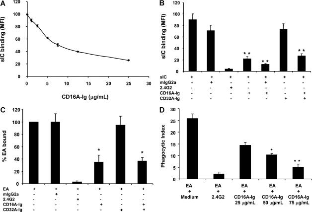
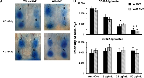
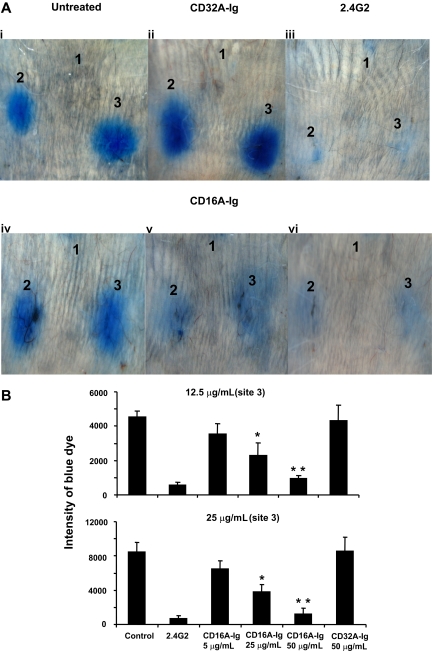
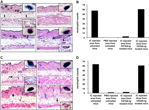
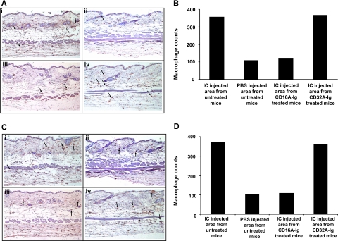
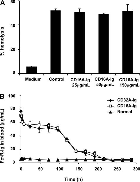
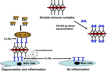
Extravasation and emigration of neutrophils to the site of inflammation are essential early steps in the initiation of many antibody-mediated autoimmune diseases. The Fc domains of cell bound autoantibodies or immune-complexes (IC) are capable of triggering the neutrophil emigration via complement and FcgammaRs-mediated mechanisms. To define the clinical relevance and the relative contribution of these 2 pathways in IC-mediated neutrophil emigration, we have neutralized the FcgammaR-binding activity of IC with a recombinant dimeric Fc receptor, CD16A-Ig, and investigated the early events of IC-induced inflammation in mice. Systemic administration of purified CD16A-Ig blocked IC-induced inflammation, mast- cell degranulation, and extravasation of neutrophils in a reversed Arthus reaction. Although the binding of CD16A-Ig to IC did not alter the complement-activating properties of IC, no evidence for complement-dependent neutrophil emigration was observed. These results suggest that interaction of IC with cells expressing FcgammaRs at the inflammatory site results in the secretion of chemoattractants, which mediate complement-independent emigration of neutrophils in this cutaneous acute inflammation model. Furthermore, blocking the interaction of IC to FcgammaRs expressed on inflammatory cells by administering high-avidity Fc fusion dimers of low-affinity FcgammaRs is an effective way of preventing IC-induced acute inflammation in autoimmune diseases.


Figures

Figure 1
Figure 1
CD16A-Ig competes with cell surface FcγRs and blocks immune-complex binding and phagocytosis by mouse macrophage cells. (A) CD16A-Ig blocked the binding of soluble IC to the mouse macrophage cell line P388D1 in a dose dependent manner. P388D1 cells were incubated with FITC-IC (soluble IC) in the presence and absence of various concentrations of CD16A-Ig and the cells were analyzed for binding of FITC-IC using flow cytometry. (B) Specificity of blocking by CD16A-Ig. P388D1 cells were incubated with soluble IC in the presence of the following reagents: anti-CD16A/32B mAb (2.4G2), CD16A-Ig and CD32A-Ig or mIgG2a. The FITC-IC binding to P388D1 cells was analyzed using standard flow cytometry. (C) CD16A-Ig blocked the binding of particulate IC to the mouse macrophage cells. P388D1 cells (50 μl of 5 × 10⁶/mL) in binding buffer (PBS/5 mM EDTA/1% BSA) were incubated with PKH labeled EA (50 μL of 1.5 × 10⁸) for 2 hours at 4°C. Binding assays were performed in the absence or presence of CD16A-Ig or CD32A-Ig, or 10 μg/mL blocking mAbs for mouse CD16A/CD32B. Monomeric mIgG2a (50 μg/mL) was used to block CD64. Blocking mAbs (2.4G2), CD16A-Ig, and CD32A-Ig were preincubated for 30 minutes at 4°C and then continuously present during their incubation with EA. EA bound to the cells were analyzed by flow cytometry. Cells incubated with PKH-labeled unopsonized-E were used as a background control. The EA binding in presence of medium was taken as 100% to calculate the percentage EA binding. Data shown are the average (mean ± SD) of 3 individual experiments. (D) CD16A-Ig blocked the phagocytosis of opsonized cells by macrophages. Phagocytosis of Alexa-EA was carried out using P388D1 cells. P388D1 cells were incubated with Alexa-EA in the absence or presence of the following blocking reagents: 2.4G2 (10 μg/mL) and CD16A-Ig (25, 50, and 75 μg/mL). The cells were then fixed and the fluorescence of phagocytosed Alexa-EA was measured using flow cytometry. Cells incubated with EA at 4°C served as the background control. The phagocytic index (PI) was calculated using the formula PI = %P × MF/100. The experiment was repeated twice. Data shown are the average of 3 individual experiments. *P < .01, **P < .001.
Figure 2
Figure 2
Local administration of CD16A-Ig at the inflammatory site blocked reversed passive Arthus reaction in mice with or without complement depletion. (A) The mice (n = 3) were injected intradermally with PBS (site 1), or various concentrations of CD16A-Ig (0 μg/mL at site 2, 5 μg/mL at site 3, 25 μg/mL at site 4, and 50 μg/mL at site 5) along with rabbit anti-Ova antibody in PBS, immediately after which ovalbumin with 1% Evan blue was injected through the tail vein. To deplete the complement, mice were twice injected intraperitoneally with cobra venom factor before the initiation of RPA. After 3h the mice were euthanized, and the dorsal side of the skin was photographed for analysis. The figure shows 3 representative mice. (B) Quantitative analysis of RPA. The dermal lesion, seen blue in the photographs, was quantified using ImageJ and KaleidaGraph softwares for groups with or without CVF treatment. Data are presented as mean plus or minus the SD from 3 individual mice. *P < .01, **P < .001.
Figure 3
Figure 3
Systemic administration of CD16A-Ig efficiently blocked the reversed passive Arthus reaction in vivo in mice without complement depletion. (A) The mice (n = 3) were injected with various concentrations of CD16A-Ig intravenously (panel iv, 5 μg/mL; panel v, 25 μg/mL; panel vi, 50 μg/mL of blood). After 1 hour, mice were injected intradermally with PBS (site 1), 12.5 μg (site 2), and 25 μg (site 3) of anti-Ova per site. RPA was initiated by injecting Ova with 1% Evan blue intravenously through tail vein. For CD32A-Ig, 50 μg of CD32A-Ig/mL of blood was injected (panel ii). The antibody (2.4G2) treated control mice (panel iii) were injected with 25 μg/mL of blood (40 μg/mice) mAb. The PBS-injected mice served as the untreated positive control (panel i). After 3 hours the mice were killed, and the dorsal side of the skin was photographed for analysis. The figure shows 3 representative mice. (B) Quantitative analysis of RPA. The dermal lesion, shown in blue in the photographs, was quantified using ImageJ and KaleidaGraph softwares for groups with or without FcγR dimer treatment. Data are presented as the mean plus or minus the SD from 3 experiments. *P < .01, **P < .001.
Figure 4
Figure 4
Histologic examination and evaluation of neutrophil infiltration in skin biopsies from anti-Ova injected areas of mice treated with or without FcγR dimers. (Ai) Skin biopsy from anti-Ova antibody–injected area in an untreated mouse with RPA (from Figure 3Ai site 3). The epidermis and dermis are essentially unremarkable. However, the subdermal fat is edematous and shows the margination of neutrophils along capillary walls (arrows; bottom inset) as well as neutrophilic infiltration into surrounding soft tissue. (Aii) Skin biopsy from PBS-injected area in an untreated mouse with RPA (from Figure 3Ai site 1). No specific pathologic changes are identified. (Aiii) Skin biopsy from anti-Ova antibody injected area in a mouse treated with 50 μg/mL CD16A-Ig before the induction of RPA (from Figure 3Avii site 3). No specific pathologic changes are identified, suggesting abrogation of the Arthus reaction. (Aiv) Skin biopsy from anti-Ova antibody injected area in a mouse treated with 50 μg/mL CD32A-Ig before the induction of RPA (from Figure 3Aii site 3). The epidermis and dermis are essentially unremarkable. However, the subdermal fat is edematous and shows neutrophilic infiltrates in soft tissue (similar to Figure 3Ai), as well as scattered foci of leukocytoclastic vasculitis (arrows; bottom inset). Top inset: Mast cell degranulation was observed in 10 different sites at 100x magnification and one of the cells is represented in the figure inset. (B) Neutrophils were counted in 10 random sites at 63x magnification and sum of the neutrophils were represented. C: To deplete the complement, mice were twice injected intraperitoneally with cobra venom factor before the initiation of RPA. The mice were injected with CD16A-Ig and CD32A-Ig intravenously (50 μg/mL of blood). After 1 hour, mice were injected intradermally with 25 μg of anti-Ova per site. (Ci) Skin biopsy from anti-Ova antibody injected area in an untreated mouse with RPA. The epidermis and dermis are essentially unremarkable. However, the subdermal fat is edematous and shows the margination of neutrophils along capillary walls (arrows; bottom inset) as well as neutrophilic infiltration into surrounding soft tissue. (Cii) Skin biopsy from PBS injected area in an untreated mouse with RPA. No specific pathologic changes are identified. (Ciii) Skin biopsy from anti-Ova antibody injected area in a mouse treated with 50 μg/mL CD16A-Ig before the induction of RPA. No specific pathologic changes are identified, suggesting abrogation of the Arthus reaction. (Civ) Skin biopsy from anti-Ova antibody injected area in a mouse treated with 50 μg/mL CD32A-Ig before the induction of RPA. The epidermis and dermis are essentially unremarkable. However, the subdermal fat is edematous and shows neutrophilic infiltrates in soft tissue, as well as scattered foci of leukocytoclastic vasculitis (arrows; bottom inset). Top inset: Mast cell degranulation was observed in 10 different sites 100x magnification and one of the cells is represented in the Figure inset. (D) Neutrophils were counted in 10 random sites at 63x magnifications and sum of the neutrophils were represented.
Figure 5
Figure 5
Immunohistochemical examination to detect the macrophages in skin biopsies from anti-Ova injected areas of mice treated with or without FcγR dimers. (A) Skin biopsies of the above mentioned sections were processed to detect the macrophages (brown cells indicated by arrow) by immunohistochemistry. (Ai) Accumulation of macrophages in anti-Ova antibody injected area of untreated mice. (Aii) Presence of resident macrophages in PBS injected area of untreated mice. (Aiii) Inhibition of macrophage accumulation in anti-Ova antibody injected area of CD16A-Ig treated mice. (Aiv) Accumulation of macrophages in anti-Ova antibody injected area of CD32A-Ig treated mice. (B) Macrophages were counted in 10 random sites at 63x magnification and the sum of the macrophages is presented. (C) Skin biopsies of the above mentioned sections from complement-depleted mice were processed to detect the macrophages (brown cells indicated by arrow) by immunohistochemistry. (Ci) Accumulation of macrophages in anti-Ova antibody injected area of untreated mice. (Cii) Presence of resident macrophages in PBS injected area of untreated mice. (Ciii) Inhibition of macrophage accumulation in anti-Ova antibody injected area of CD16A-Ig treated mice. (Civ) Accumulation of macrophages in anti-Ova antibody injected area of CD32A-Ig treated mice. (D) Macrophages were counted in 10 random sites at 63x magnification and the sum of the macrophages is presented.
Figure 6
Figure 6
Effect of CD16A-Ig on complement-mediated hemolytic activity. (A) Rabbit anti-DNP-IgG opsonized rRBCs were incubated with or without various concentrations of CD16A-Ig and then 1:5 diluted mouse serum was added. Hemolytic activity was then carried out as described in the text. Cells treated with buffer alone served as a control. Cells lysed with water represented 100% hemolysis. The supernatant was read at 450nm. Data are representative of 3 independent experiments. (B) Clearance of FcγR-Ig dimers from circulation. A group of mice (n = 3) were injected with dimers and blood samples were collected at various time points and diluted in PBS/EDTA. Plasma was collected to detect the FcγR-Ig dimers by sandwich ELISA. Plates were coated with 50 μL of 10 μg/mL anti-hCD16A (CLBFcgran-1) and anti-hCD32A (IV.3) mAbs overnight at 4°C. The wells were then blocked with PBS/5mM EDTA/1% BSA. After washing, 50 μL of the plasma samples were added into the wells and incubated for another 1 hour. The wells were washed and 50 μL of HRP-conjugated anti–human Fc specific antibody were added and incubated for another 1 hour. HRP substrate was added to the washed wells and read at 450 nm. The plasma from a group of normal mice injected with PBS served as a specificity control. As positive controls, purified CD16A-Ig and CD32A-Ig were used while BSA coated wells served as negative controls. Purified CD32A-Ig was used as a standard to quantify the level of dimers in the blood. Data are means plus or minus SD of triplicates.
Figure 7
Figure 7
A hypothetical model depicting the action of in vivo administered CD16A-Ig in Arthus reaction. Mφ = macrophages, Nφ = neutrophils.

Similar articles

Cited by

References

    1. Centers for Disease Control. The most frequent causes of disability among Americans aged 18 years or older. MMWR. 2001;50:120–125. - PubMed
    1. Schwartz RS. Autoimmunity and autoimmune diseases. In: Paul WE, editor. Fundamental Immunology. New York: Raven Press, Ltd; 1993. pp. 1033–1097.
    1. Abbas AK, Lichtman AH, Pober JS. Cellular and Molecular Immunology. Philadelphia PA: W.B. Saunders; 1997. Immune-mediated tissue injury and disease. pp. 423–438.
    1. Guo RF, Ward PA. Role of C5a in inflammatory responses. Annu Rev Immunol. 2005;23:821–852. - PubMed
    1. Müller-Eberhard HJ. Molecular organisation of the complement system. Annu Rev Biochem. 1988;57:321–347. - PubMed

Publication types

MeSH terms