. 2008 Jan 15;177(2):208-18.
doi: 10.1164/rccm.200612-1890OC. Epub 2007 Oct 25.

IFN-gamma production during initial infection determines the outcome of reinfection with respiratory syncytial virus

Affiliations

IFN-gamma production during initial infection determines the outcome of reinfection with respiratory syncytial virus

Young-Mok Lee et al. Am J Respir Crit Care Med. .

Abstract

Rationale: Severe respiratory syncytial virus (RSV) bronchiolitis has been associated with deficient IFN-gamma production in humans, but the role of this cytokine in determining the outcome of reinfection is unknown.

Objectives: To define the role of IFN-gamma in the development of RSV-mediated airway hyperresponsiveness (AHR) and lung histopathology in mice.

Methods: Wild-type (WT) and IFN-gamma knockout mice were infected with RSV in the newborn or weaning stages and reinfected 5 weeks later. Airway responses were assessed on Day 6 after the primary or secondary infection.

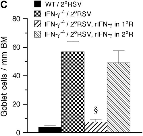
Measurements and main results: Both WT and IFN-gamma knockout mice developed similar levels of AHR and airway inflammation after primary infection. After reinfection, IFN-gamma knockout mice, but not WT mice, developed AHR, airway eosinophilia, and mucus hyperproduction. Intranasal administration of IFN-gamma during primary infection but not during reinfection prevented the development of these altered airway responses on reinfection in IFN-gamma knockout mice. Adoptive transfer of WT T cells into IFN-gamma knockout mice before primary infection restored IFN-gamma production in the lungs and prevented the development of altered airway responses on reinfection. Treatment of mice with IFN-gamma during primary neonatal infection prevented the enhancement of AHR and the development of airway eosinophilia and mucus hyperproduction on reinfection.

Conclusions: IFN-gamma production during primary RSV infection is critical to the development of protection against AHR and lung histopathology on reinfection. Provision of IFN-gamma during primary infection in infancy may be a potential therapeutic approach to alter the course of RSV-mediated long-term sequelae.


Figures

<b>Figure 1.</b>
Figure 1.
Airway responsiveness (A), the inflammatory profile in bronchoalveolar lavage fluid (B), lung histopathology (C), and mucus goblet cell numbers (D) after primary respiratory syncytial virus (RSV) infection in wild-type (WT) and IFN-γ−/− mice. Groups of mice were infected with RSV (n = 8/group) or sham inoculated (n = 6/group). Responses were assessed on Day 6 after infection. Both mouse groups developed significant airway hyperresponsiveness (A) with lymphocyte accumulation in the airways (B), and exhibited peribronchial airway tissue inflammation (C, arrowhead) and mucus production (C, arrows, and D). *Statistical difference between the groups (P < 0.05). Scale bar in C represents 100 μm. BM = basement membrane; Eos = eosinophil; Lym = lymphocyte; Mac = macrophage; Neu = neutrophil; 1°RSV = primary RSV infection.
<b>Figure 2.</b>
Figure 2.
Airway responsiveness (A), the inflammatory profile in bronchoalveolar lavage (BAL) fluid (B), lung histopathology (C), and mucus goblet cell numbers (D) after reinfection in wild-type (WT) and IFN-γ−/− mice. Mice were initially infected with respiratory syncytial virus (RSV) (n = 8/group) and reinfected 5 weeks later; controls received sham inoculation (n = 6/group). Airway responses were assessed on Day 6 after reinfection. After reinfection, IFN-γ−/− mice developed significant airway hyperresponsiveness (A), associated with BAL eosinophilia (B) and enhanced airway mucus production (C, arrows, and D). By contrast, WT mice did not develop these altered responses after reinfection, despite increased airway inflammation. *Statistical difference between the groups (P < 0.05). Scale bar in C represents 100 μm. BM = basement membrane; Eos = eosinophil; Lym = lymphocyte; Mac = macrophage; Neu = neutrophil; 2°RSV = secondary RSV infection.
<b>Figure 3.</b>
Figure 3.
IFN-γ–producing lung T cells and natural killer (NK) cells. (A) Flow cytometry scatter plot showing IFN-γ–positive cells within gated CD4+ and CD8+ lung cell populations. (B) Numbers of IFN-γ–producing CD4 and CD8 T cells, and NK cells. CD4+ and CD8+ T cells were the predominant source of IFN-γ in the lung of respiratory syncytial virus (RSV)–infected wild-type mice. The number of these cells was significantly increased after reinfection of these mice. *Statistical difference between primary and secondary infection (P < 0.05).
<b>Figure 4.</b>
Figure 4.
Effect of treatment with IFN-γ on airway responsiveness to reinfection in IFN-γ−/− mice. IFN-γ−/− mice were administered recombinant IFN-γ (rIFN-γ) during either the primary or the secondary respiratory syncytial virus (RSV) infection. Airway responses were assessed on Day 6 after reinfection. Compared with untreated IFN-γ−/− mice, mice administered rIFN-γ during the primary infection, but not during the secondary infection, developed a marked reduction in airway hyperresponsiveness (A), bronchoalveolar lavage (BAL) eosinophilia (B), mucus goblet cell metaplasia (C), and BAL Th2 (IL-5 and IL-13) cytokine levels (D) on reinfection. *Significant difference between the groups (P < 0.05); §significant difference compared with IFN-γ−/−/2°RSV group (P < 0.05). MCh = methacholine; WT = wild-type. See Figure 2 legend for definition of other abbreviations.
<b>Figure 5.</b>
Figure 5.
Effect of adoptive T-cell transfer on airway responsiveness to reinfection in IFN-γ−/− mice. IFN-γ–sufficient T cells, isolated from uninfected wild-type (WT) mice, were adoptively transferred into recipient IFN-γ−/− mice, before primary or secondary respiratory syncytial virus (RSV) infection. Airway responses were assessed on Day 6 after reinfection. (A) Airway responsiveness to inhaled methacholine (MCh); (B) bronchoalveolar lavage (BAL) cellularity; (C) mucus goblet cell metaplasia; (D) BAL cytokine levels; (E) in vitro IFN-γ production by isolated lung leukocytes. Transfer of IFN-γ–sufficient T cells before either primary or secondary RSV infection restored significant IFN-γ production in the lungs of recipient IFN-γ−/− mice (D, E). However, the development of airway hyperresponsiveness (A), BAL eosinophilia (B), and mucus goblet cell metaplasia (C) in these animals was only inhibited when IFN-γ–sufficient T cells were transferred before the primary but not the secondary RSV infection. *Significant difference between the groups (P < 0.05); §significant difference compared with IFN-γ−/−/2°RSV group (P < 0.05). BM = basement membrane; Eos = eosinophil; Lym = lymphocyte; Mac = macrophage; Neu = neutrophil; 1°R, 1°RSV = primary RSV infection; 2°R, 2°RSV = secondary RSV infection.
<b>Figure 6.</b>
Figure 6.
Effect of treatment with IFN-γ on airway responsiveness to reinfection in newborn mice. Newborn BALB/c mice were treated with recombinant IFN-γ (rIFN-γ), administered during primary neonatal infection or during reinfection. Airway responses were assessed on Day 6 after re-infection. (A) Airway responsiveness to inhaled methacholine (MCh); (B) bronchoalveolar lavage (BAL) cellularity; (C) lung histopathology; (D) mucus goblet cell metaplasia; (E) BAL cytokine levels; (F) lung viral titers; (G) in vitro cytokine production by peribronchial lymph node (PBLN) mononuclear cells. Treatment with rIFN-γ during primary neonatal infection, but not during reinfection, prevented the enhancement of airway hyperresponsiveness (A) and inhibited the development of airway eosinophilia (B), mucus hyperproduction (C, D), BAL Th2 cytokine levels (E), and Th2 cytokine production by PBLN cells on reinfection. IFN-γ levels (E, G) and lung viral titers (F) were not altered by IFN-γ treatment. *Significant difference compared with sham group (P < 0.05); §significant difference compared with 2°RSV group (P < 0.05). 1°RSV = primary RSV infection; 2°RSV = secondary RSV infection.

References

    1. Glezen WP, Taber LH, Frank AL, Kasel JA. Risk of primary infection and reinfection with respiratory syncytial virus. Am J Dis Child 1986;140:543–546. - PubMed
    1. Hall CB. Respiratory syncytial virus: a continuing culprit and conundrum. J Pediatr 1999;135:2–7. - PubMed
    1. Nicholson KG, Kent J, Ireland DC. Respiratory viruses and exacerbations of asthma in adults. BMJ 1993;307:982–986. - PMC - PubMed
    1. Falsey AR, Walsh EE. Relationship of serum antibody to risk of respiratory syncytial virus infection in elderly adults. J Infect Dis 1998;177:463–466. - PubMed
    1. Stein RT, Sherrill D, Morgan WJ, Holberg CJ, Halonen M, Taussig LM, Wright AL, Martinez FD. Respiratory syncytial virus in early life and risk of wheeze and allergy by age 13 years. Lancet 1999;354:541–545. - PubMed

Publication types

MeSH terms

Substances