

Soluble factors released by activated cytotoxic T lymphocytes interfere with death receptor pathways in neuroblastoma
- PMID: 17962944
- PMCID: PMC11031004
- DOI: 10.1007/s00262-007-0412-2
Soluble factors released by activated cytotoxic T lymphocytes interfere with death receptor pathways in neuroblastoma
Abstract
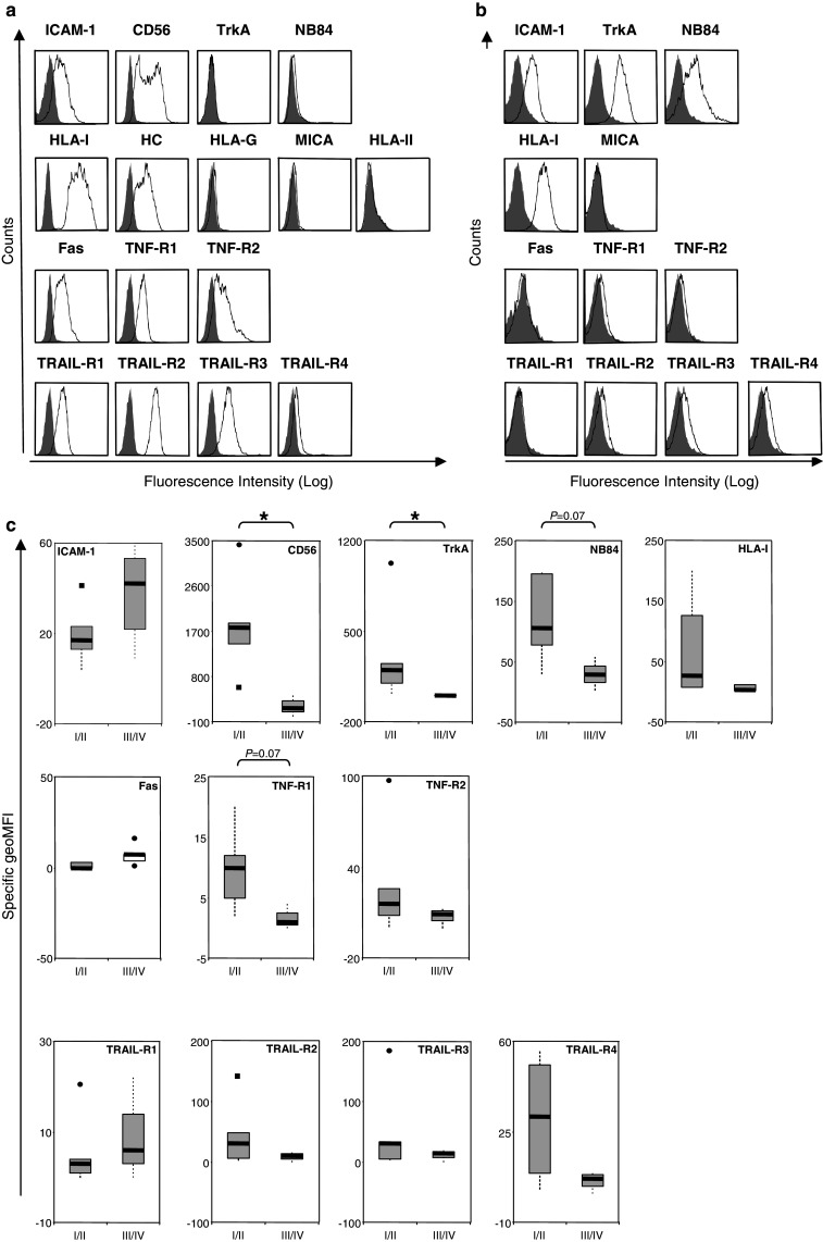
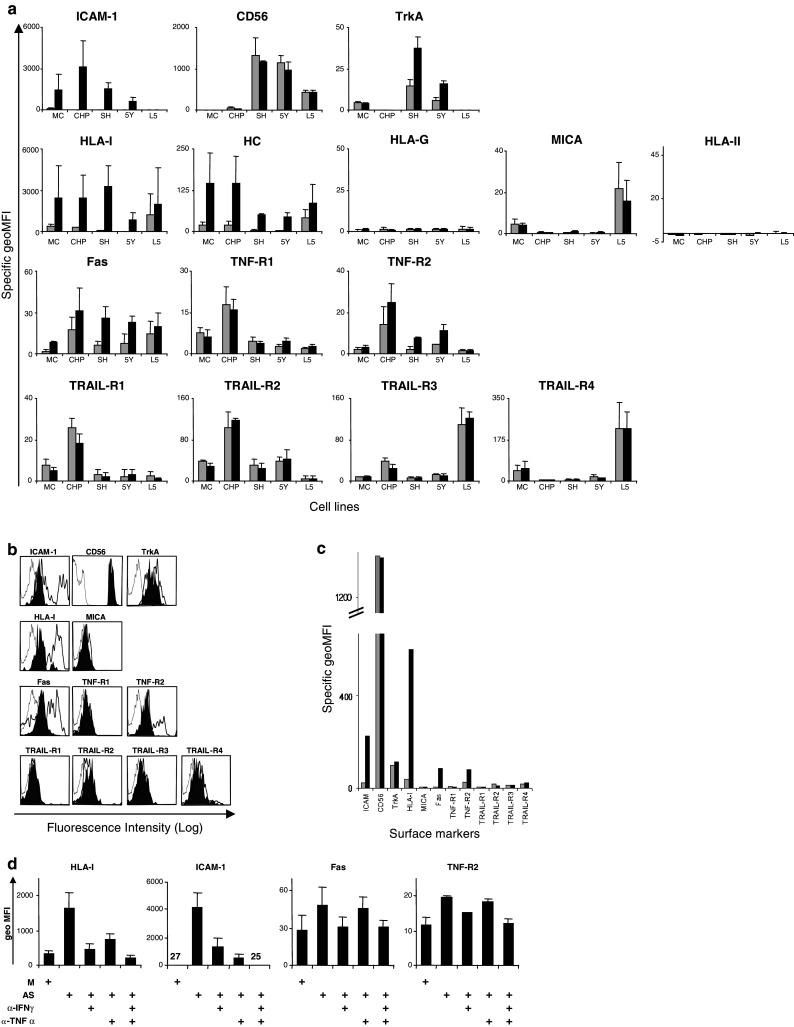
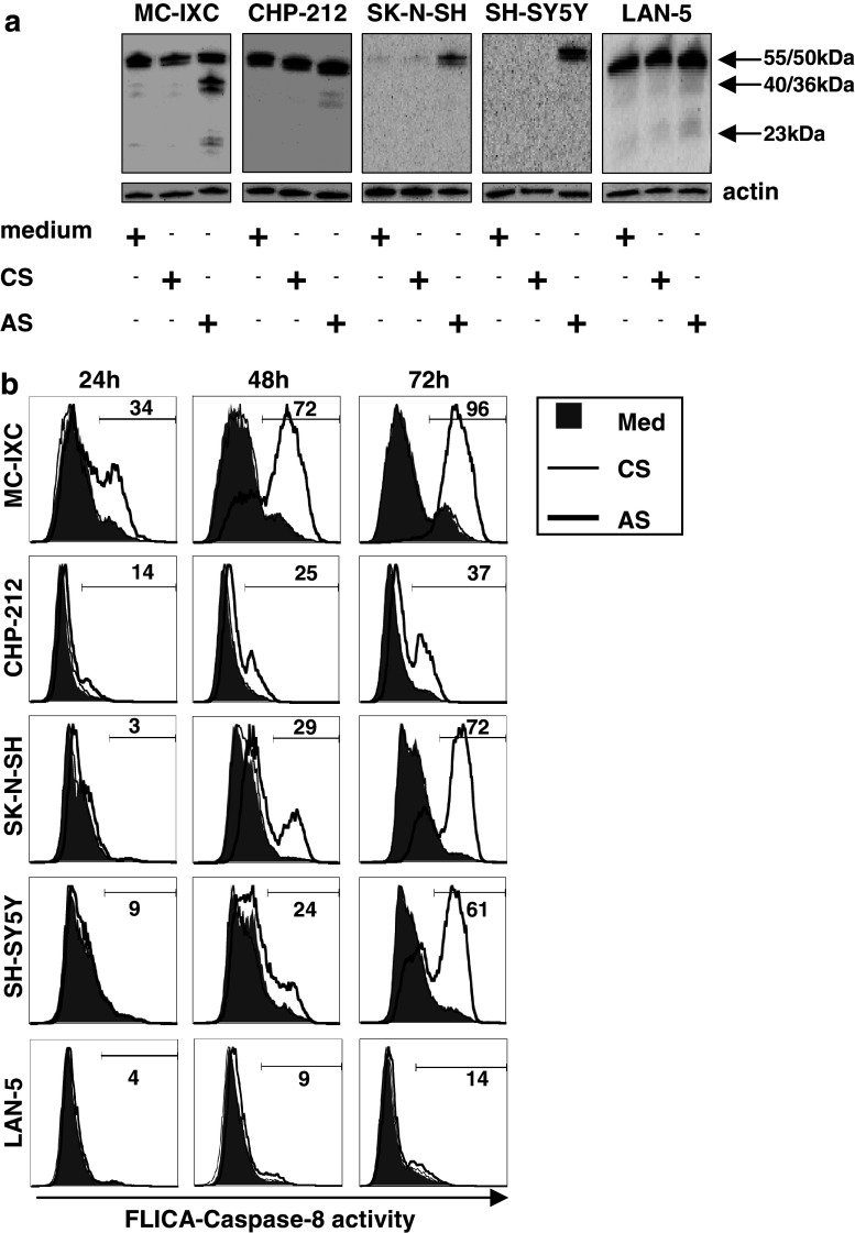
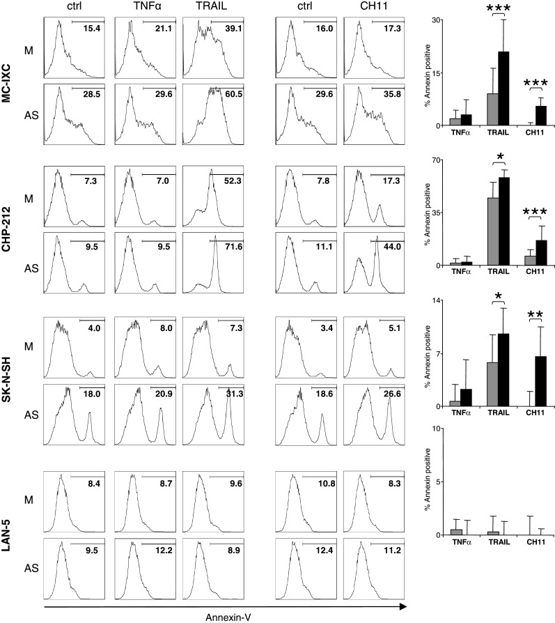
Neuroblastoma (NB) is often described as an unfavorable target for both HLA-restricted and death receptor-mediated elimination by cytotoxic T lymphocytes (CTLs) due to low or absent HLA class I and caspase-8 expression. We investigated the effects of soluble factors released by CTLs activated by TCR triggering (named as activated supernatant; AS) on the levels and composition of cell surface molecules involved in HLA-restricted and HLA-independent NB cell recognition (surface immune phenotype). Using a panel of long-term propagated NB cell lines and freshly isolated primary human NB cells, we analyzed surface expression of the (1) cognate receptors for TNFalpha, Fas and TRAIL; (2) HLA class I and II heterodimers; (3) adhesion molecules; (4) the intracellular expression and activation of caspase-8, as well as (5) the susceptibility of NB cells to death receptor-mediated killing prior to and after exposure to AS. The exposure of NB cells to soluble factors released by activated CTLs skewed the surface immune phenotype of both long term cultured and primary NB cells, induced the expression and activation of caspase-8 and increased the susceptibility of tumor cells to lysis by TRAIL and Fas-agonistic antibody. Blocking experiments identified IFNgamma and TNFalpha as main factors responsible for modulating the surface antigens of NB cells by AS. Our data suggest that recruitment of CTLs activated on third party targets into the vicinity of the NB tumor mass, may override the "silent" immune phenotype of NB cells via the action of soluble factors.
Figures
Similar articles
-
Cytotoxic T lymphocytes induce caspase-dependent and -independent cell death in neuroblastomas in a MHC-nonrestricted fashion.J Immunol. 2006 Dec 1;177(11):7540-50. doi: 10.4049/jimmunol.177.11.7540. J Immunol. 2006. PMID: 17114423
-
IRF1 and NF-kB restore MHC class I-restricted tumor antigen processing and presentation to cytotoxic T cells in aggressive neuroblastoma.PLoS One. 2012;7(10):e46928. doi: 10.1371/journal.pone.0046928. Epub 2012 Oct 5. PLoS One. 2012. PMID: 23071666 Free PMC article.
-
Differentiation-related expression of adhesion molecules and receptors on human neuroblastoma tissues, cell lines and variants.Int J Cancer. 1992 Aug 19;52(1):85-91. doi: 10.1002/ijc.2910520116. Int J Cancer. 1992. PMID: 1354203
-
Immunogenicity of human neuroblastoma.Ann N Y Acad Sci. 2004 Dec;1028:69-80. doi: 10.1196/annals.1322.008. Ann N Y Acad Sci. 2004. PMID: 15650233 Review.
-
Mechanisms of immune evasion of human neuroblastoma.Cancer Lett. 2005 Oct 18;228(1-2):155-61. doi: 10.1016/j.canlet.2004.11.064. Cancer Lett. 2005. PMID: 15923080 Review.
Cited by
-
Multiple Interactions Between Cancer Cells and the Tumor Microenvironment Modulate TRAIL Signaling: Implications for TRAIL Receptor Targeted Therapy.Front Immunol. 2019 Jul 3;10:1530. doi: 10.3389/fimmu.2019.01530. eCollection 2019. Front Immunol. 2019. PMID: 31333662 Free PMC article. Review.
-
The microenvironment of human neuroblastoma supports the activation of tumor-associated T lymphocytes.Oncoimmunology. 2013 Mar 1;2(3):e23618. doi: 10.4161/onci.23618. Oncoimmunology. 2013. PMID: 23802089 Free PMC article.
-
Bi-specific TCR-anti CD3 redirected T-cell targeting of NY-ESO-1- and LAGE-1-positive tumors.Cancer Immunol Immunother. 2013 Apr;62(4):773-85. doi: 10.1007/s00262-012-1384-4. Epub 2012 Dec 22. Cancer Immunol Immunother. 2013. PMID: 23263452 Free PMC article.
-
Bispecific Antibodies for Multiple Myeloma: A Review of Targets, Drugs, Clinical Trials, and Future Directions.Front Immunol. 2020 Apr 24;11:501. doi: 10.3389/fimmu.2020.00501. eCollection 2020. Front Immunol. 2020. PMID: 32391000 Free PMC article. Review.
-
T cells in the microenvironment of solid pediatric tumors: the case of neuroblastoma.Front Immunol. 2025 Feb 28;16:1544137. doi: 10.3389/fimmu.2025.1544137. eCollection 2025. Front Immunol. 2025. PMID: 40092980 Free PMC article. Review.
References
-
- Blaheta RA, Daher FH, Michaelis M, Hasenberg C, Weich EM, Jonas D, Kotchetkov R, Doerr HW, Cinatl J., Jr Chemoresistance induces enhanced adhesion and transendothelial penetration of neuroblastoma cells by down-regulating NCAM surface expression. BMC Cancer. 2006;6:294. doi: 10.1186/1471-2407-6-294. - DOI - PMC - PubMed
-
- Blaheta RA, Hundemer M, Mayer G, Vogel JU, Kornhuber B, Cinatl J, Markus BH, Driever PH, Cinatl J., Jr Expression level of neural cell adhesion molecule (NCAM) inversely correlates with the ability of neuroblastoma cells to adhere to endothelium in vitro. Cell Commun Adhes. 2002;9:131–147. doi: 10.1080/15419060214520. - DOI - PubMed
-
- Brodeur GM, Pritchard J, Berthold F, Carlsen NL, Castel V, Castelberry RP, De Bernardi B, Evans AE, Favrot M, Hedborg F, et al. Revisions of the international criteria for neuroblastoma diagnosis, staging, and response to treatment. J Clin Oncol. 1993;11:1466–1477. - PubMed
Publication types
MeSH terms
Substances
LinkOut - more resources
Full Text Sources
Medical
Research Materials
Miscellaneous
