. 2008 May;57(5):701-18.
doi: 10.1007/s00262-007-0410-4. Epub 2007 Oct 26.

Anti-inflammatory pretreatment enables an efficient dendritic cell-based immunotherapy against established tumors

Affiliations

Anti-inflammatory pretreatment enables an efficient dendritic cell-based immunotherapy against established tumors

Paula Chiarella et al. Cancer Immunol Immunother. 2008 May.

Abstract

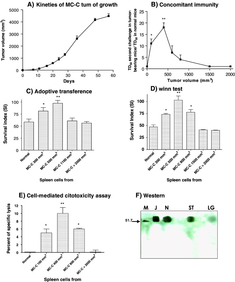
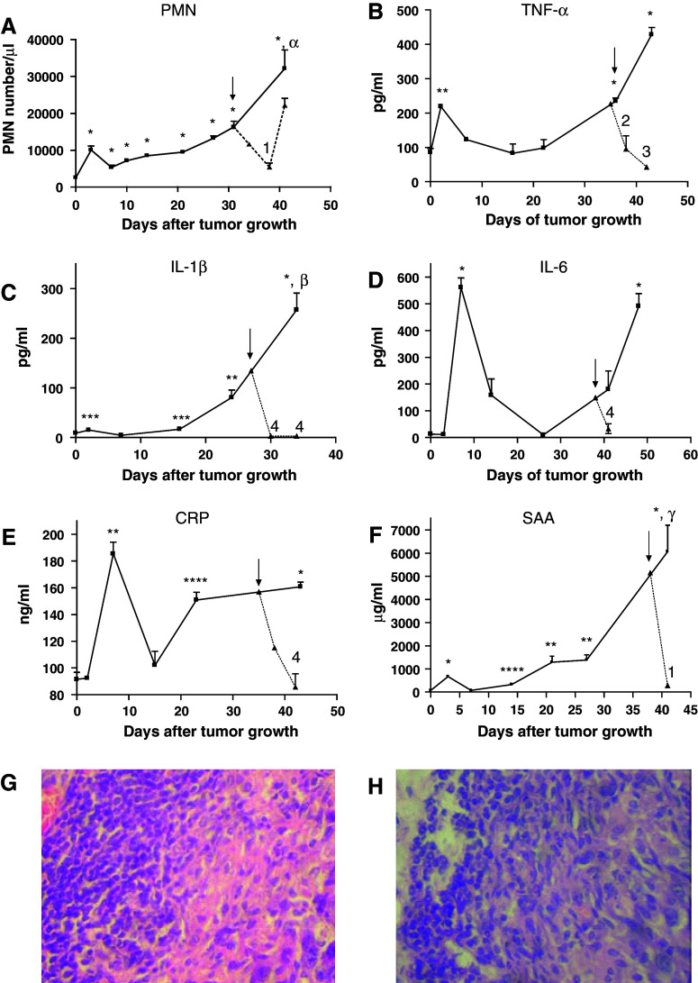
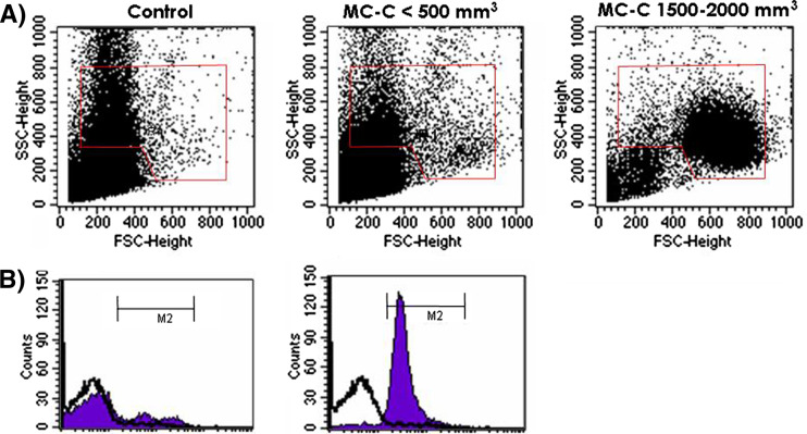
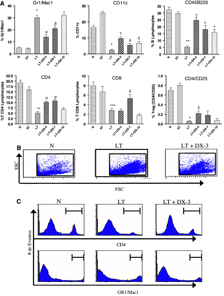
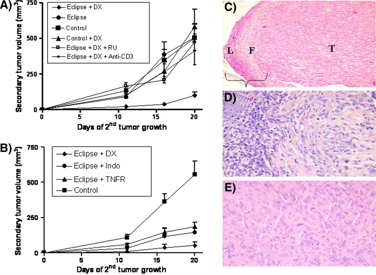
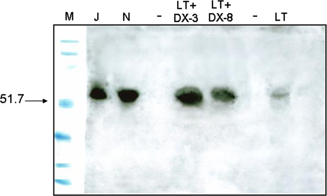
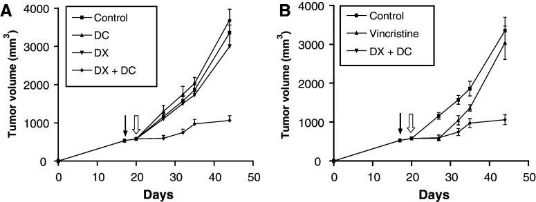
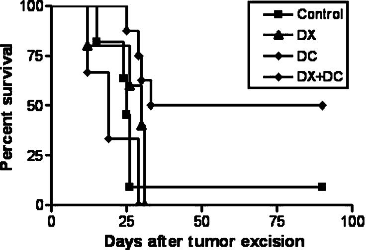
Although animals can be immunized against the growth of some tumor implants, most of the attempts to use immunotherapy to cause the regression of animal and human tumors once they have become established have been disappointing even when strongly immunogenic tumors were used as target. In this paper, we demonstrate that the failure to achieve an efficient immunological treatment against an established strongly immunogenic murine fibrosarcoma was paralleled with the emergence of a state of immunological unresponsiveness (immunological eclipse) against tumor antigens observed when the tumor surpassed the critical size of 500 mm(3). In turn, the onset of the immunological eclipse was coincidental with the onset of a systemic inflammatory condition characterized by a high number of circulating and splenic polymorphonucleated neutrophils (PMN) displaying activation and Gr1(+)Mac1(+) phenotype and an increasing serum concentration of the pro-inflammatory cytokines TNF-alpha, IL-1beta and IL-6 cytokines and C-reactive protein (CRP) and serum A amyloid (SAA) phase acute proteins. Treatment of tumor-bearing mice with a single low dose (0.75 mg/kg) of the synthetic corticoid dexamethasone (DX) significantly reduced all the systemic inflammatory parameters and simultaneously reversed the immunological eclipse, as evidenced by the restoration of specific T-cell-dependent concomitant immunity, ability of spleen cells to transfer anti-tumor activity and recovery of T-cell signal transduction molecules. Two other anti-inflammatory treatments by using indomethacin or dimeric TNF-alpha receptor, also partially reversed the immunological eclipse although the effect was not as striking as that observed with DX. The reversion of the immunological eclipse was not enough on its own to inhibit the primary growing tumor. However, when we used the two-step strategy of inoculating DX to reverse the eclipse and then dendritic cells loaded with tumor antigens (DC) as an immunization booster, a significant inhibition of the growth of both established tumors and remnant tumor cells after excision of large established tumors was observed, despite the fact that the vaccination alone (DC) had no effect or even enhanced tumor growth in certain circumstances. The two-step strategy of tumor immunotherapy that we present is based on the rationale that it is necessary to eliminate or ameliorate the immunological eclipse as a precondition to allow an otherwise ineffective anti-tumor immunological therapy to have a chance to be successful.


Figures

Fig. 1
Fig. 1
a Growth of s.c. MC-C tumor initiated (on day 0) with 5 × 105 MC-C tumor cells. Each point represents the mean ± standard error (SE) of 12 mice. b Concomitant immunity expressed, in the ordinate, as the ratio between TD50 of secondary MC-C tumor in MC-C tumor-bearing mice/TD50 in control mice. Abscissa indicates the primary tumor volume at the day of the second challenge. Each point represents the mean ± SE of five experiments. * P < 0.002 as compared with control mice and mice bearing MC-C tumors measuring 1,500 and 2,000 mm3; P < 0.05 as compared with mice bearing MC-C tumors measuring 800 mm3. ** P < 0.001 as compared with control mice and mice bearing MC-C tumors measuring 800, 1,500 and 2,000 mm3; P < 0.02 and P < 0.05 as compared with mice bearing MC-C tumors measuring 600 mm3 and 100 mm3, respectively. c In the adoptive transference assay, normal mice received 1 × 108 spleen cells by the i.p. route, from normal mice or mice bearing MC-C tumors with different sizes; 2 h later mice were challenged with 5 × 105 MC-C tumor cells by the s.c. route. Each bar represents the mean ± SE of six experiments. Ordinate: Survival index of recipient mice = survival time/t/n, where t = number of mice that died of tumor and n = number of mice inoculated. * P < 0.01 as compared with normal mice and mice bearing tumors measuring 1,100 and >2,000 mm3. ** P < 0.001 as compared with normal mice and mice bearing tumors measuring 1,100 and >2,000. d In the Winn test, 50 × 106 spleen cells from normal mice or mice bearing MC-C tumors with different sizes were mixed in vitro with 5 × 105 MC-C tumor cells and then the mixture was s.c. inoculated in naive mice. Each bar represents the mean ± SE of five experiments. Ordinate Survival index of mice inoculated. * P < 0.001 as compared with normal mice and mice bearing MC-C tumors measuring 1,500 and >2,000 mm3. ** P < 0.001 as compared with normal mice and mice bearing MC-C tumors measuring 1,500 and >2,000 mm3; P < 0.002 and P < 0.01 as compared with mice bearing tumors measuring 300 mm3 and 800 mm3, respectively. e Cytotoxic activity of 2 × 106 spleen cells from normal mice or mice bearing MC-C tumors with different sizes against 2 × 104 51Cr-labeled MC-C tumor cells. Each bar represents the mean ± SE of four experiments. * P < 0.01 as compared with normal mice and mice bearing MC-C tumors >2,000 mm3. ** P < 0.001 as compared with normal mice and mice bearing MC-C tumors >2,000 mm3 and P < 0.05 as compared with mouse bearing MC-C tumor measuring 100 and 800 mm3. f Levels of p56lck in T splenocytes throughout MC-C tumor growth. Cell lysates from enriched T cells (4 × 106 cell equivalent/lane) were separated on a 10% SDS-PAGE gel, transferred to a polyvinylidene difluoride membrane and immunoblotted with an anti-p56lck antibody, followed by a secondary antibody conjugated to peroxidase and developed via enhanced chemiluminescence. These results are representative of those obtained in two similar experiments. Lanes: M markers of molecular weight in Kilo Daltons, J Jurkat cells (positive control of p56lck expression), N, T splenocytes from normal mice, ST, T splenocytes from mice bearing small tumors (<500 mm3), LT T splenocytes from mice bearing large tumors (1,500–2,000 mm3)
Fig. 2
Fig. 2
Number of circulating polymorphonuclear neutrophils (PMN) per μl (a) and serum concentration of the pro-inflammatory cytokines TNF-α (b), IL-1β (c), IL-6 (d) and the phase acute proteins CRP (e) and SAA (f) throughout MC-C tumor growth (solid lines). Dotted lines represent the values observed after treatment with a single dose (0.75 mg/Kg) of dexamethasone by the intraperitoneal route; day of treatment is indicated by arrow. Each point represents the mean ± SE of 2–4 determinations for the cytokines and phase acute proteins and the mean ± SE of 3–10 determinations for the PMN cell counts. Comparison with normal values: * P < 0.001, ** P < 0.01, *** P < 0.05, **** P < 0.02. Comparison between the second peak (days 34–48) and the first peak (days 2–7) of systemic inflammation: α P < 0.002, β P < 0.001; γ P < 0.01. Comparison between dexamethasone-treated and untreated mice: 1 P < 0,05, 2 P < 0.002, 3 P < 0.001, 4 P < 0.01. Peritumoral infiltrate preferentially composed by lymphocytes in a small (<500 mm3) tumor (g) or preferentially composed by PMN in a large (1,500–2,000 mm3) tumor (h) (×400)
Fig. 3
Fig. 3
a Change in phenotype of circulating PMN throughout MC-C tumor growth. b Pattern of activation of circulating PMN from mice bearing a large MC-C tumor (1,500–2,000 mm3 size; black area, graphic on the right) as compared with normal mice (white area) and mice bearing a small MC-C tumor (<500 mm3; black area, graphic on the left) as determined by a brightly fluorescent FL-1 product of dihydrorhodamine 123 upon reactive oxygen species generation. Similar results were obtained in three additional experiments
Fig. 4
Fig. 4
a Change in phenotype of splenocytes throughout MC-C tumor growth and following DX treatment. Splenocytes from normal mice (N), mice bearing a small tumor (size < 500 mm3; ST) and mice bearing a large tumor (size 1,500–2,000 mm3) untreated (LT) or 3, 7 and 10 days after treatment with dexamethasone (LT + DX-3, LT + DX-7 and LT + DX-10) were studied. Splenocytes from all the groups were stained with 100 ng of fluoresceinated anti-Gr1 and anti-Mac1, anti-CD11c, anti-CD45 B220, anti-CD4, anti-CD8 and anti-CD 4/CD25. The percentage of cells staining with the irrelevant isotype-matched controls has been subtracted from the values obtained with the specific antibodies. Fluorescence was analyzed via flow cytometry. Bars represent the percentage of each cellular type as compared with the total number of splenic nucleated cells. Comparison between LT versus N: * P < 0.001; ** P < 0.01; *** P < 0.05. Comparison between LT + DX-3 or LT + DX-7 versus LT: α P < 0.002. β P < 0.02; γ P < 0.001; δ P < 0.05; ε P < 0.01. These values represent the mean ± ES of four similar experiments. Absolute number of splenocytes per spleen was similar in LT [(5.53 ± 0.80) × 108, n = 4 experiments] and LT + DX-3, LT + DX-7 and LT + DX-10 [(5.37 ± 0.82) × 108, n = 12]. On the other hand, a significantly lower number of splenocytes per spleen was seen in N [(1.33 ± 0.17) × 108, n = 4, P < 0.01 vs. LT] and ST [(2.05 ± 0.37) × 108, n = 4, P < 0.05 vs. LT ]. b Dot blot showing, in one representative experiment, the change in phenotype of splenocytes from LT as compared with LT + DX-3 and N mice. c Histograms from the representative experiment shown in b, showing splenic CD4+T lymphocytes and Gr1+/Mac1+ PMN from N, LT and LT + DX-3 mice
Fig. 5
Fig. 5
a Reversion of the immunological eclipse mediated by dexamethasone (DX) as evidenced by the restoration of the capacity of mice bearing a large MC-C tumor to restrain the growth of a secondary MC-C tumor implant (concomitant immunity). The figure shows the volume of the secondary tumor (ordinate) as a function of the days after the secondary tumor implant (abscissa). Mice bearing a s.c. MC-C tumor 1,500–2,000 mm3 tumor size, which had received DX 3 days earlier, were challenged with a secondary s.c. implant of 5 × 105 MC-C tumor cells in the contralateral flank (eclipse + DX, n = 14). Untreated tumor bearing mice (eclipse, n = 16) as well as untreated (control, n = 16) and DX-treated normal mice (control + DX, n = 10) challenged with 5 × 105 MC-C tumor cells served as controls. Growth of secondary tumor implant was also determined in tumor-bearing mice which received DX plus the glucocorticoid antagonist RU-486 (eclipse + DX + RU; n = 3) or DX plus anti-CD3 antibody (eclipse + DX + anti-CD3, n = 6). Comparison of Eclipse + DX with the other groups: Day 11: P < 0.01 versus eclipse and control + DX; P < 0.001 versus control and eclipse + DX + RU; P < 0.02 versus eclipse + DX + anti-CD3. Day 16: P < 0.001 versus all the other groups. Day 20: P < 0.01 versus control; P < 0.002 versus eclipse and control + DX; P < 0.001 versus eclipse + DX + anti-CD3 and eclipse + DX + RU. b Reversion of the immunological eclipse mediated by dexamethasone (eclipse + DX), indomethacin (eclipse + indo) and tumor necrosis factor receptor (eclipse + TNFR) as evidenced by the restoration of the capacity of mice bearing a large MC-C tumor (1,500–2,000 mm3) to restrain the growth of a secondary tumor s.c. implant of 5 × 105 MC-C tumor cells in the contralateral flank (concomitant immunity). Control group was represented by normal mice challenged with a s.c. implant of 5 × 105 MC-C tumor cells. The figure shows the volume of the secondary tumor (ordinate) as a function of the days after the secondary tumor implant (abscissa). Comparison between eclipse + DX versus control: day 11: P < 0.01; day 16: P < 0.001; day 20: P < 0.002. Comparison between eclipse + DX versus eclipse + TNFR: days 11, 16 and 20: P < 0.01. Comparison between Eclipse + DX versus eclipse + indo; day 20: P < 0.05. Comparison between eclipse + TNFR and eclipse + indo versus control: days 16 and 20: P < 0.05. c Secondary MC-C tumor challenge prevented to grow in a dexamethasone-treated mice bearing a large primary MC-C tumor (day 25 after secondary tumor implant). Note a dense lymphocyte infiltration (L) followed by a dense fibrotic area (F) surrounding a tumor core (T) (H–E, ×100). d Magnification of c showing an immunological reaction with abundant mononuclear host cells infiltrating the tumor (HE, ×400). e Secondary MC-C tumor challenge growing in untreated mice bearing a large primary MC-C tumor (day 25 after secondary tumor implant). See viable tumor cells displaying mitosis and without lymphocyte infiltration (H–E, ×400)
Fig. 6
Fig. 6
Normalization or cuasi-normalization of the levels of p56lck in splenic T cells from mice bearing a large MC-C tumor (1,500–2,000 mm3), 3 and 8 days after DX treatment. For more technical details see Fig. 1f. Lanes: M markers of molecular weight in Kilo Daltons; J Jurkat cells (positive control of p56lck expression), N T splenocytes from normal mice, LT + DX-3 splenocytes from mice bearing large tumors pretreated with DX 3 days earlier, LT + DX-8 splenocytes from mice bearing large tumors pretreated with DX 8 days earlier, LT T splenocytes from mice bearing large tumors non-pretreated with DX
Fig. 7
Fig. 7
a Two-step immunotherapy against an established MC-C tumor using DX treatment followed by dendritic cell vaccination. Mice bearing an MC-C tumor (about 550 mm3 of tumor size) received DX and 3 days later (when tumor size was about 600 mm3) dendritic cells (DC) pulsed with MC-C tumor lysate (DX + DC, n = 6). Tumor growth was evaluated in this group as well as in tumor bearing mice receiving DX only (DX, n = 6), DC only (DC, n = 8) or none (control, n = 11). Comparison of DX + DC with the others groups: day 27: P < 0.001 versus control, P < 0.01 versus DC; and P < 0.02 versus DX; day 32: P < 0.001 versus control, P < 0.01 versus DC and DX; day 35: P < 0.01 versus control, P < 0.001 versus DC and P < 0.05 versus DX; day 44: P < 0.001 versus control and DC and P < 0.01 versus DX. Comparison between DC versus control and DX: day 27: P < 0.05. Arrow: day of DX inoculation, open arrow day of DC inoculation. b Comparison between the therapeutic effects of the two-step immunotherapy (DX + DC) and conventional chemotherapy by using vincristine against an established MC-C tumor. Although vincristine-treated mice (vincristine, n = 10) as well as DX, DC and DX + DC-treated and control mice (control) were studied simultaneously, for clarity, data from vincristine were shown in b instead of a. For comparative purposes, data from DX + DC-treated and control mice shown in a were reproduced in b. Comparison between vincristine versus control: P < 0.001, P < 0.01 and P < 0.05 at days 27, 32, and 35, respectively. Comparison between vincristine versus DX + DC: P < 0.02 and P < 0.01 at days 35 and 44, respectively. Arrow Day of DX inoculation. Open arrow Day of DC or vincristine inoculation
Fig. 8
Fig. 8
Percent survival of mice after surgical excision of MC-C tumors measuring 2,500 mm3. Death of mice (if occurred) was associated with local tumor recurrences. The figure shows the percentage of the survivors of the different groups (ordinate) as a function of the days after surgery (abscissa). Control (n = 11): tumor-excised mice without additional treatment. DX (n = 10): mice receiving dexamethasone (DX) 3 days before surgery. DC (n = 6): mice receiving dendritic cells loaded with MC-C tumor lysate (DC) 2 days after surgery. DX + DC (n = 8): mice receiving DX and DC, 3 days before and 2 days after surgery, respectively. Comparison of DX + DC versus control and DC, P < 0.01; versus DX, P < 0.05. The mice displaying no-recurrence which had received the two-step immunotherapeutic protocol (DX + DC, n = 4 of 8) or none (control, n = 1 of 11) remained disease-free until day 180 after surgery, when the study was terminated

Similar articles

Cited by

References

    1. Abraham SM, Lawrence T, Kleiman A, Warden P, Medghalchi M, Tuckermann J, Saklatvala J, Clark AR. Antiinflammatory effects of dexamethasone are partly dependent on induction of dual specificity phosphatase I. J Exp Med. 2006;203:1883–1889. doi: 10.1084/jem.20060336. - DOI - PMC - PubMed
    1. Adler AJ. Mechanisms of T cell tolerance and suppression in cancer mediated by tumor-associated antigens and hormones. Curr Cancer Drug Target. 2007;7:3–14. doi: 10.2174/156800907780006931. - DOI - PubMed
    1. Arbyn M, Dillner J. Review of current knowledge on HPV vaccination: an appendix to the European guidelines for quality assurance in cervical cancer screening. J Clin Virol. 2007;38:189–197. doi: 10.1016/j.jcv.2006.12.009. - DOI - PubMed
    1. Bustuoabad OD, Ruggiero RA, di Gianni P, Lombardi MG, Belli C, Camerano GV, Dran G, Schere-Levy C, Costa H, Isturiz MA, Narvaitz M, van Rooijen N, Bustuoabad VA, Meiss RP. Tumor transition zone: a putative new morphological and functional hallmark of tumor aggressiveness. Oncol Res. 2005;15:169–182. - PubMed
    1. Bronte V, Wang M, Overwijk WW, Surman DR, Pericle F, Rosenberg SA, Restifo NP. Apoptotic death of CD8+ T lymphocytes after immunization: induction of a suppressive population of Mac1+/Gr1+ cells. J Immunol. 1998;161:5313–5320. - PMC - PubMed

Publication types