. 2008 May 1;71(2):575-86.
doi: 10.1002/prot.21787.

Influence of oligomerization on the dynamics of G-protein coupled receptors as assessed by normal mode analysis

Affiliations

Influence of oligomerization on the dynamics of G-protein coupled receptors as assessed by normal mode analysis

Masha Y Niv et al. Proteins. .

Abstract

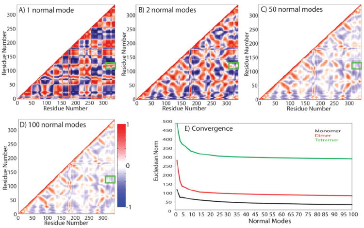
The recently discovered impact of oligomerization on G-protein coupled receptor (GPCR) function further complicates the already challenging goal of unraveling the molecular and dynamic mechanisms of these receptors. To help understand the effect of oligomerization on the dynamics of GPCRs, we have compared the motion of monomeric, dimeric, and tetrameric arrangements of the prototypic GPCR rhodopsin, using an approximate-yet powerful-normal mode analysis (NMA) technique termed elastic network model (ENM). Moreover, we have used ENM to discriminate between putative dynamic mechanisms likely to account for the recently observed conformational rearrangement of the TM4,5-TM4,5 dimerization interface of GPCRs that occurs upon activation. Our results indicate: (1) significant perturbation of the normal modes (NMs) of the rhodopsin monomer upon oligomerization, which is mainly manifested at interfacial regions; (2) increased positive correlation among the transmembrane domains (TMs) and between the extracellular loop (EL) and TM regions of the rhodopsin protomer; (3) highest interresidue positive correlation at the interfaces between protomers; and (4) experimentally testable hypotheses of differential motional changes within different putative oligomeric arrangements.


Figures

Figure 1
Figure 1
(A) Intracellular view of the 1N3M tetrameric arrangement. Protomer A is shown in red, B in cyan, C in green, and D in blue. (B) Activated interface model obtained by clockwise rotations of contacting TM4 helices along their own helical axes; (C) Activated interface model obtained by protomer displacement (i.e. clockwise rotation of each protomer involved in intradimeric interaction along the membrane axis; and (D) Activated interface model obtained by protomer exchange (i.e. clockwise rotation of each protomer involved in intradimeric interaction followed by their sliding within the array).
Figure 2
Figure 2
Fractions of (A) translational and (B) rotational motions of the rhodopsin monomer in the low frequency nonzero modes of the 1N3M tetrameric arrangement (gren line) and the three putative activated arrangements of the TM4,5-TM4,5 interface (gray line for TM4 rotation, blue line for protomer exchange, and red line for protomer displacement).
Figure 3
Figure 3
(A) B-factors calculated according to Eq. (5) of Materials and Methods for the first 50 lowest normal modes of rhodopsin monomer, dimer, and tetramer. (B) Vertical view of protomers A and B colored by the fluctuations calculated for the dimer, ranging from red (high fluctuations) to blue (no fluctuations). The Cα atoms of residues 140 to 150 in IL2 region are shown in VDW representation, prepared with VMD.
Figure 4
Figure 4
Convergence of covariance matrices of rhodopsin monomer. High correlation is shown in red, high anti-correlation in blue, and noncorrelated regions in white. Covariance is calculated according to Eq. (6) of Materials and Methods for: (A) one lowest nonzero frequency normal mode; (B) two lowest nonzero frequency normal modes; (C) 50 lowest nonzero frequency modes; and (D) 100 lowest nonzero frequency modes. Green boxes highlight one of the regions (TM3/helix8 and C-terminus) that is positively correlated in normal Mode 1, but anticorrelated when more modes are included in the calculation. (E) Eucledian norm of correlation matrices calculated using an increasing number of normal modes to estimate convergence. Covariance matrices were calculated with MatLab version 7.
Figure 5
Figure 5
Interresidue correlations in (A) rhodopsin monomer, (B) protomer A of rhodopsin dimer, and (C) protomer A of rhodopsin tetramer. Covariance matrices [Eq. (6) of Materials and Methods] were calculated with MatLab version 7 using 50 lowest frequency normal modes.
Figure 6
Figure 6
Interresidue correlations between (A) protomers A and B within the rhodopsin dimer, (B) protomers A and B within the tetramer, and (C) protomers A and C within the tetramer. Covariance matrices were calculated as for Figure 5.
Figure 7
Figure 7
Contribution of lowest frequency normal modes of the rhodopsin tetramer to the conformational rearrangements of the interface of GPCR oligomerization deriving from: (A) TM4 rotation, (B) protomer displacement, and (C) protomer exchange. Overlap (black line) and Cumulative (red line) were calculated according to Eqs. (3) and (4), respectively (see Materials and Methods).
Figure 8
Figure 8
Residue fluctuations of protomer A within the tetrameric arrangements of rhodopsin.

Similar articles

Cited by

References

    1. Muller G. Towards 3D structures of G protein-coupled receptors: a multidisciplinary approach. Curr Med Chem. 2000;7:861–888. - PubMed
    1. George SR, O’Dowd BF, Lee SP. G-protein-coupled receptor oligomerization and its potential for drug discovery. Nat Rev Drug Discov. 2002;1:808–820. - PubMed
    1. Filizola M, Weinstein H. The structure and dynamics of GPCR oligomers: a new focus in models of cell-signaling mechanisms and drug design. Curr Opin Drug Discov Dev. 2005;8:577–584. - PubMed
    1. Milligan G. G-protein-coupled receptor heterodimers: pharmacology, function and relevance to drug discovery. Drug Discov Today. 2006;11:541–549. - PubMed
    1. Liang Y, Fotiadis D, Filipek S, Saperstein DA, Palczewski K, Engel A. Organization of the G protein-coupled receptors rhodopsin and opsin in native membranes. J Biol Chem. 2003;278:21655–21662. - PMC - PubMed

Publication types

LinkOut - more resources