Different modulation of the cortical silent period by two phases of short interval intracortical inhibition
- PMID: 17963336
- PMCID: PMC2628145
- DOI: 10.3349/ymj.2007.48.5.795
Different modulation of the cortical silent period by two phases of short interval intracortical inhibition
Abstract
Purpose: To investigate the influence of 2 phases of short interval intracortical inhibition (SICI) on the cortical silent period (SP).
Materials and methods: Single- and paired-pulse transcranial magnetic stimulations (TMSs) at 1 and 2.5ms interstimulus intervals (ISIs) were applied to the left motor cortex in 12 healthy subjects while their right hand muscles were moderately activated. Conditioning stimulation intensity was 90% of the active motor threshold (AMT). Test stimulation intensities were 120, 140, 160, 180, 200, 220, 240, 260% of the AMT and at 100% of the maximal stimulator output, the order of which was arranged randomly. The rectified electromyography area of motor evoked potential (MEP) and duration of the SP were measured off-line using a computerized program.
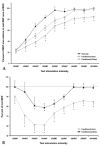
Results: At high-test stimulation intensities, MEP areas were saturated in both single- and paired-pulse stimulations, except that saturated MEPs were smaller for the paired-pulse TMS at 1ms ISI than for the other conditions. As the test stimulation intensity increased, SP was progressively prolonged in both single- and paired-pulse stimulations but was shorter in paired-pulse than single-pulse TMS. Overall, the ratio of SP duration/MEP area was comparable between single- and paired-pulse TMS except for the paired-pulse TMS at 1 ms ISI with a test stimulation intensity at 140-180% of the AMT, in which the ratio was significantly higher than in the single pulse TMS.
Conclusion: These results suggest that 2 phases of SICI modulate MEP saturation and SP duration differently and provide additional evidence supporting the view that 2 phases of SICI are mediated by different inhibitory mechanisms.
Figures


Similar articles
-
Modulation of the cortical silent period elicited by single- and paired-pulse transcranial magnetic stimulation.BMC Neurosci. 2013 Apr 2;14:43. doi: 10.1186/1471-2202-14-43. BMC Neurosci. 2013. PMID: 23547559 Free PMC article.
-
Conditioning the cortical silent period with paired transcranial magnetic stimulation.Brain Stimul. 2013 Jul;6(4):541-4. doi: 10.1016/j.brs.2012.09.011. Epub 2012 Oct 13. Brain Stimul. 2013. PMID: 23092703
-
Intracortical inhibition in the human trigeminal motor system.Clin Neurophysiol. 2007 Aug;118(8):1785-93. doi: 10.1016/j.clinph.2007.05.003. Epub 2007 Jun 15. Clin Neurophysiol. 2007. PMID: 17574911
-
Spread of electrical activity at cortical level after repetitive magnetic stimulation in normal subjects.Exp Brain Res. 2002 Nov;147(2):186-92. doi: 10.1007/s00221-002-1237-z. Epub 2002 Sep 20. Exp Brain Res. 2002. PMID: 12410333
-
Effects of low-frequency whole-body vibration on motor-evoked potentials in healthy men.Exp Physiol. 2009 Jan;94(1):103-16. doi: 10.1113/expphysiol.2008.042689. Epub 2008 Jul 25. Exp Physiol. 2009. PMID: 18658234
Cited by
-
The effect of test TMS intensity on short-interval intracortical inhibition in different excitability states.Exp Brain Res. 2009 Feb;193(2):267-74. doi: 10.1007/s00221-008-1620-5. Epub 2008 Oct 31. Exp Brain Res. 2009. PMID: 18974984
-
Modulation of motor cortex excitability in obsessive-compulsive disorder: an exploratory study on the relations of neurophysiology measures with clinical outcome.Psychiatry Res. 2013 Dec 30;210(3):1026-32. doi: 10.1016/j.psychres.2013.08.054. Epub 2013 Sep 21. Psychiatry Res. 2013. PMID: 24064461 Free PMC article. Clinical Trial.
-
Modulation of the cortical silent period elicited by single- and paired-pulse transcranial magnetic stimulation.BMC Neurosci. 2013 Apr 2;14:43. doi: 10.1186/1471-2202-14-43. BMC Neurosci. 2013. PMID: 23547559 Free PMC article.
References
-
- Hallett M. Transcranial magnetic stimulation: a useful tool for clinical neurophysiology. Ann Neurol. 1996;40:344–345. - PubMed
-
- Fuhr P, Agostino R, Hallett M. Spinal motor neuron excitability during the silent period after cortical stimulation. Electroencephalogr Clin Neurophysiol. 1991;81:257–262. - PubMed
-
- Fisher RJ, Nakamura Y, Bestmann S, Rothwell JC, Bostock H. Two phases of intracortical inhibition revealed by transcranial magnetic threshold tracking. Exp Brain Res. 2002;143:240–248. - PubMed
Publication types
MeSH terms
LinkOut - more resources
Full Text Sources
Research Materials

