. 2007 Oct 26:8:97.
doi: 10.1186/1471-2199-8-97.

Extensive ssDNA end formation at DNA double-strand breaks in non-homologous end-joining deficient cells during the S phase

Affiliations

Extensive ssDNA end formation at DNA double-strand breaks in non-homologous end-joining deficient cells during the S phase

Karin H Karlsson et al. BMC Mol Biol. .

Abstract

Background: Efficient and correct repair of DNA damage, especially DNA double-strand breaks, is critical for cellular survival. Defects in the DNA repair may lead to cell death or genomic instability and development of cancer. Non-homologous end-joining (NHEJ) is the major repair pathway for DNA double-strand breaks in mammalian cells. The ability of other repair pathways, such as homologous recombination, to compensate for loss of NHEJ and the ways in which contributions of different pathways are regulated are far from fully understood.

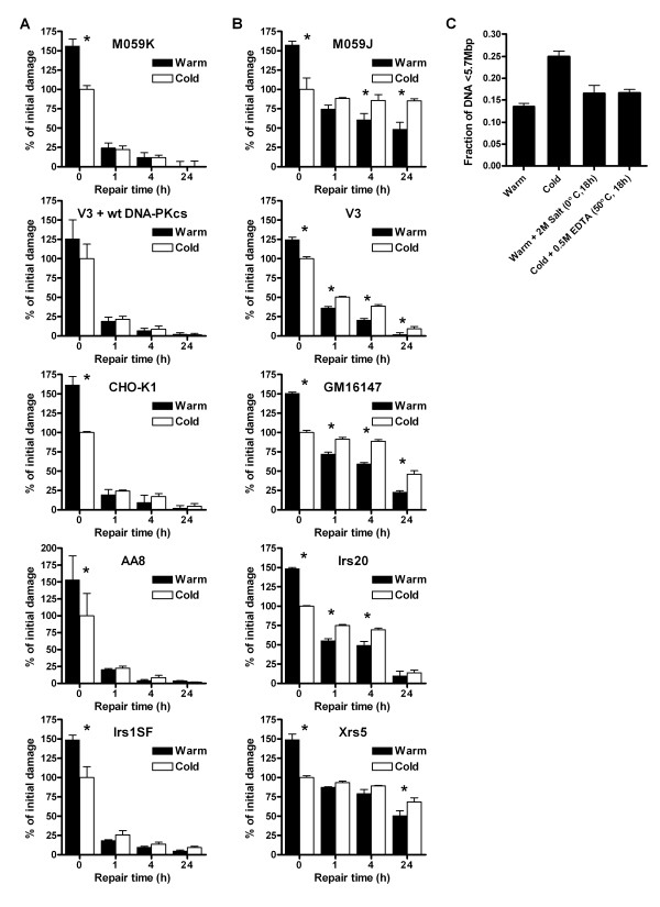
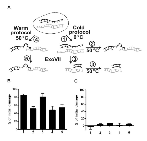
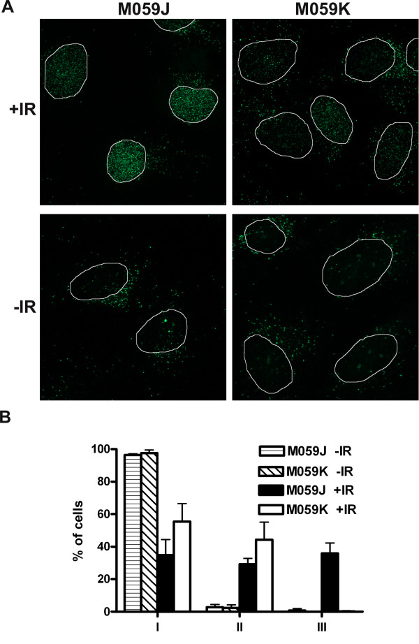
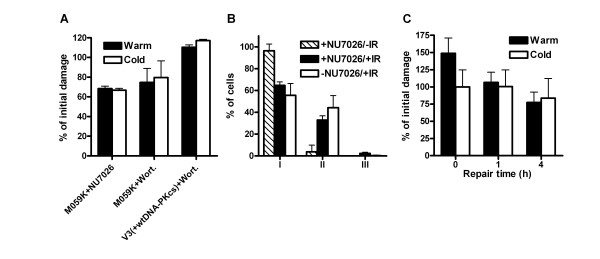
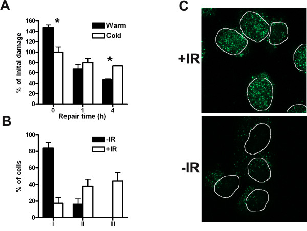
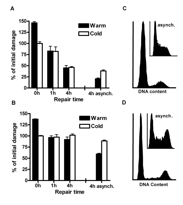
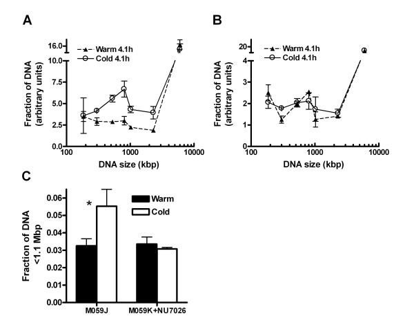
Results: In this report we demonstrate that long single-stranded DNA (ssDNA) ends are formed at radiation-induced DNA double-strand breaks in NHEJ deficient cells. At repair times > or = 1 h, processing of unrejoined DNA double-strand breaks generated extensive ssDNA at the DNA ends in cells lacking the NHEJ protein complexes DNA-dependent protein kinase (DNA-PK) or DNA Ligase IV/XRCC4. The ssDNA formation was cell cycle dependent, since no ssDNA ends were observed in G1-synchronized NHEJ deficient cells. Furthermore, in wild type cells irradiated in the presence of DNA-PKcs (catalytic subunit of DNA-PK) inhibitors, or in DNA-PKcs deficient cells complemented with DNA-PKcs mutated in six autophosphorylation sites (ABCDE), no ssDNA was formed. The ssDNA generation also greatly influences DNA double-strand break quantification by pulsed-field gel electrophoresis, resulting in overestimation of the DNA double-strand break repair capability in NHEJ deficient cells when standard protocols for preparing naked DNA (i. e., lysis at 50 degrees C) are used.

Conclusion: We provide evidence that DNA Ligase IV/XRCC4 recruitment by DNA-PK to DNA double-strand breaks prevents the formation of long ssDNA ends at double-strand breaks during the S phase, indicating that NHEJ components may downregulate an alternative repair process where ssDNA ends are required.


Figures

Figure 1
Figure 1
Heat reduced mobility of unrejoined DNA fragments in NHEJ deficient cells. (A) NHEJ proficient cells (M059K, V3+wt DNA-PKcs, CHO-K1, AA8 and Irs1SF (XRCC3 mutant)) or (B) NHEJ deficient cells (M059J (DNA-PKcs mutant), V3 (DNA-PKcs mutant), Irs20 (DNA-PKcs mutant), GM16147 (XRCC4 mutant) and Xrs5 (Ku80 mutant)) were irradiated and were allowed to repair the DNA for up to 24 h. Naked DNA was produced either by a warm or cold DNA extraction protocol and was separated by PFGE. The amount of damage detected at t = 0 h in cells lysed by the cold protocol was set to 100% (cont. on opposite page). The damage above this level detected by the warm DNA extraction protocol at 0 h consists of non-true DSBs due to the inclusion of artifactual DSBs at 50°C; these non-true DSBs were repaired within 1 h after irradiation. (C) 24 h after irradiation M059J (NHEJ deficient) cells were lysed with four different protocols: 1. Warm DNA extraction (50°C, 18 h); 2. Cold DNA extraction (0°C, 2 × 18 h); 3. Warm DNA extraction (50°C, 18 h) + 2 M Salt (0°C, 18 h); 4. Cold DNA extraction (0°C, 2 × 18 h) + 0.5 M EDTA (50°C, 18 h). Error bars represent the SD of at least three experiments. *Denotes significant difference with paired t-test (p < 0.05) between warm and cold extraction.
Figure 2
Figure 2
ssDNA end formation at DSBs in NHEJ deficient cells. Cells 24 h after irradiation were treated in five different ways (A): 1. Cold DNA extraction; 2. Cold DNA extraction + 18 h at 50°C; 3. Cold DNA extraction + ExoVII + 18 h at 50°C; 4. Warm DNA extraction; 5. Warm DNA extraction + ExoVII. In (B) and (C) the relative amount of initial damage in the NHEJ deficient M059J cell line and the NHEJ proficient M059K cell line, respectively are shown after the different treatments outlined in A. The level of damage at 0 h with the cold DNA extraction protocol was set to 100%. The error bars represent the SD of three experiments (M059J) or the maximum deviations of two experiments (M059K).
Figure 3
Figure 3
ssDNA (BrdU foci) in NHEJ deficient cells. M059J (NHEJ deficient) and M059K (NHEJ proficient) cells were fixed 4 h after irradiation. (A) BrdU corresponding to ssDNA was detected with immunofluorescence, and (B) cells were scored as belonging to one of the following categories: I. Weak staining in the whole nucleus; II. Increased staining in the nucleus compared to I, dots are present in the whole nucleus; III. Strong staining in the nucleus, many dots with high intensity in the whole nucleus. At least 100 cells in three experiments were scored. Error bars represent the SD.
Figure 4
Figure 4
Inhibition of DNA-PKcs autophosphorylation does not result in ssDNA formation. (A) The catalytic activity of DNA-PKcs was inhibited by 50 μM NU7026 or 50 μM wortmannin in M059K cells and V3+wild type DNA-PKcs cells. At 4 h after irradiation the DNA was extracted either with the warm or the cold protocol and the number of DNA fragments was detected by PFGE. The number of DNA fragments detected at 0 h by the cold protocol was set to 100%. The error bars represent the maximum deviation of two experiments (M059K+NU7026), the standard error of the mean of three experiments (M059K+Wortmannin), and the maximum deviation of duplicates from one experiment (V3+wt DNA-PKcs). (B) BrdU corresponding to ssDNA was detected 4 h after irradiation in M059K cells treated with 50 μM NU7026 and cells were scored as in Figure 3. Irradiated M059K cells without NU7026 are plotted for comparison. At least 100 cells in three experiments were scored. (C) V3 cells expressing DNA-PKcs with six autophosphorylation mutated sites (ABCDE) were treated as in (A) after irradiation. Error bars represent the SD of at least three experiments.
Figure 5
Figure 5
Inhibition of the catalytic activity of DNA-PKcs in XRCC4 deficient cells does not prevent the processing of DSB ends into ssDNA. (A) GM16147 (XRCC4 deficient) cells were incubated with 50 μM NU7026 and the DNA was extracted with the warm or cold protocol at different repair times after irradiation. The number of DNA fragments was determined by PFGE and normalized to the number of DNA fragments at 0 h by the cold protocol. Error bars represent the SD of three experiments. *Denotes significant difference with paired t-test (p < 0.05) between warm and cold extraction. (B and C) BrdU corresponding to ssDNA was detected 4 h after irradiation in GM16147 cells treated with 50 μM NU7026 and cells were scored as belonging to one of the following categories: I. Weak BrdU staining in the whole nucleus; II. Increased BrdU staining in the nucleus compared to I or > 20 strong dots in the nucleus; III. Strong dots in the whole nucleus and background staining in the whole nucleus. At least 100 cells in three experiments were scored. Error bars represent the SD.
Figure 6
Figure 6
G1 synchronization of NHEJ deficient cells prevents the formation of ssDNA at DSBs. (A) G1-synchronized V3 (DNA-PKcs deficient) and (B) GM16147 (XRCC4 deficient) cells were irradiated and at different repair times the DNA was extracted with the warm or cold protocol. The number of DNA fragments was determined by PFGE and the number of DNA fragments detected at 0 h by the cold protocol was set to 100%. The corresponding remaining damage at 4 h in asynchronous cells is plotted for comparison. (C) The V3 and (D) GM16147 cell cycle distributions at the time of irradiation. The corresponding distributions in the asynchronous cell population are seen in the insert. Error bars represent the SD of three experiments.
Figure 7
Figure 7
ssDNA ends at DSBs in S-phase cells lacking DNA-PKcs. DNA-PKcs deficient cells (M059J) and DNA-PKcs inhibited cells (M059K+NU7026) were pulsed with 3H-thymidine 1 h before irradiation with 80 Gy. The size distributions of DNA fragments containing incorporated 3H-thymidine (i.e. DNA from cells in the S phase during the 3H incubation) in M059J and M059K+NU7026 cells at 4.1 h are shown in (A) and (B), respectively. The DNA was extracted by the warm or cold protocol. The amount of fragments < 1.1 Mbp was summed and the total fraction of S-phase DNA < 1.1 Mbp, extracted by the warm and cold protocol, in M059J and M059K+NU7026 was plotted for 4.1 h (C). Error bars represent the SEM from three experiments (M059J) or the maximum error from two experiments (M059K+NU7026).* Denotes a significant difference with the t-test (p < 0.025).

Similar articles

Cited by

References

    1. Hefferin ML, Tomkinson AE. Mechanism of DNA double-strand break repair by non-homologous end joining. DNA Repair (Amst) 2005;4:639–648. - PubMed
    1. Weterings E, van Gent DC. The mechanism of non-homologous end-joining: a synopsis of synapsis. DNA Repair (Amst) 2004;3:1425–1435. doi: 10.1016/j.dnarep.2004.06.003. - DOI - PubMed
    1. DeFazio LG, Stansel RM, Griffith JD, Chu G. Synapsis of DNA ends by DNA-dependent protein kinase. EMBO J. 2002;21:3192–3200. doi: 10.1093/emboj/cdf299. - DOI - PMC - PubMed
    1. Kurimasa A, Kumano S, Boubnov NV, Story MD, Tung CS, Peterson SR, Chen DJ. Requirement for the kinase activity of human DNA-dependent protein kinase catalytic subunit in DNA strand break rejoining. Mol Cell Biol. 1999;19:3877–3884. - PMC - PubMed
    1. Chan DW, Chen BP-C, Prithivirajsingh S, Kurimasa A, Story MD, Qin J, Chen DJ. Autophosphorylation of the DNA-dependent protein kinase catalytic subunit is required for rejoining of DNA double-strand breaks. Genes Dev. 2002;16:2333–2338. doi: 10.1101/gad.1015202. - DOI - PMC - PubMed

Publication types

MeSH terms