

Diagnostic utility of p501s (prostein) in comparison to prostate specific antigen (PSA) for the detection of metastatic prostatic adenocarcinoma
- PMID: 17963516
- PMCID: PMC2174437
- DOI: 10.1186/1746-1596-2-41
Diagnostic utility of p501s (prostein) in comparison to prostate specific antigen (PSA) for the detection of metastatic prostatic adenocarcinoma
Abstract
Background: Immunohistochemical detection of prostate specific antigen (PSA) is widely used to identify metastatic prostatic adenocarcinoma. However, PSA may not be expressed in some poorly differentiated prostatic carcinomas and its immunoreactivity has been found in some non-prostatic tissues. P501s (prostein) is a prostate-specific marker that is expressed in the cytoplasm of benign and malignant prostatic glandular cells. It has not been detected in any other normal or malignant tissues. The purpose of this study was to evaluate the expression of P501s in metastatic prostatic adenocarcinoma and compare its expression with PSA.
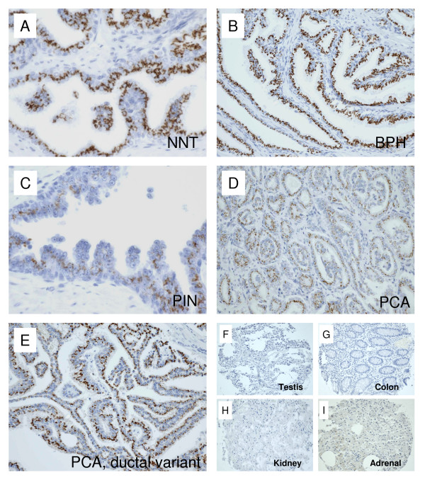
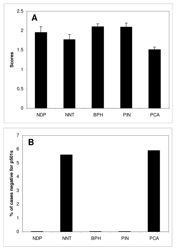
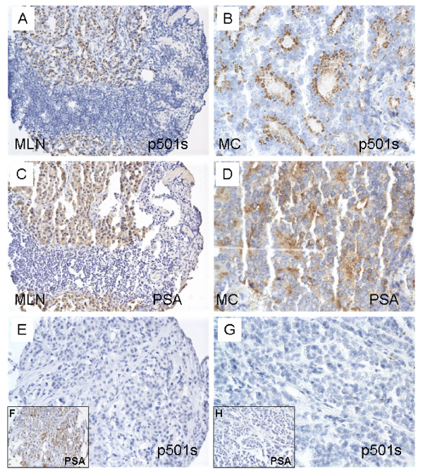
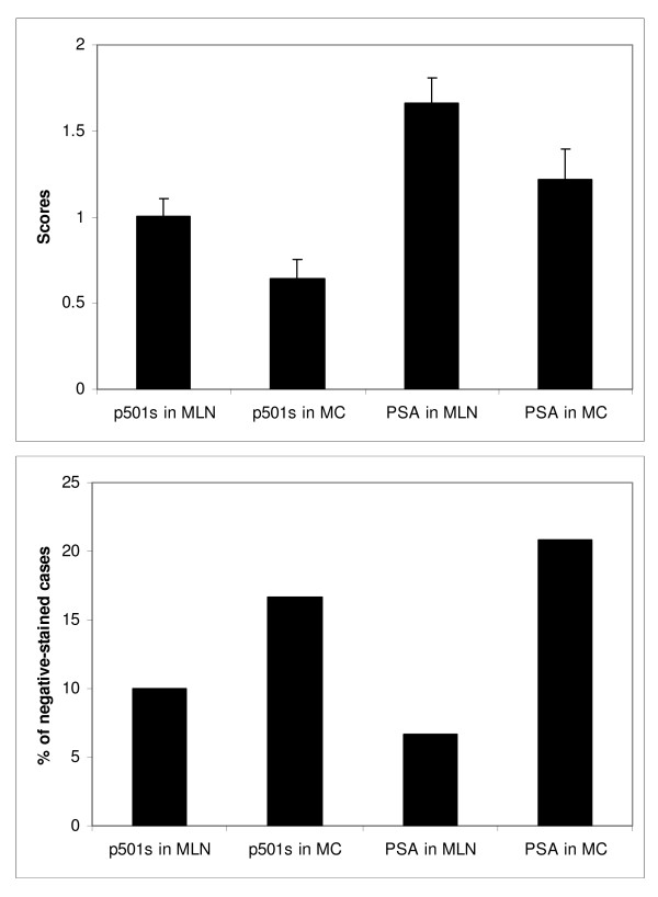
Methods: Immunohistochemical stains with anti-P501s antibodies were performed on 5-micron sections of tissue microarray (TMA) specimens. The TMA is constructed with normal donor prostates (NDP), prostatic adenocarcinoma (PRCA), non-neoplastic prostatic tissues adjacent to malignant glands (NAT), benign prostatic hyperplasia (BPH), high-grade prostatic neoplasia (PIN), metastatic adenocarcinoma to lymph nodes (MLN), metastatic adenocarcinoma to other sites (MC), and samples of benign testis, colon, adrenal and kidney. The two groups of metastatic lesions were also subjected to stains with antibodies to PSA. A composite score (ranging from 0 to 3) was assigned to score intensity of staining.
Results: Granular staining pattern of p501s was seen in all benign glands (score = 1.77 - 2.1) and malignant acini (score = 1.52) at the apical aspect of cytoplasm, predominantly adjacent to the nuclei. No staining was observed in controls including testis, colon, adrenal and kidney. The MLN group received a score of 1.0, with 10% of cases negative for p501s. The MC cases had a score of 0.64, with 16.7% of case showing loss of p501s expression. Although the metastatic lesions demonstrated similar rate of negative expression with PSA antibody, only 2 MC cases (3.3%) showed simultaneous negative stains for both P501S and PSA.
Conclusion: P501s is an organ specific marker for benign and malignant prostatic epithelial cells. Its characteristic cytoplasmic stain pattern provides an additional valuable immunomarker for detection of metastatic prostatic malignancy, even though the intensity of its expression is reduced, as in the case with PSA. Simultaneous stains with P501S and PSA will greatly improve the detection rate and identify a significant majority of the metastases.
Figures
Similar articles
-
The role of P501S and PSA in the diagnosis of metastatic adenocarcinoma of the prostate.Am J Surg Pathol. 2007 Sep;31(9):1351-5. doi: 10.1097/PAS.0b013e3180536678. Am J Surg Pathol. 2007. PMID: 17721190
-
Immunohistochemical expression of prostatic antigens in adenocarcinoma and villous adenoma of the urinary bladder.Am J Surg Pathol. 2008 Sep;32(9):1322-6. doi: 10.1097/PAS.0b013e3181656ca0. Am J Surg Pathol. 2008. PMID: 18670358
-
Immunohistochemical differentiation of high-grade prostate carcinoma from urothelial carcinoma.Am J Surg Pathol. 2007 Aug;31(8):1246-55. doi: 10.1097/PAS.0b013e31802f5d33. Am J Surg Pathol. 2007. PMID: 17667550
-
Best practices recommendations in the application of immunohistochemistry in the prostate: report from the International Society of Urologic Pathology consensus conference.Am J Surg Pathol. 2014 Aug;38(8):e6-e19. doi: 10.1097/PAS.0000000000000238. Am J Surg Pathol. 2014. PMID: 25029122
-
Immunohistochemistry in diagnostic surgical pathology of the prostate.Semin Diagn Pathol. 2005 Feb;22(1):88-104. doi: 10.1053/j.semdp.2005.11.001. Semin Diagn Pathol. 2005. PMID: 16512601 Review.
Cited by
-
Diagnostic utility of p63/P501S double sequential immunohistochemical staining in differentiating urothelial carcinoma from prostate carcinoma.Diagn Pathol. 2011 Jul 21;6:67. doi: 10.1186/1746-1596-6-67. Diagn Pathol. 2011. PMID: 21777423 Free PMC article.
-
Update on histopathological evaluation of lymphadenectomy specimens from prostate cancer patients.World J Urol. 2017 Apr;35(4):517-526. doi: 10.1007/s00345-015-1752-8. Epub 2015 Dec 22. World J Urol. 2017. PMID: 26694187 Review.
-
Sensitivity of HOXB13 as a Diagnostic Immunohistochemical Marker of Prostatic Origin in Prostate Cancer Metastases: Comparison to PSA, Prostein, Androgen Receptor, ERG, NKX3.1, PSAP, and PSMA.Int J Mol Sci. 2017 May 29;18(6):1151. doi: 10.3390/ijms18061151. Int J Mol Sci. 2017. PMID: 28555048 Free PMC article.
-
Recent Advances in Nanomedicine for the Diagnosis and Treatment of Prostate Cancer Bone Metastasis.Molecules. 2021 Jan 13;26(2):384. doi: 10.3390/molecules26020384. Molecules. 2021. PMID: 33450939 Free PMC article. Review.
-
NKX3.1 and PSMA are sensitive diagnostic markers for prostatic carcinoma in bone metastasis after decalcification of specimens.Am J Clin Exp Urol. 2018 Oct 20;6(5):182-188. eCollection 2018. Am J Clin Exp Urol. 2018. PMID: 30510970 Free PMC article.
References
-
- J.I. Epstein FA, W.C. Allsbrook Jr., S. Bastacky, L. Boccon-Gibod, A.M. De Marzo, L. Egevad, M. Furusato,, Hamper UM. Acinar adenocarcinoma. In: John N. Eble GSJIEIAS, editor. WHO Classification of Tumours: Pathology and genetics of tumours of the urinary system and male genital organs. Lyon , IARC Press; 2004. p. 162.
-
- Landis SH, Murray T, Bolden S, Wingo PA. Cancer statistics, 1999. CA Cancer J Clin. 1999;49:8–31, 1. - PubMed
-
- Epstein JI, Walsh PC, Brendler CB. Radical prostatectomy for impalpable prostate cancer: the Johns Hopkins experience with tumors found on transurethral resection (stages T1A and T1B) and on needle biopsy (stage T1C) J Urol. 1994;152:1721–1729. - PubMed
LinkOut - more resources
Full Text Sources
Other Literature Sources
Research Materials
Miscellaneous
