

Post-duplication charge evolution of phosphoglucose isomerases in teleost fishes through weak selection on many amino acid sites
- PMID: 17963532
- PMCID: PMC2176064
- DOI: 10.1186/1471-2148-7-204
Post-duplication charge evolution of phosphoglucose isomerases in teleost fishes through weak selection on many amino acid sites
Abstract
Background: The partitioning of ancestral functions among duplicated genes by neutral evolution, or subfunctionalization, has been considered the primary process for the evolution of novel proteins (neofunctionalization). Nonetheless, how a subfunctionalized protein can evolve into a more adaptive protein is poorly understood, mainly due to the limitations of current analytical methods, which can detect only strong selection for amino acid substitutions involved in adaptive molecular evolution. In this study, we employed a comparative evolutionary approach to this question, focusing on differences in the structural properties of a protein, specifically the electric charge, encoded by fish-specific duplicated phosphoglucose isomerase (Pgi) genes.
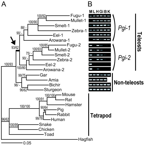
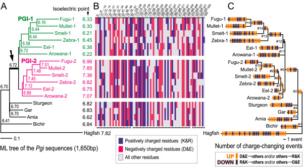
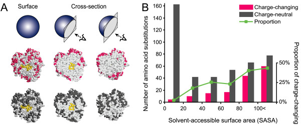
Results: Full-length cDNA cloning, RT-PCR based gene expression analyses, and comparative sequence analyses showed that after subfunctionalization with respect to the expression organ of duplicate Pgi genes, the net electric charge of the PGI-1 protein expressed mainly in internal tissues became more negative, and that of PGI-2 expressed mainly in muscular tissues became more positive. The difference in net protein charge was attributable not to specific amino acid sites but to the sum of various amino acid sites located on the surface of the PGI molecule.
Conclusion: This finding suggests that the surface charge evolution of PGI proteins was not driven by strong selection on individual amino acid sites leading to permanent fixation of a particular residue, but rather was driven by weak selection on a large number of amino acid sites and consequently by steady directional and/or purifying selection on the overall structural properties of the protein, which is derived from many modifiable sites. The mode of molecular evolution presented here may be relevant to various cases of adaptive modification in proteins, such as hydrophobic properties, molecular size, and electric charge.
Figures
Similar articles
-
Phosphoglucose isomerases of hagfish, zebrafish, gray mullet, toad, and snake, with reference to the evolution of the genes in vertebrates.Mol Biol Evol. 2002 Apr;19(4):367-74. doi: 10.1093/oxfordjournals.molbev.a004092. Mol Biol Evol. 2002. PMID: 11919278
-
Electric charge divergence in proteins: insights into the evolution of their three-dimensional properties.Gene. 2009 Jul 15;441(1-2):3-11. doi: 10.1016/j.gene.2008.06.026. Epub 2008 Jul 2. Gene. 2009. PMID: 18652881
-
From DNA to fitness differences: sequences and structures of adaptive variants of Colias phosphoglucose isomerase (PGI).Mol Biol Evol. 2006 Mar;23(3):499-512. doi: 10.1093/molbev/msj062. Epub 2005 Nov 16. Mol Biol Evol. 2006. PMID: 16292000 Free PMC article.
-
Molecular evolution of Na+ channels in teleost fishes.Integr Zool. 2009 Mar;4(1):64-74. doi: 10.1111/j.1749-4877.2008.00136.x. Integr Zool. 2009. PMID: 21392277 Review.
-
Evolution of the duplicated intracellular lipid-binding protein genes of teleost fishes.Mol Genet Genomics. 2017 Aug;292(4):699-727. doi: 10.1007/s00438-017-1313-5. Epub 2017 Apr 7. Mol Genet Genomics. 2017. PMID: 28389698 Review.
Cited by
-
Molecular evolution of teleost neural isozymes.J Mol Evol. 2012 Dec;75(5-6):198-213. doi: 10.1007/s00239-012-9532-1. Epub 2012 Nov 25. J Mol Evol. 2012. PMID: 23183893
-
Whole-genome duplication and the functional diversification of teleost fish hemoglobins.Mol Biol Evol. 2013 Jan;30(1):140-53. doi: 10.1093/molbev/mss212. Epub 2012 Sep 4. Mol Biol Evol. 2013. PMID: 22949522 Free PMC article.
-
Chromosomal-Level Assembly of the Asian Seabass Genome Using Long Sequence Reads and Multi-layered Scaffolding.PLoS Genet. 2016 Apr 15;12(4):e1005954. doi: 10.1371/journal.pgen.1005954. eCollection 2016 Apr. PLoS Genet. 2016. PMID: 27082250 Free PMC article.
-
Evolution of multiple phosphodiesterase isoforms in stickleback involved in cAMP signal transduction pathway.BMC Syst Biol. 2009 Feb 20;3:23. doi: 10.1186/1752-0509-3-23. BMC Syst Biol. 2009. PMID: 19232106 Free PMC article.
-
When a foreign gene meets its native counterpart: computational biophysics analysis of two PgiC loci in the grass Festuca ovina.Sci Rep. 2020 Oct 30;10(1):18752. doi: 10.1038/s41598-020-75650-0. Sci Rep. 2020. PMID: 33127989 Free PMC article.
References
-
- Ohno S. Evolution by gene duplication. New York: Springer-Verlag; 1970.
Publication types
MeSH terms
Substances
LinkOut - more resources
Full Text Sources
