. 2007 Oct 26;28(2):214-27.
doi: 10.1016/j.molcel.2007.08.029.

Pyrin activates the ASC pyroptosome in response to engagement by autoinflammatory PSTPIP1 mutants

Affiliations

Pyrin activates the ASC pyroptosome in response to engagement by autoinflammatory PSTPIP1 mutants

Je-Wook Yu et al. Mol Cell. .

Abstract

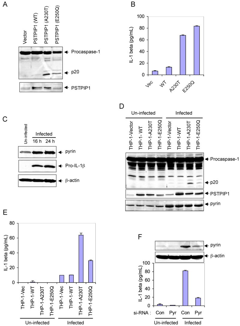
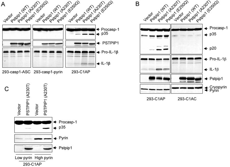
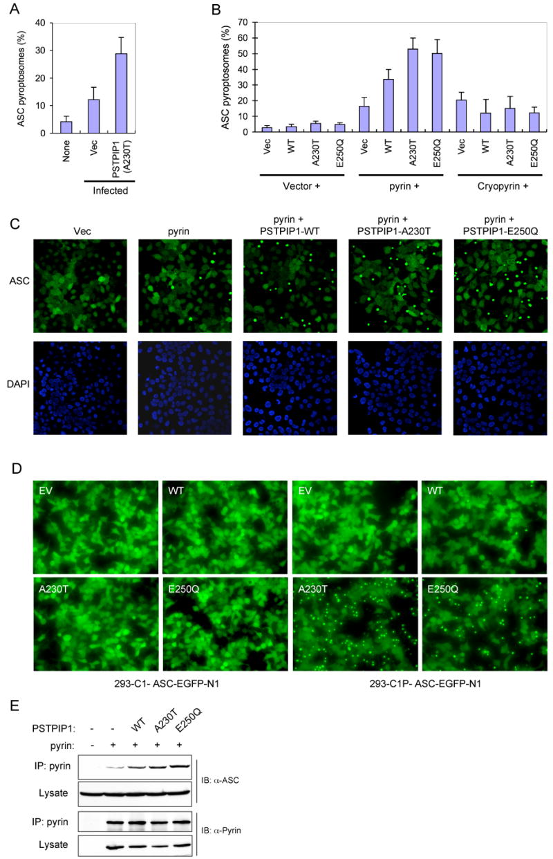
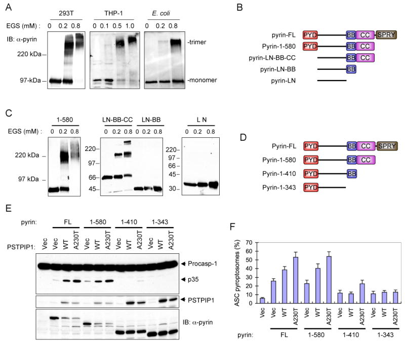
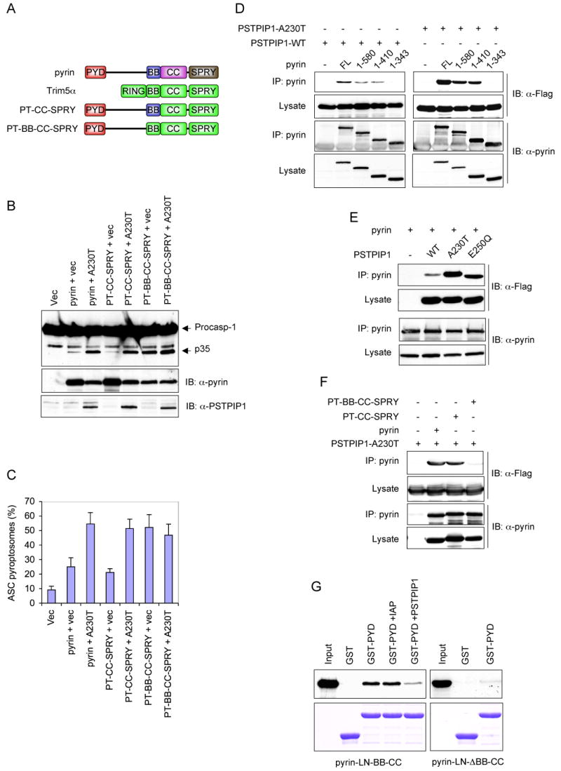
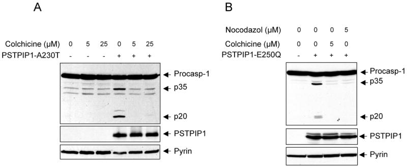
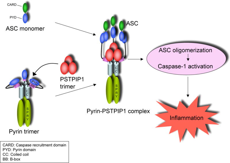
The molecular mechanism by which mutations in the cytoskeleton-organizing protein PSTPIP1 cause the autoinflammatory PAPA syndrome is still elusive. Here, we demonstrate that PSTPIP1 requires the familial Mediterranean fever protein pyrin to assemble the ASC pyroptosome, a molecular platform that recruits and activates caspase-1. We provide evidence that pyrin is a cytosolic receptor for PSTPIP1. Pyrin exists as a homotrimer in an autoinhibited state due to intramolecular interactions between its pyrin domain (PYD) and B-box. Ligation by PSTPIP1, which is also a homotrimer, activates pyrin by unmasking its PYD, thereby allowing it to interact with ASC and facilitate ASC oligomerization into an active ASC pyroptosome. Because of their high binding affinity to pyrin's B-box, PAPA-associated PSTPIP1 mutants were found to be more effective than WT PSTPIP1 in inducing pyrin activation. Therefore, constitutive ligation and activation of pyrin by mutant PSTPIP1 proteins explain the autoinflammatory phenotype seen in PAPA syndrome.


Figures

Figure 1
Figure 1. PAPA-associated PSTPIP1 mutants induce caspase-1 activation in THP-1 cells
(A) Western blot analyses showing caspase-1 and PSTPIP1 in THP-1 cells 24h post-infection with an empty MSCV retroviral vector, or MSCV retroviruses encoding the indicated WT or mutant PSTPIP1 proteins. (B) IL-1β in the culture media of the cells described in A (mean ± SD; n = 3). (C) Western blot analyses showing pyrin, pro-IL-1β and β-actin in un-infected or empty MSCV retrovirus-infected THP-1 cells at 16 and 24h post-infection. (D) Western blot analyses showing caspase-1, PSTPIP1 and pyrin in the indicated stable THP-1 cells, which were left without infection (un-infected) or infected with empty MSCV retrovirus for 24h. (E) IL-1β in the culture media of the cells described in D (mean ± SD; n = 3). (F) Stable THP-1-A230T cells were transfected with control non-specific (Con) or pyrin-specific (Pyr) siRNAs as indicated. The cells were then left untreated (Un-infected) or infected with an empty MSCV retrovirus for 24h. The secreted IL-1β in the culture media of these cells is shown (mean ± SD; n = 3). The upper panels show western blots of Pyrin and β-actin in these cells.
Figure 2
Figure 2. Pyrin is required for activation of caspase-1 by PSTPIP1
(A-C) The indicated stable 293T cell lines were transfected with the indicated plasmids. Caspase-1 and IL-1β processing were determined as in “Experimental Procedures”.
Figure 3
Figure 3. PSTPIP1 induces pyrin-dependent ASC oligomerization
(A) Percentages of ASC pyroptosomes in THP-1-ASC-GFP cells, which were left without infection (none) or infected with an empty MSCV (Vec) or PSTPIP1-A230T-encoding (A230T) retroviral vectors (mean ± SD; n = 5). (B) Percentages of ASC pyroptosomes in 293-ASC-EGFP-N1 cells which were co-transfected with an empty vector (1st to 4th columns) or plasmids encoding pyrin (5th to 8th columns) or cryopyrin (9th to 12th columns) together with an empty vector (Vec), or the indicated PSTPIP1 plasmids (WT, A230T, E250Q) (mean ± SD; n = 5). (C) Fluorescence confocal micrographs showing ASC-GFP (green) or DAPI-stained nuclei (Blue) in 293-ASC-EGFP-N1 cells 24h after transfection with empty vector or the indicated plasmids. (D) Fluorescence confocal micrographs showing ASC-GFP in 293-caspase-1-ASC-EGFP-N1 cells or 293-C1P-ASC-EGFP-N1 cells 24h after transfection with empty vector (EV) or the indicated PSTPIP1 expression plasmids (WT, A230T, and E250Q). (E) Pyrin was immunoprecipitated (IP) from 293-ASC cell lysates 24h after co-transfection with pcDNA-pyrin-myc-His plasmid (+) together with constructs for the indicated WT or mutant PSTPIP1 proteins. The lysates and IPs were fractionated by SDS-PAGE and immunoblotted (IB) with ASC or pyrin antibodies.
Figure 4
Figure 4. Pyrin is a homotrimer
(A) Western blots showing pyrin from 293T, THP-1 or E. coli after cross-linking with the indicated concentrations of EGS. (B) Schematic representations of the domain structure of the full-length pyrin (FL) and the truncated pyrin mutants used in C below. PYD, pyrin domain; BB, B-Box; CC, coiled-coil; SPRY, domain in SPIa and Ryanodine receptor. (C) Western blots showing the indicated bacterially-expressed truncated pyrin mutants after cross-linking with the indicated concentrations of EGS. (D) Schematic representations of the three C-terminal-truncated pyrin mutants (1-580, 1-410 and 1-343) used in E below. (E) Western blots showing caspase-1, PSTPIP1 and pyrin in 293-caspase-1-ASC cells 28h after transfection with an empty vector (1st lane), or co-transfection with the indicated pyrin expression plasmids together with an empty vector (Vec), or the indicated PSTPIP1 plasmids (WT, A230T, E250Q). The decrease in pyrin expression (FL and 1-580) in the presence of PSTPIP1 is due to cell death and cleavage of pyrin by the activated caspase-1. Our data show that caspase-1 cleaves pyrin into smaller fragments (see supplementary Fig. 5) (F) Percentages of ASC pyroptosomes in 293-ASC-EGFP-N1 cells similarly transfected as in E (mean ± SD; n = 5).
Figure 5
Figure 5. The B-box regulates the activity of pyrin
(A) Schematic representations of the domain structure of pyrin and Trim5α, and the chimeric pyrin-Trim5α mutants used in B, C and F below. (B) Western blots showing caspase-1, PSTPIP1 and pyrin or chimeric pyrin mutants in 293-caspase-1-ASC cells 28h after transfection with an empty vector (1st lane), or co-transfection with the indicated pyrin or chimeric pyrin-Trim5α mutants plus an empty vector (vec) or PSTPIP1-A230T mutant plasmid (A230). (C) Percentages of ASC pyroptosomes in 293-ASC-EGFP-N1 cells similarly transfected as in B (mean ± SD; n = 5). (D-F) Pyrin or pyrin mutants were immunoprecipitated (IP) from HEK293 cell lysates 24h after transfection with the indicated plasmids for full-length (FL) pyrin or pyrin mutants plus the indicated Flag-tagged PSTPIP1 plasmids. The lysates and IPs were fractionated by SDS-PAGE and immunoblotted (IB) with anti-pyrin or anti-Flag (detects PSTPIP1) antibodies. (G) The PYD of pyrin interacts with its B-box. GST or GST-PYD fusion protein (pyrin PYD residues 1-100) were incubated with 35S-labeled pyrin-LN-BB-CC (left panels) or pyrin-LN-ΔBB-CC (right panels) in the absence (2nd and 3rd lanes) or the presence of dIAP as a non-specific control (4th lane) or PSTPIP1 A230T (5th lane). The bound proteins were fractionated by SDS-PAGE and detected by autoradiography (top panels). The corresponding immobilized GST and GST-PYD proteins are shown in the lower panels.
Figure 6
Figure 6. Microtubule disrupting agents inhibit pyrin-induced caspase-1 activation
(A and B) Western blots showing caspase-1, PSTPIP1 and pyrin in 293-C1AP cells 24h after transfection with an empty vector or the indicated PSTPIP1 plasmids in the presence of the indicated concentrations of colchicine or nocodazol.
Figure 7
Figure 7. Mechanism of activation of pyrin by PSTPIP1
See discussion for details.

References

    1. Badour K, Zhang J, Shi F, McGavin MK, Rampersad V, Hardy LA, Field D, Siminovitch KA. The Wiskott-Aldrich syndrome protein acts downstream of CD2 and the CD2AP and PSTPIP1 adaptors to promote formation of the immunological synapse. Immunity. 2003;18:141–154. - PubMed
    1. Bao Q, Shi Y. Apoptosome: a platform for the activation of initiator caspases. Cell Death Differ. 2007;14:56–65. - PubMed
    1. Cao T, Borden KL, Freemont PS, Etkin LD. Involvement of the rfp tripartite motif in protein–protein interactions and subcellular distribution. J Cell Sci. 1997;110:1563–1571. - PubMed
    1. Centola M, Wood G, Frucht DM, Galon J, Aringer M, Farrell C, Kingma DW, Horwitz ME, Mansfield E, Holland SM, et al. The gene for familial Mediterranean fever, MEFV, is expressed in early leukocyte development and is regulated in response to inflammatory mediators. Blood. 2000;95:3223–3231. - PubMed
    1. Chae JJ, Centola M, Aksentijevich I, Dutra A, Tran M, Wood G, Nagaraju K, Kingma DW, Liu PP, Kastner DL. Isolation, genomic organization, and expression analysis of the mouse and rat homologs of MEFV, the gene for familial mediterranean fever. Mamm Genome. 2000;11:428–435. - PubMed

Publication types

MeSH terms