. 2007 Oct 26;28(2):240-52.
doi: 10.1016/j.molcel.2007.09.026.

U2 snRNP binds intronless histone pre-mRNAs to facilitate U7-snRNP-dependent 3' end formation

Affiliations

U2 snRNP binds intronless histone pre-mRNAs to facilitate U7-snRNP-dependent 3' end formation

Kyle Friend et al. Mol Cell. .

Abstract

In metazoa, pre-mRNA 3' end formation occurs via two pathways: cleavage/polyadenylation for the majority of RNA polymerase II transcripts and U7-snRNP-dependent cleavage for replication-dependent histone pre-mRNAs. An RNA element derived from a replication-dependent histone gene affects multiple steps of pre-mRNA processing. Here, we demonstrate that a portion of this RNA element, present in the majority of histone mRNAs, stimulates U7-snRNP-dependent cleavage. Surprisingly, this element binds U2 snRNP, although it is derived from an intronless mRNA. Specifically, SF3b, a U2 and U12-snRNP component, contacts the RNA element both in vitro and in vivo in conjunction with hPrp43, a DEAH-box helicase. Tethering either U2 or U12 snRNP to histone pre-mRNA substrates stimulates U7-snRNP-dependent cleavage in vitro and in vivo. Finally, we show that U2 snRNP associates with histone pre-mRNAs in vivo. We conclude that U2 snRNP plays a nonsplicing role in histone mRNA maturation.


Figures

Figure 1
Figure 1. An RNA Element Derived from a Replication-Dependent Histone mRNA Stimulates 3′-end Formation
(A) The U7-snRNP (derived from mouse histone H3 mRNA) and CPA (SV40 late) substrates are schematized with sites of cleavage indicated by arrowheads. The 22-nt RNA element (W) and a control element (C), with changed nucleotides indicated by arrows, have been described (Huang and Steitz, 2001). The 7-nt conserved motif is underlined. (B and C) Time courses of U7-snRNP-dependent cleavage of two U7-snRNP substrates (H3 and H2a) containing four copies of the 22-nt W element or the C element were performed in HeLa nuclear extract (Kolev and Steitz, 2005). The H3 substrate (Pre→Pro) is shown in B; processing of both substrates was quantitated (error bars represent standard deviations; panel C) as the ratio of the 5′ product to total RNA. We verified that the substrates are cleaved in a U7-snRNP-dependent manner (data not shown; Bond et al., 1991; Huang et al., 2003). (D and E) Processing time courses of the SV40 late polyadenylation substrate (Pre) with four copies of either the 22-nt W element or the C element were performed in HeLa nuclear extract (Ryan et al., 2004); note the shorter time required for comparable extent of cleavage in E versus C). In D, cordycepin was added to inhibit polyadenylation of the cleavage product (Pro). E shows quantitations, as in panel C, for both the uncoupled and coupled CPA reactions.
Figure 2
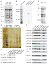
Figure 2. U2 snRNP Binds the 22-nt RNA Element In Vitro and In Vivo
(A)RNAs containing four copies of the 22-nt (W) or control (C) element were UV cross-linked under CPA conditions (Ryan et al., 2004). RNAs were synthesized in the presence of 32P-labeled CTP or GTP as indicated, and cross-linked proteins were separated by 5–15% gradient SDS-PAGE. The 22-nt RNA element (W) cross-linked specifically to three proteins (*) compared to the control RNA (C). SRp20 and 9G8, previously characterized as cross-linking to the RNA element (Huang and Steitz, 2001), are detected for both RNAs labeled at C-residues (†). (B) A CTP-labeled RNA containing four copies of the 22-nt element (W) was incubated and UV-irradiated as in A; immunoprecipitations were performed with the indicated antibodies followed by 5–15% gradient SDS-PAGE. (C) CTP-labeled RNA containing the complete H2a 101-nt RNA element 5′ to the H3-derived U7-snRNP substrate was injected into GVs and UV-irradiated after 0 or 1 hr; cross-linked proteins were separated by 12% SDS-PAGE. Proteins visible at zero time most likely represent a pre-processing heterogeneous RNP, as described for in vitro CPA (Skolnik-David et al., 1987) and serve as a control. A specific 150 kDa cross-linked protein was observed (*), but could not be identified by α-SF3b155 immunoprecipitation because the signal was too weak. (D) Affinity purification was performed using the biotinylated 22-nt RNA element (W, in four copies), the mutated control (C, in four copies), or streptavidin beads (S) alone. Supernatants (Sups), proteins retained on the beads after RNase elution (Beads), and the eluted proteins (Eluates) were separated by 5–15% gradient SDS-PAGE. Proteins enriched in the purification with W relative to C are indicated (*), as are proteins identified by mass spectrometric analysis (•). (E) Western blots were performed on affinity-purified samples to confirm and extend the mass spectrometry results. U2B″ and U2A′ were also enriched in W versus C (data not shown). (F) Northern blotting identified U2 snRNA in the eluate from the W affinity purification.
Figure 3
Figure 3. Isolated SF3b/hPrp43 Requires the 7-nt RNA Motif for Binding
(A) Nuclear extract digested with micrococcal nuclease (MN) or mock treated was incubated with [α-32P]-CTP-labeled RNA containing four copies of the 22-nt RNA element and UV-irradiated. The profile of cross-linked proteins separated by 8% SDS-PAGE indicated comparable cross-linking of the 150 and 93 kDa proteins (*). (B) A Northern blot was performed to confirm destruction of U2 snRNA by MN-treatment. (C) Glycerol gradient fractions 2–7 (top-bottom) from the SF3b/hPrp43 purification were analyzed by 8% SDS-PAGE and silver stained. SF3b155, SF3b145, SF3b130, and hPrp43 are visible in fractions 5–7 (*), whereas SF3a components peak in fraction 3 (SF3a120 is indicated, †). A similarly sized band to hPrp43 is seen in fractions 2–4, but must be a different protein (see D below). (D) Fractions containing SF3b (fraction 6) and SF3a (fraction 3) were compared by Western blotting for SF3b155, SF3a120, p14, and hPrp43. Fraction 3 does not contain detectable levels of hPrp43. (E) Purified SF3b/hPrp43 was tested for cross-linking in vitro to the H2a 101-nt element (W) and to mutant RNAs with either the 22-nt RNA element (Δ22) or conserved 7-nt motif (Δ7) deleted or a 37-nt RNA containing the 7-nt motif flanked by different sequences (7nt; see Supplemental Data). Three independent experiments showed >10-fold stronger cross-linking to W than to either mutant. (F) Purified SF3b/hPrp43 was cross-linked in vitro to the radiolabeled H2a 101-nt RNA element in the presence of the indicated unlabeled competitor RNAs at low (1x) and high (10x) concentrations. Comparable results were obtained in three independent experiments. The band at ~80 kDa is variable, probably a breakdown product.
Figure 4
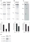
Figure 4. Tethering Either U2 or U12 snRNP Stimulates U7-snRNP-Dependent Cleavage In Vitro
A single copy of a 10-nt sequence complementary to the 5′-end of U1 or of U11 snRNA (U1AS, U11AS) or the branchpoint recognition region of U2 or of U12 snRNA (U2AS, U12AS) was substituted for 4x22nt in the H3 and H2a U7-snRNP substrates schematized in Figure 1A. (A and B) A processing time course of the antisense (AS) element-containing H3-derived substrates in HeLa nuclear extract is shown in A. Quantitations (error bars represent standard deviations) for both the H2a and H3 substrates are in B. (C and D) In vitro processing of the U2 or U12 tethering construct was monitored in extracts with U1, U2, or U12 snRNAs depleted by DNA-directed RNase H treatment. M is a mock-treated sample. Representative processing reactions are shown in C; the fraction processed at the 3-hr timepoint (error bars represent standard deviations) is graphed in D. (E) Northern blotting determined the knockdown efficiencies for the DNA-directed RNase H-treated extracts used in C, compared to untreated (NE) and mock-treated (M) extracts. 3′ fragments of U1 and U2 snRNAs generated by RNase H are visualized, but the 3′ U12 snRNA fragment ran off the gel (data not shown). Knockdown efficiencies were U1 snRNA (98%), U2 snRNA (90%), and U12 snRNA (94%).
Figure 5
Figure 5. Tethering either U2 or U12 snRNP Stimulates U7-snRNP-Dependent Cleavage In Vivo
(A) The U2 or U12 snRNA antisense element-containing transcripts (ASPre; from Figure 4) were microinjected into Xenopus GVs, and U7-snRNP-dependent cleavage (ASPro) was monitored after 1 hr. AS processing was normalized (below) to a different, co-microinjected control H2a-derived U7-snRNP substrate (CPre and CPro; efficiency set to 1). U2 and U12 tethering constructs displayed enhanced processing compared to a U11 antisense element-containing construct (from Figure 4) where error bars represent standard deviations (p<0.001). (B and C) Processing of the U2AS-H3 and U12AS-H3 constructs were monitored in GVs where either U1, U2, or U12 snRNA was knocked down by DNA-directed RNase H activity. Results are quantitated as in panel A (p<0.001 for U2AS and p<0.005 for U12AS). (D) Knockdown (KD) efficiency was determined relative to untreated GVs by Northern blotting for U1 and U2 snRNAs and RT-PCR for U12 snRNA. The Xenopus laevis homolog of U11 snRNA has not yet been identified, so it could not be knocked down.
Figure 6
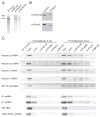
Figure 6. Intact U2 snRNP Associates with Histone Pre-mRNAs In Vivo
(A) A labeled U7-snRNP substrate containing the H2a 101-nt RNA element was microinjected into Xenopus GVs and incubated for 1 hr. After UV-irradiation, extracts were prepared and immunoprecipitated with α-hPrp43 or α-SF3b155 antibodies; α-IBP160 antibody served as a control. Pre-mRNA (Pre) enrichment of ~2-fold over processed product (Pro) was observed in the α-hPrp43 and α-SF3b155 precipitations in two separate experiments. (B) Western blots with the indicated antibodies were performed on GV extract and HeLa nuclear extract, separated by SDS-PAGE. (C) Immunoprecipitations using various α-U2 snRNP antibodies and α-IBP160 as a control were performed on extracts prepared with (+) or without (−) prior formaldehyde fixation of the HeLa cells. Histone mRNAs and RPL15 mRNA were detected via RT-PCR. The snRNAs, 7SK, and mgU2-25/61 scaRNA were detected by Northern blotting.
Figure 7
Figure 7. Model for Interactions between U2 snRNP and Histone Pre-mRNAs to Stimulate U7-snRNP-dependent Cleavage
The 7-nt conserved motif, histone 3′ stem-loop, and histone downstream element (HDE) that base-pairs with U7 snRNA are schematized. Stimulation of cleavage is proposed to occur when SF3b130 and SF3b49 mediate contact between U2 snRNP and CPSF, which contains the putative endonuclease for U7-snRNP-dependent cleavage, CPSF73 (Dominski et al., 2005; Kolev and Steitz, 2005). In contrast to the stimulation of CPA, where U2 snRNA-branchpoint interactions are critical (Kyburz et al., 2006), here the stimulatory RNA element is recognized by protein (but can be substituted by RNA-RNA) interactions.

Comment in

Similar articles

Cited by

References

    1. Arenas JE, Abelson JN. Prp43: An RNA helicase-like factor involved in spliceosome disassembly. Proc Natl Acad Sci USA. 1997;94:11798–11802. - PMC - PubMed
    1. Barnard DC, Ryan K, Manley JL, Richter JD. Symplekin and xGLD-2 are required for CPEB-mediated cytoplasmic polyadenylation. Cell. 2004;119:641–651. - PubMed
    1. Behrens SE, Tyc K, Kastner B, Reichelt J, Lührmann R. Small nuclear ribonucleoprotein (RNP) U2 contains numerous additional proteins and has a bipartite RNP structure under splicing conditions. Mol Cell Biol. 1993;13:307–319. - PMC - PubMed
    1. Blanchette M, Labourier E, Green RE, Brenner SE, Rio DC. Genome-wide analysis reveals an unexpected function for the Drosophila splicing factor U2AF50 in the nuclear export of intronless mRNAs. Mol Cell. 2004;14:775–786. - PubMed
    1. Bond UM, Yario TA, Steitz JA. Multiple processing-defective mutations in a mammalian histone pre-mRNA are suppressed by compensatory changes in U7 RNA both in vivo and in vitro. Genes Dev. 1991;5:1709–1722. - PubMed

Publication types

MeSH terms

LinkOut - more resources