

Redox-dependent impairment of vascular function in sickle cell disease
- PMID: 17964418
- PMCID: PMC2139908
- DOI: 10.1016/j.freeradbiomed.2007.08.014
Redox-dependent impairment of vascular function in sickle cell disease
Abstract
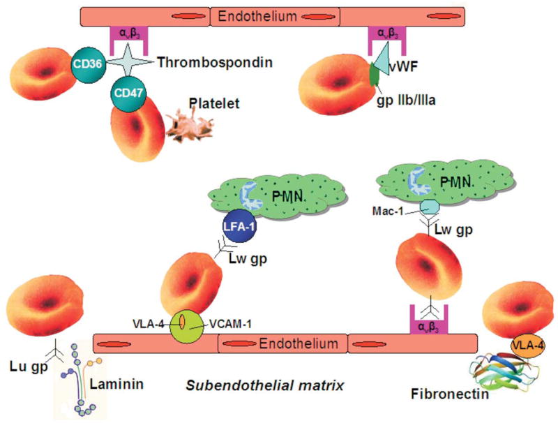
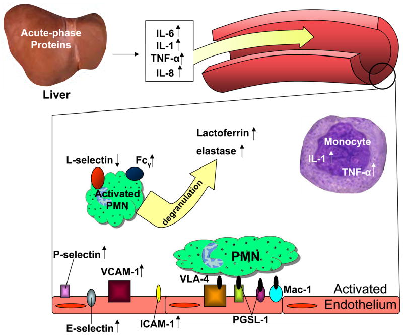
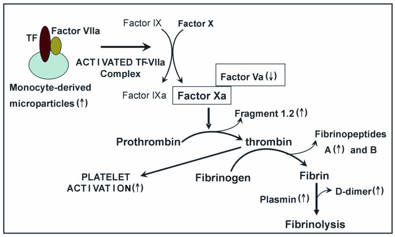
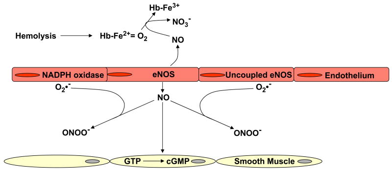
The vascular pathophysiology of sickle cell disease (SCD) is influenced by many factors, including adhesiveness of red and white blood cells to endothelium, increased coagulation, and homeostatic perturbation. The vascular endothelium is central to disease pathogenesis because it displays adhesion molecules for blood cells, balances procoagulant and anticoagulant properties of the vessel wall, and regulates vascular homeostasis by synthesizing vasoconstricting and vasodilating substances. The occurrence of intermittent vascular occlusion in SCD leads to reperfusion injury associated with granulocyte accumulation and enhanced production of reactive oxygen species. The participation of nitric oxide (NO) in oxidative reactions causes a reduction in NO bioavailability and contributes to vascular dysfunction in SCD. Therapeutic strategies designed to counteract endothelial, inflammatory, and oxidative abnormalities may reduce the frequency of hospitalization and blood transfusion, the incidence of pain, and the occurrence of acute chest syndrome and pulmonary hypertension in patients with SCD.
Figures
References
-
- Pauling L, Itano HA, Singer SJ, Wells IC. Sickle cell anemia a molecular disease. Science. 1949;110:543–548. - PubMed
-
- Ingram VM. Gene mutations in human haemoglobin: the chemical difference between normal and sickle cell haemoglobin. Nature. 1957;180:326–328. - PubMed
-
- Dykes G, Crepeau RH, Edelstein SJ. Three-dimensional reconstruction of the fibres of sickle cell haemoglobin. Nature. 1978;272:506–510. - PubMed
-
- Eaton WA, Hofrichter J. Sickle cell hemoglobin polymerization. Adv Protein Chem. 1990;40:63–279. - PubMed
-
- Burtis CA, Ashwood ER, editors. Teitz Fundamentals of Clinical Chemistry. 4. Saunders Company; Philadelphia, PA: 1996.
Publication types
MeSH terms
Substances
Grants and funding
LinkOut - more resources
Full Text Sources
Other Literature Sources
Medical
