

A functional magnetic resonance imaging predictor of treatment response to venlafaxine in generalized anxiety disorder
- PMID: 17964548
- PMCID: PMC2654286
- DOI: 10.1016/j.biopsych.2007.08.019
A functional magnetic resonance imaging predictor of treatment response to venlafaxine in generalized anxiety disorder
Abstract
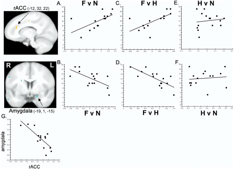
Background: Functional magnetic resonance imaging (fMRI) holds promise as a noninvasive means of identifying neural responses that can be used to predict treatment response before beginning a drug trial. Imaging paradigms employing facial expressions as presented stimuli have been shown to activate the amygdala and anterior cingulate cortex (ACC). Here, we sought to determine whether pretreatment amygdala and rostral ACC (rACC) reactivity to facial expressions could predict treatment outcomes in patients with generalized anxiety disorder (GAD).
Methods: Fifteen subjects (12 female subjects) with GAD participated in an open-label venlafaxine treatment trial. Functional magnetic resonance imaging responses to facial expressions of emotion collected before subjects began treatment were compared with changes in anxiety following 8 weeks of venlafaxine administration. In addition, the magnitude of fMRI responses of subjects with GAD were compared with that of 15 control subjects (12 female subjects) who did not have GAD and did not receive venlafaxine treatment.
Results: The magnitude of treatment response was predicted by greater pretreatment reactivity to fearful faces in rACC and lesser reactivity in the amygdala. These individual differences in pretreatment rACC and amygdala reactivity within the GAD group were observed despite the fact that 1) the overall magnitude of pretreatment rACC and amygdala reactivity did not differ between subjects with GAD and control subjects and 2) there was no main effect of treatment on rACC-amygdala reactivity in the GAD group.
Conclusions: These findings show that this pattern of rACC-amygdala responsivity could prove useful as a predictor of venlafaxine treatment response in patients with GAD.
Figures
Similar articles
-
The neural substrates of affective processing in depressed patients treated with venlafaxine.Am J Psychiatry. 2003 Jan;160(1):64-75. doi: 10.1176/appi.ajp.160.1.64. Am J Psychiatry. 2003. PMID: 12505803
-
fMRI of fearful facial affect recognition in panic disorder: the cingulate gyrus-amygdala connection.J Affect Disord. 2006 Aug;94(1-3):173-81. doi: 10.1016/j.jad.2006.04.007. Epub 2006 Jun 16. J Affect Disord. 2006. PMID: 16782207
-
Pattern of symptom improvement following treatment with venlafaxine XR in patients with generalized anxiety disorder.J Clin Psychiatry. 2001 Nov;62(11):888-93. doi: 10.4088/jcp.v62n1109. J Clin Psychiatry. 2001. PMID: 11775049 Clinical Trial.
-
The functional anatomy, neurochemistry, and pharmacology of anxiety.J Clin Psychiatry. 1999;60 Suppl 22:12-7. J Clin Psychiatry. 1999. PMID: 10634350 Review.
-
A non-inferiority comparison of duloxetine and venlafaxine in the treatment of adult patients with generalized anxiety disorder.J Psychopharmacol. 2008 Jun;22(4):417-25. doi: 10.1177/0269881108091588. J Psychopharmacol. 2008. PMID: 18635722
Cited by
-
Early life sleep disruption is a risk factor for increased ethanol drinking after acute footshock stress in prairie voles.Behav Neurosci. 2020 Oct;134(5):424-434. doi: 10.1037/bne0000410. Epub 2020 Jul 23. Behav Neurosci. 2020. PMID: 32700922 Free PMC article.
-
Uncertainty and anticipation in anxiety: an integrated neurobiological and psychological perspective.Nat Rev Neurosci. 2013 Jul;14(7):488-501. doi: 10.1038/nrn3524. Nat Rev Neurosci. 2013. PMID: 23783199 Free PMC article. Review.
-
The neurobiology of anxiety disorders: brain imaging, genetics, and psychoneuroendocrinology.Psychiatr Clin North Am. 2009 Sep;32(3):549-75. doi: 10.1016/j.psc.2009.05.004. Psychiatr Clin North Am. 2009. PMID: 19716990 Free PMC article. Review.
-
Genetic polymorphisms in monoamine systems and outcome of cognitive behavior therapy for social anxiety disorder.PLoS One. 2013 Nov 15;8(11):e79015. doi: 10.1371/journal.pone.0079015. eCollection 2013. PLoS One. 2013. PMID: 24260145 Free PMC article. Clinical Trial.
-
Dynamic effective connectivity among large-scale brain networks mediates risk of anxiety.Hum Brain Mapp. 2023 Jun 15;44(9):3730-3743. doi: 10.1002/hbm.26308. Epub 2023 Apr 12. Hum Brain Mapp. 2023. PMID: 37042391 Free PMC article.
References
-
- LeDoux JE. The Emotional Brain. Simon-Schuster; New York: 1996.
-
- Davis M, Whalen PJ. The amygdala: Vigilance and emotion. Mol Psychiatry. 2001;6:13–34. - PubMed
-
- Whalen PJ. Fear, vigilance and ambiguity: Initial neuroimaging studies of the human amygdala. Curr Dir Psychol Sci. 1998;7:177–188.
-
- Pandya DN, Van Hoeseon GW, Mesulam M-M. Efferent connections of the cingulate gyrus in the rhesus monkey. Exp Brain Res. 1981;42:319–330. - PubMed
-
- Amaral DG, Price JL, Pitkänen A, Carmichael ST. Anatomical organization of the primate amygdaloid complex. In: Aggleton JP, editor. The Amygdala: Neurobiological Aspects of Emotion, Memory and Mental Dys-function. Wiley-Liss; New York: 1992. pp. 1–66.
Publication types
MeSH terms
Substances
Grants and funding
LinkOut - more resources
Full Text Sources
Other Literature Sources
Medical
