

Brainstem regions involved in the expiration reflex. A c-fos study in anesthetized cats
- PMID: 17964550
- PMCID: PMC2701351
- DOI: 10.1016/j.brainres.2007.09.064
Brainstem regions involved in the expiration reflex. A c-fos study in anesthetized cats
Abstract
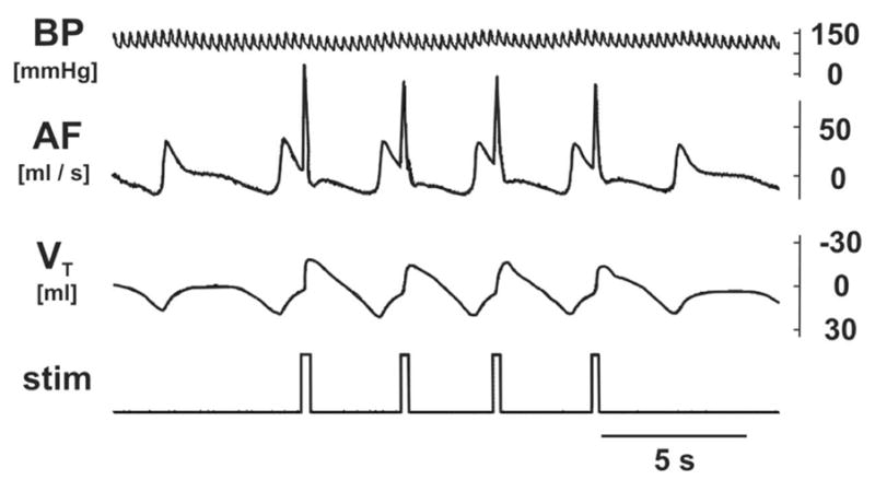
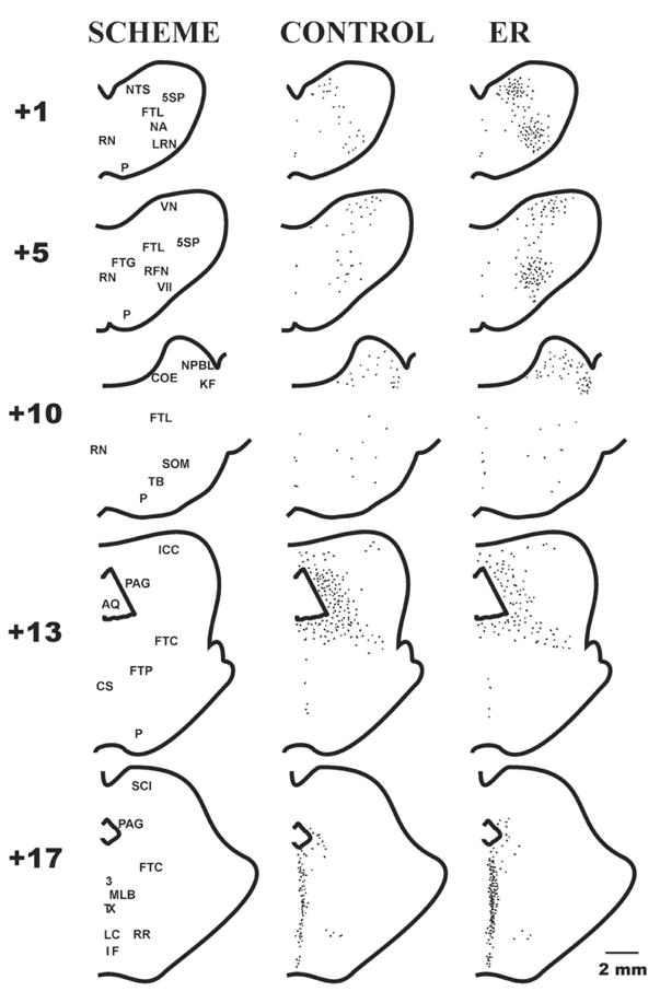
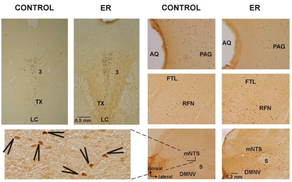
Expression of the immediate-early gene c-fos, a marker of neuronal activation, was employed to localize brainstem neuronal populations functionally related to the expiration reflex (ER). Twelve spontaneously breathing, non-decerebrate, pentobarbital anesthetized cats were used. The level of Fos-like immunoreactivity (FLI) in 6 animals with repetitive ERs mechanically induced from the glottis (296+/-9 ERs) was compared to FLI in 6 control non-stimulated cats. Respiratory rate, arterial blood pressure, and end tidal CO(2) concentration remained stable during the experiment. In the medulla, increased FLI was found in the region of nucleus tractus solitarii (p<0.001), in the ventrolateral medulla along with the lateral tegmental field (p<0.01), and in the vestibular nuclei (p<0.01). In the pons, increased FLI was detected in the caudal extensions of the lateral parabrachial and Kölliker-Fuse nuclei (p<0.05). Within the rostral mesencephalon, FLI was enhanced in the midline area (p<0.05). A lower level of ER-related FLI compared to control animals was detected in the pontine raphe region (p<0.05) and the lateral division of mesencephalic periaqueductal gray (p<0.05). The results suggest that the ER is coordinated by a complex long loop of medullary-pontine-mesencephalic neuronal circuits, some of which may differ from those of other respiratory reflexes. The FLI related to the expulsive behavior ER differs from that induced by laryngeal stimulation and laryngeal adductor responses, particularly in ventrolateral medulla and mesencephalon.
Figures
Similar articles
-
Brainstem areas involved in the aspiration reflex: c-Fos study in anesthetized cats.Physiol Res. 2004;53(6):703-17. Physiol Res. 2004. PMID: 15588140
-
Brainstem circuitry of tracheal-bronchial cough: c-fos study in anesthetized cats.Respir Physiol Neurobiol. 2008 Feb 29;160(3):289-300. doi: 10.1016/j.resp.2007.10.014. Epub 2007 Oct 30. Respir Physiol Neurobiol. 2008. PMID: 18055277 Free PMC article.
-
Directionally specific changes in arterial pressure induce differential patterns of fos expression in discrete areas of the rat brainstem: a double-labeling study for Fos and catecholamines.J Comp Neurol. 1994 Nov 1;349(1):36-50. doi: 10.1002/cne.903490104. J Comp Neurol. 1994. PMID: 7852625
-
Midbrain control of breathing and blood pressure: The role of periaqueductal gray matter and mesencephalic collicular neuronal microcircuit oscillators.Eur J Neurosci. 2020 Oct;52(8):3879-3902. doi: 10.1111/ejn.14727. Epub 2020 May 1. Eur J Neurosci. 2020. Retraction in: Eur J Neurosci. 2021 Oct;54(7):6685. doi: 10.1111/ejn.15484. PMID: 32227408 Retracted. Review.
-
Production of reflex cough by brainstem respiratory networks.Pulm Pharmacol Ther. 2004;17(6):369-76. doi: 10.1016/j.pupt.2004.09.022. Pulm Pharmacol Ther. 2004. PMID: 15564078 Review.
Cited by
-
Serotonergic mechanisms on breathing modulation in the rat locus coeruleus.Pflugers Arch. 2010 Feb;459(3):357-68. doi: 10.1007/s00424-009-0741-4. Epub 2009 Oct 21. Pflugers Arch. 2010. PMID: 19844739
-
Neurochemical properties of the synapses between the parabrachial nucleus-derived CGRP-positive axonal terminals and the GABAergic neurons in the lateral capsular division of central nucleus of amygdala.Mol Neurobiol. 2015 Feb;51(1):105-18. doi: 10.1007/s12035-014-8713-x. Epub 2014 May 4. Mol Neurobiol. 2015. PMID: 24794145
-
The Kölliker-Fuse nucleus: a review of animal studies and the implications for cranial nerve function in humans.Eur Arch Otorhinolaryngol. 2016 Nov;273(11):3505-3510. doi: 10.1007/s00405-015-3861-9. Epub 2015 Dec 19. Eur Arch Otorhinolaryngol. 2016. PMID: 26688431 Review.
References
-
- Ambalavanar R, Tanaka Y, Damirjian M, Ludlow CL. Laryngeal Afferent Stimulation Enhances Fos Immunoreactivity in Periaqueductal Gray in the Cat. J Comp Neurol. 1999;409:411–423. - PubMed
-
- Baekey DM, Morris KF, Nuding SC, Segers LS, Lindsey BG, Shannon R. Medullary raphe neuron activity is altered during fictive cough in the decerebrate cat. J Appl Physiol. 2003;94:93–100. - PubMed
-
- Baekey DM, Morris KF, Nuding SC, Segers LS, Lindsey BG, Shannon R. Ventrolateral medullary respiratory network participation in the expiration reflex in the cat. J Appl Physiol. 2004;96:2057–2072. - PubMed
-
- Bandler R, Shipley MT. Columnar organization in the midbrain periaqueductal gray: modules for emotional expression? Trends Neurosci. 1994;17:379–389. - PubMed
Publication types
MeSH terms
Substances
Grants and funding
LinkOut - more resources
Full Text Sources
Miscellaneous
