Kainate receptors are primarily postsynaptic to SP-containing axon terminals in the trigeminal dorsal horn
- PMID: 17964552
- PMCID: PMC2238683
- DOI: 10.1016/j.brainres.2007.09.070
Kainate receptors are primarily postsynaptic to SP-containing axon terminals in the trigeminal dorsal horn
Abstract
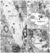
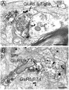
Kainate receptors (KARs) are involved in the modulation and transmission of nociceptive information from peripheral afferents to neurons in the spinal cord and trigeminal dorsal horns. KARs are found at both pre- and postsynaptic sites in the dorsal horn. We hypothesized that KARs and Substance P (SP), a modulatory neuropeptide that is used as a marker of nociceptive afferents, have a complex interactive relationship. To determine the cellular relationship and connectivity between KARs and SP afferents, we used electron microscopic dual immunocytochemical analysis to examine the ultrastructural localization of KAR subunits GluR5, 6 and 7 (GluR5,6,7) in relation to SP within laminae I and II in the rat trigeminal dorsal horn. KARs were distributed both postsynaptically in dendrites and somata (51% of GluR5,6,7 immunoreactive (-ir) profiles) and presynaptically in axons and axon terminals (45%). We also found GluR5,6,7-ir glial profiles (5%). The majority of SP-ir profiles were presynaptic axons and axon terminals. SP-ir dendritic profiles were rare, yet 23% contained GluR5,6,7 immunoreactivity. GluR5,6,7 and SP were also colocalized at presynaptic sites (18% of GluR5,6,7-ir axons and axon terminals contained SP; while 11% of SP-ir axons and axon terminals contained GluR5,6,7). The most common interaction between KARs and SP we observed was GluR5,6,7-ir dendrites contacted by SP-ir axon terminals; 54% of the dendritic targets of SP-ir axon terminals were GluR5,6,7-ir. These results provide anatomical evidence that KARs primarily mediate nociceptive transmission postsynaptic to SP-containing afferents and may also modulate the presynaptic release of SP and glutamate in trigeminal dorsal horn.
Figures


Similar articles
-
Dual ultrastructural localization of mu-opiate receptors and substance p in the dorsal horn.Synapse. 2000 Apr;36(1):12-20. doi: 10.1002/(SICI)1098-2396(200004)36:1<12::AID-SYN2>3.0.CO;2-E. Synapse. 2000. PMID: 10700022
-
mu-Opioid receptors often colocalize with the substance P receptor (NK1) in the trigeminal dorsal horn.J Neurosci. 2000 Jun 1;20(11):4345-54. doi: 10.1523/JNEUROSCI.20-11-04345.2000. J Neurosci. 2000. PMID: 10818170 Free PMC article.
-
The N-methyl-D-aspartate (NMDA) receptor is postsynaptic to substance P-containing axon terminals in the rat superficial dorsal horn.Brain Res. 1997 Oct 24;772(1-2):71-81. doi: 10.1016/s0006-8993(97)00637-9. Brain Res. 1997. PMID: 9406957
-
Ionotropic glutamate receptors are expressed in GABAergic terminals in the rat superficial dorsal horn.J Comp Neurol. 2005 May 30;486(2):169-78. doi: 10.1002/cne.20525. J Comp Neurol. 2005. PMID: 15844209
-
Molecular Anatomy of Synaptic and Extrasynaptic Neurotransmission Between Nociceptive Primary Afferents and Spinal Dorsal Horn Neurons.Int J Mol Sci. 2025 Mar 6;26(5):2356. doi: 10.3390/ijms26052356. Int J Mol Sci. 2025. PMID: 40076973 Free PMC article. Review.
Cited by
-
Peripheral nerve injury increases glutamate-evoked calcium mobilization in adult spinal cord neurons.Mol Pain. 2012 Jul 28;8:56. doi: 10.1186/1744-8069-8-56. Mol Pain. 2012. PMID: 22839304 Free PMC article.
-
Modulation of nociceptive dural input to the trigeminocervical complex through GluK1 kainate receptors.Pain. 2015 Mar;156(3):439-450. doi: 10.1097/01.j.pain.0000460325.25762.c0. Pain. 2015. PMID: 25679470 Free PMC article.
-
Glutamate receptor phosphorylation and trafficking in pain plasticity in spinal cord dorsal horn.Eur J Neurosci. 2010 Jul;32(2):278-89. doi: 10.1111/j.1460-9568.2010.07351.x. Epub 2010 Jul 11. Eur J Neurosci. 2010. PMID: 20629726 Free PMC article. Review.
-
The Trigeminal Sensory System and Orofacial Pain.Int J Mol Sci. 2024 Oct 21;25(20):11306. doi: 10.3390/ijms252011306. Int J Mol Sci. 2024. PMID: 39457088 Free PMC article. Review.
-
Ionotropic glutamate receptors in spinal nociceptive processing.Mol Neurobiol. 2009 Dec;40(3):260-88. doi: 10.1007/s12035-009-8086-8. Epub 2009 Oct 31. Mol Neurobiol. 2009. PMID: 19876771 Review.
References
-
- Advokat C, Rutherford D. Selective antinociceptive effect of excitatory amino acid antagonists in intact and acute spinal rats. Pharmacol Biochem Behav. 1995;51:855–860. - PubMed
-
- Afrah AW, Fiska A, Gjerstad J, Gustafsson H, Tjolsen A, Olgart L, Stiller CO, Hole K, Brodin E. Spinal substance P release in vivo during the induction of long-term potentiation in dorsal horn neurons. Pain. 2002;96:49–55. - PubMed
-
- Aicher SA, Mitchell JL, Swanson KC, Zadina JE. Endomorphin-2 axon terminals contact mu-opioid receptor-containing dendrites in trigeminal dorsal horn. Brain Res. 2003;977:190–198. - PubMed
-
- Aicher SA, Sharma S, Cheng PY, Liu-Chen LY, Pickel VM. Dual ultrastructural localization of mu-opiate receptors and substance p in the dorsal horn. Synapse. 2000b;36:12–20. - PubMed
Publication types
MeSH terms
Substances
Grants and funding
LinkOut - more resources
Full Text Sources
Miscellaneous

