. 2007 Dec 5:1183:32-42.
doi: 10.1016/j.brainres.2007.09.051. Epub 2007 Oct 26.

Pro-NGF, sortilin, and p75NTR: potential mediators of injury-induced apoptosis in the mouse dorsal root ganglion

Affiliations

Pro-NGF, sortilin, and p75NTR: potential mediators of injury-induced apoptosis in the mouse dorsal root ganglion

Melinda G Arnett et al. Brain Res. .

Abstract

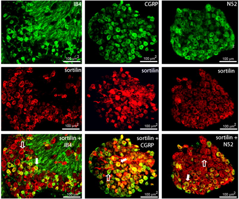
The nerve growth factor precursor (pro-NGF) may function as a death-inducing ligand that mediates its apoptotic effects via p75NTR. Pro-NGF-induced apoptosis is postulated to be dependent upon membrane expression of the sortilin receptor, which interacts with p75NTR to promote a high-affinity binding site for pro-NGF. Here, we explore the expression of pro-NGF, sortilin and p75NTR in the mouse lumbar dorsal root ganglion (DRG) to understand the potential for this trimeric signaling complex to function in injury-induced neuronal death of DRG neurons. Our results reveal the expression of all 3 components within the DRG and that a subpopulation of neurons coexpresses sortilin and p75NTR. Following sciatic nerve transection, the expression of these proteins appears insensitive to injury; however, the majority of small p75NTR-sortilin coexpressing neurons are lost 25 days after sciatic nerve transection. These results propose pro-NGF-induced, p75NTR-sortilin-mediated neuronal death as a critical aspect of nerve injury-induced death in the DRG.


Figures

Figure 1
Figure 1
Immunocytochemistry was performed on L4/5 DRG of uninjured and injured C57BL/6 mice to identify neurons that express sortilin (red, top panels) or p75NTR (green, middle panels). Top left) On average, 73% of neurons express sortilin in uninjured mice. Top right) Similarly, 72.8% of neurons express sortilin 3 days following sciatic nerve transection. Middle left) 66.1% of neurons express p75NTR in uninjured mice. Middle right) Similarly, 67.2% of neurons express p75NTR three days after sciatic nerve transection. Bottom left) Merged image illustrating the overlap of sortilin and p75 with no injury (left) and following injury (right) Scale bars equal 100μm for each panel.
Figure 2
Figure 2
Immunoblotting analysis was performed using L4/5 DRG removed 3 days following no injury (lane 1) or sciatic nerve transection (lane 2). A) A polyclonal antibody against sortilin recognized a band of 95 kDa that was determined to be the sortilin receptor. All protein loading was normalized to cyclophilin A (18 kDa). An unpaired t-test performed on band intensity revealed no significant difference in sortilin expression between injured and uninjured groups (p = 0.8643), suggesting that sortilin expression is not altered in DRG neurons following sciatic nerve injury. B) Immunoblotting analysis was performed using L4/5 DRG removed 3 days following no injury (lane 1) or sciatic nerve transection (lane 2). A polyclonal antibody designed to detect p75NTR recognized a band of 75 kDa that was identified as the p75 receptor. Bands of ~55, ~135, and ~155 kDa represent possible cleaved or post-translationally modified forms of p75NTR, respectively. All protein loading was normalized to cyclophilin A (18 kDa). An unpaired t-test performed on band intensity revealed no significant difference in p75NTR expression between injured and uninjured groups (p = 0.7836), suggesting that p75NTR expression is not altered in DRG neurons following sciatic nerve injury.
Figure 3
Figure 3
Immunocytochemistry was performed on L4/5 DRG from uninjured C57BL/6 mice using a primary polyclonal sortilin (red) antibody in conjunction with the neuronal markers (green) IB4, CGRP, and N52. 13.6% of sortilin-expressing neurons coexpress IB4. Alternatively, 31.7% of IB4-expressing neurons coexpress the sortilin receptor. 20.6% of sortilin-expressing neurons coexpress CGRP while 44.1% of CGRP-expressing neurons coexpress sortilin. 18.2% of sortilin-expressing neurons coexpress N52; 52.8% of N52-expresing neurons coexpress sortilin. Filled arrows represent neurons that coexpress sortilin and a specific neuronal marker; open arrows identify neurons that express sortilin in the absence of neuronal markers.
Figure 4
Figure 4
A) Immunoblotting analysis was performed using L4/5 DRG from the following groups of mice: no injury (lane 1), three day post-sciatic nerve crush (lane 2), or transection (lane 3). A polyclonal antibody specific for the pro-domain of proNGF detected multiple bands of ~35, ~40, ~44, and ~80 kDa. The ~ 35 kDa band was determined to identify the unglycosylated NGF precursor. All higher molecular weight bands likely represent post-translationally modified forms of proNGF. Statistical analysis using ANOVA revealed no significant difference in band intensity amongst groups (p = 0.999), suggesting that proNGF expression is not altered in response to injury in the DRG. A polyclonal antibody specific for mature NGF detected bands of similar size, including a band of ~ 91 kDa (see panel C). Because this band was not detectable when using the pro-NGF antibody, this band was determined to not be of true NGF origin. B) Commercially prepared NGF (20ng/lane) was used to test the specificity of the mature antibody. C) Mature NGF (12 kDa) was not present in detectable concentrations. No significant change in peptide expression was detected by ANOVA between treatment groups (p = 0.9868). All protein loading was normalized to cyclophilin A (18kDa).
Figure 5
Figure 5
Size-frequency histogram illustrating the distribution of neuronal area (μm2) in uninjured (black bars) and sciatic nerve transected (grey bars) L4/5 DRG 25-days following sciatic nerve axotomy. Small neurons (0–200μm2) comprise over half (58.03%) of the L4/5 DRG neuronal population in uninjured mice (black bars). This population of neurons is completely lost 25-days following sciatic nerve transection (grey bars).

Similar articles

Cited by

References

    1. Beattie MS, Harrington AW, Lee R, Kim JY, Boyce SL, Longo FM, Bresnahan JC, Hempstead BL, Yoon SO. ProNGF induces p75-mediated death of oligodendrocytes following spinal cord injury. Neuron. 2002;36:375–386. - PMC - PubMed
    1. Chen Y, Dicou E, Djakiew D. Characterization of nerve growth factor precursor protein expression in rat round spermatids and the trophic effects of nerve growth factor in the maintenance of Sertoli cell viability. Molecular and Cellular Endocrinology. 1997;127:129–136. - PubMed
    1. Christianson JA, Ryals JM, McCarson KE, Wright DE. Beneficial actions of neurotrophin treatment on diabetes-induced hypoalgesia in mice. J Pain. 2003;4:493–504. - PubMed
    1. Christianson JA, Ryals JM, McCarson KE, Wright DE. Beneficial actions of neurotrophin treatment on diabetes-induced hypoalgesia in mice. J Pain. 2003;4:493–504. - PubMed
    1. Crockett DP, Harris SL, Egger MD. Neurotrophin receptor (p75) in the trigeminal thalamus of the rat: development, response to injury, transient vibrissa-related patterning, and retrograde transport. The Anatomical Record. 2000;259:446–460. - PubMed

MeSH terms