. 2007 Dec 1;312(1):435-47.
doi: 10.1016/j.ydbio.2007.09.050. Epub 2007 Oct 4.

Smad4 is required to regulate the fate of cranial neural crest cells

Affiliations

Smad4 is required to regulate the fate of cranial neural crest cells

Seung O Ko et al. Dev Biol. .

Abstract

Smad4 is the central mediator for TGF-beta/BMP signals, which are involved in regulating cranial neural crest (CNC) cell formation, migration, proliferation and fate determination. It is unclear whether TGF-beta/BMP signals utilize Smad-dependent or -independent pathways to control the development of CNC cells. To investigate the functional significance of Smad4 in regulating CNC cells, we generated mice with neural crest specific inactivation of the Smad4 gene. Our study shows that Smad4 is not required for the migration of CNC cells, but is required in neural crest cells for the development of the cardiac outflow tract. Smad4 is essential in mediating BMP signaling in the CNC-derived ectomesenchyme during early stages of tooth development because conditional inactivation of Smad4 in neural crest derived cells results in incisor and molar development arrested at the dental lamina stage. Furthermore, Smad-mediated TGF-beta/BMP signaling controls the homeobox gene patterning of oral/aboral and proximal/distal domains within the first branchial arch. At the cellular level, a Smad4-mediated downstream target gene(s) is required for the survival of CNC cells in the proximal domain of the first branchial arch. Smad4 mutant mice show underdevelopment of the first branchial arch and midline fusion defects. Taken together, our data show that TGF-beta/BMP signals rely on Smad-dependent pathways in the ectomesenchyme to mediate epithelial-mesenchymal interactions that control craniofacial organogenesis.


Figures

Fig. 1
Fig. 1. Wnt1-Cre;Smad4fl/fl embryos exhibit severe defects
(A,B) Yolk sac blood vessels in wild type (A) and Wnt1-Cre;Smad4fl/fl embryos (B) at E12.0. Arrows indicate the apparent decreased blood supply in the Wnt1-Cre;Smad4fl/fl yolk sac. (C-J) Wild type and Wnt1-Cre;Smad4fl/fl embryos at E12.0 after removal of the yolk sac (C,D), intact E12.0 embryos (E,F), and E11.5 embryos (G-J). Wnt1-Cre;Smad4fl/fl embryos show underdevelopment of the 1st branchial arch (arrows in E,F,I,J) and failure of fusion in the middle of the frontonasal process and the mandibular process of the first branchial arch (asterisks in E,F,I,J). MX, maxillary process of the first branchial arch; MN, mandibular process of the first branchial arch. Scale bars in A-D:0.5mm, E-J:1.0mm.
Fig. 2
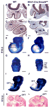
Fig. 2. Validation of Smad4 localization and contribution of cranial and cardiac neural crest cells in Wnt1-Cre;Smad4fl/fl embryos
(A-D) Immunohistochemistry of Smad4 in coronal sections of E10.5 wild type (A,C) and Wnt1-Cre;Smad4fl/fl (B,D) embryos. Smad4 is expressed ubiquitously in the facial region of wild type embryos, including the frontonasal process, first and second branchial arches. Smad4 expression is no longer detectable specifically in the neural crest derived ectomesenchyme of Wnt1-Cre;Smad4fl/fl embryos. Epithelial and mesodermal cells (arrows) are positively stained with anti-Smad4. (E-J) Whole mount LacZ staining visualizes migrating neural crest cells in Wnt1-Cre;R26R and Wnt1-Cre;R26R;Smad4fl/fl at E10.5. Cranial neural crest cells are normally distributed in the facial region including the frontonasal process, first branchial arch, 2nd branchial arch and rudimentary third branchial arch in both Wnt1-Cre;R26R (E,G) and Wnt1-Cre;R26R;Smad4fl/fl (F,H) embryos. (I,J) Cardiac neural crest cell migration to the outflow tract (OFT) of the heart is clearly reduced in Wnt1-Cre;Smad4fl/fl embryos. (K,L) H&E staining of horizontal sections from E12.5 wild type (K) and Wnt1-Cre;Smad4fl/fl (L) embryos. The OFT showed defective development with malformed cushion in Wnt1-Cre;Smad4fl/fl(L). Arrows indicate OFT of the heart. FNP, frontonasal process; MX, maxillary process of the first branchial arch; MN, mandibular process of the first branchial arch; 2BA, second branchial arch; 3BA, second branchial arch; OFT, outflow tract. Scale bars in A,C-L: 0.1mm, B:0.2mm.
Fig. 3
Fig. 3. Neural crest-derived cranial ganglia in Wnt1-Cre;Smad4fl/fl embryos
Immunostaining for neurofilaments with the 2H3 antibody in wild type and Wnt1-Cre;Smad4fl/fl embryos at E10.5. The patterning of the cranial ganglia in Wnt1-Cre;Smad4fl/fl embryos (B,D) is indistinguishable from wild type (A,C). However, the trigeminal ganglion (arrows in B) is slightly reduced in size and the distal projections into the trigeminal (arrow heads in B) and facial nerves (arrows in C, D) are shortened in Wnt1-Cre;Smad4fl/fl embryos. III, oculomoter nerve; V, trigeminal nerve; VII, facial nerve; IX, glossopharyngeal nerve; X, vagus nerve. Scale bars: 1mm.
Fig. 4
Fig. 4. Tooth development in Wnt1-Cre;Smad4fl/fl embryos
(A-D) H&E staining of coronal sections from E11.5 wild type (A,C) and Wnt1-Cre;Smad4fl/fl (B,D) embryos. Arrows indicate epithelial thickening at incisor and molar tooth developing sites. (E-H) H&E staining of the first branchial arch explants from E11.5 wild type (E,G) and Wnt1-Cre;Smad4fl/fl (F,H) embryos following two days of organ culture. In wild type samples, the early bud stage of tooth development (arrow in E) and Meckel's cartilage development are detectable. In Wnt1-Cre;Smad4fl/fl explants, tooth germ-like structures beyond the dental lamina stage are not detectable and Meckel's cartilage is underdeveloped. (I-M) Wild type and Wnt1-Cre;Smad4fl/fl first branchial arch were cultivated for either 6 days or 4 weeks after kidney capsule transplantation. The cap stage of tooth development (arrows in I,K) and normal cusp patterning (L) is detectable in wild type, however tooth germ-like structures are not detectable in Wnt1-Cre;Smad4fl/fl. MC, Meckel's cartilage; INC, incisor tooth; MO, molar tooth. Scale bars: 0.1mm.
Fig. 5
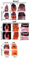
Fig. 5. Fgf8 expression pattern is altered in the oral epithelium of Wnt1-Cre;Smad4fl/fl embryos
(A-F) Whole mount in situ hybridization analysis of Bmp4 during early pattering of the oral epithelium in wild type (A,C) and Wnt1-Cre;Smad4fl/fl (B,D) embryos. Bmp4 is expressed in the distal oral epithelium at E9.5, E10.5 (arrows in A-D) and its expression increases in the underlying ectomesenchyme at E11.5 (arrows in E,F). (G-N) Whole mount in situ hybridization analysis of Fgf8. Fgf8 expression is localized in the proximal oral epithelium of wild type embryos at E9.5 and E10.5 (arrows in G,I). Fgf8 expression expands into the distal oral epithelium in Wnt1-Cre;Smad4fl/fl embryos at E10.5 (arrow in H,J). Fgf8 is expressed in the proximal oral epithelium of K14-Cre;Smad4fl/fl embryos in a pattern indistinguishable from wild type at E11.0 (arrows in K,M). (L,N) Immunohistochemistry of Smad4 in coronal sections of E11.0 wild type (L) and K14-Cre;Smad4fl/fl (N) embryos. Smad4 is expressed ubiquitously in the first branchal arch including epithelium (arrow heads in L), Smad4 expression is no longer detectable specifically in the epithelium in K14-Cre;Smad4fl/fl embryos (arrows in N). (O,P) Pax9 expression in Wnt1-Cre;Smad4fl/fl embryos is detectable in the mesenchyme at E11.5, diffusely expanded from its wild type pattern. (Q,R) Shh expression in Wnt1-Cre;Smad4fl/fl embryos is detectable in the epithelium at E11.0, comparable to its wild type pattern. Open arrow heads indicate upper incisors, open arrows indicate upper molars, arrow heads indicate lower incisors and arrows indicate lower molars in O-R. Scale bars: 0.5mm.
Fig. 6
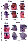
Fig. 6. Alteration of ectomesenchyme patterning in the first branchial arch of Wnt1-Cre;Smad4fl/fl embryos
Whole mount in situ hybridization analysis of Msx1 (A-D), Msx2 (E,F), Dlx5 (G,H), Barx1 (I-L), Pax9 (M,N), Shh (O,P) and ActivinβA (Q,R) in wild type and Wnt1-Cre;Smad4fl/fl embryos. (A-F) Msx1 expression in wild type embryos at E10.5 and E11.5 (arrows in A,C), is not detectable in the mandibular mesenchyme of Wnt1-Cre;Smad4fl/fl embryos (open arrows in B,D). Msx2 expression in wild type at E9.5 (arrows in E) is not detectable in the mandibular mesenchyme of Wnt1-Cre;Smad4fl/fl embryos (open arrow in F). (G,H) Dlx5 expression in Wnt1-Cre;Smad4fl/fl embryos is detectable in the distal mesenchyme at E10.5 (arrows in H), expanded from its wild type pattern (G). (I-L) Barx1 expression in Wnt1-Cre;Smad4fl/fl embryos at E10.5 is expanded into the distal mesenchyme (arrowheads in J), relative to its expression in wild type embryos (I,K) and restricted to the oral region (arrowheads in J, open arrow in L). Scale bar: 0.5mm.
Fig. 7
Fig. 7. Alteration of ectomesenchyme patterning during mandibular arch patterning of Wnt1-Cre;Smad4fl/fl embryos
Whole mount in situ hybridization analyses of Gsc (A-D), Lhx6 (E-H), Alx4 (I,J), and Alx3 (K,L) in wild type and Wnt1-Cre;Smad4fl/fl embryos. Gsc is expressed in the aboral region of the wild type mandibular arch at E10.5 (arrows in A,C). Lhx6 is expressed in the oral region of the wild type mandibular arch at E10.5 (arrows in E,G). Alx4 is expressed in the distal mesenchymal region of the mandibular arch at E10.5 (arrows in I). Arrows in K denote the Alx3 expression region that is restricted to the most distal region of the wild type mandibular arch at E11.5. Gsc, Alx4, and Alx3 expression (open arrows) is not detectable in Wnt1-Cre;Smad4fl/fl embryos, although Alx3 expression in the medial nasal process is detectable (arrowheads in K,L). Lhx6 expression is clearly visible in Wnt1-Cre;Smad4fl/fl embryos (arrows in F,H). Scale bars in A-H: 0.5mm, I-L: 0.6mm.
Fig. 8
Fig. 8. Increased apoptosis in Wnt1-Cre;Smad4fl/fl embryos
TUNEL assays to detect apoptosis in wild type and Wnt1-Cre;Smad4fl/fl embryos. At E10.5, apoptotic cells are not detectable in the first branchial arch and the frontonasal process of wild type embryos (A,C,E). At E11.5, a small number of apoptotic cells are detectable in the region of the first branchial arch (arrow heads in G,I,K). In Wnt1-Cre;Smad4fl/fl embryos, a large number of apoptotic cells in the mesenchyme of the first branchial arch and frontonasal process are detectable at both E10.5 and E11.5 (B,D,F,H,J,L). Note that the increased apoptotic cells observed in the mandibular arch are restricted to the aboral and proximal region (arrows in D,F,J,L). Arrowheads in F indicate apoptotic cells in the second branchial arch. FNP, frontonasal process; MX, maxillary process of the first branchial arch; MN, mandibular process of the first branchial arch; 1BA, first branchial arch; 2BA, second branchial arch; DBA, distal region of the first branchial arch. Scale bars: 0.1mm.

Similar articles

Cited by

References

    1. Allen SP, Borgadi JP, Barlow AJ, Mir SA, Qayyum SR, Verbeek FJ, Anderson RH, Francis-West PH, Brown NA, Richardson MK. Misexpression of noggin leads to septal defects in the outflow tract of the chick heart. Dev Biol. 2001;235:98–109. - PubMed
    1. Baker CV, Bronner-Fraser M. Vertebrate cranial placodes I. Embryonic induction. Dev Biol. 2001;232:1–61. - PubMed
    1. Barlow LA. Cranial nerve development: placodal neurons ride the crest. Curr Biol. 2002;12:R171–R173. - PubMed
    1. Beverdam A, Brouwer A, Reijnen M, Korving J, Meijlink F. Severe nasal clefting and abnormal embryonic apoptosis in Alx3/Alx4 double mutant mice. Development. 2001;128:3975–3986. - PubMed
    1. Chai Y, Jiang X, Ito Y, Bringas P, Jr, Han J, Rowitch DH, Soriano P, McMahon AP, Sucov HM. Fate of the mammalian cranial neural crest during tooth and mandibular morphogenesis. Development. 2000;127:1671–1679. - PubMed

Publication types

MeSH terms