

Short term triiodo-L-thyronine treatment inhibits cardiac myocyte apoptosis in border area after myocardial infarction in rats
- PMID: 17964598
- PMCID: PMC2235814
- DOI: 10.1016/j.yjmcc.2007.09.009
Short term triiodo-L-thyronine treatment inhibits cardiac myocyte apoptosis in border area after myocardial infarction in rats
Abstract
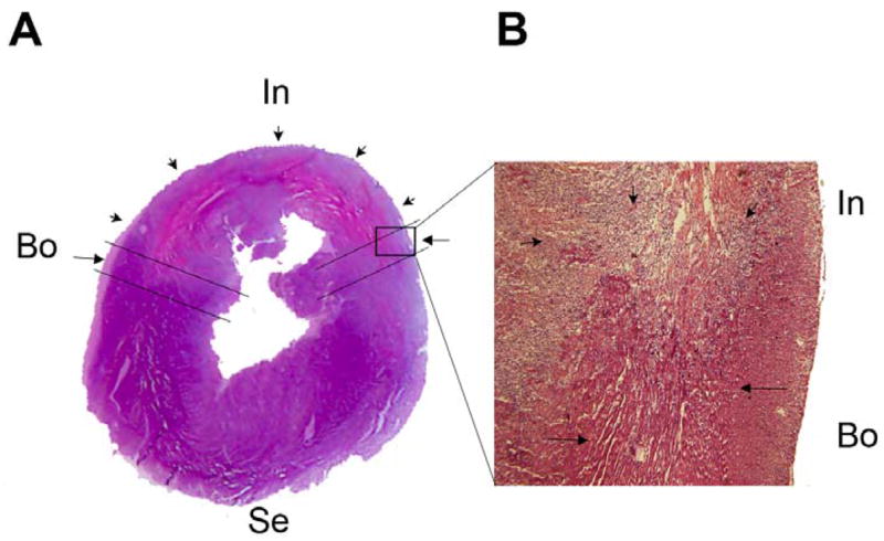
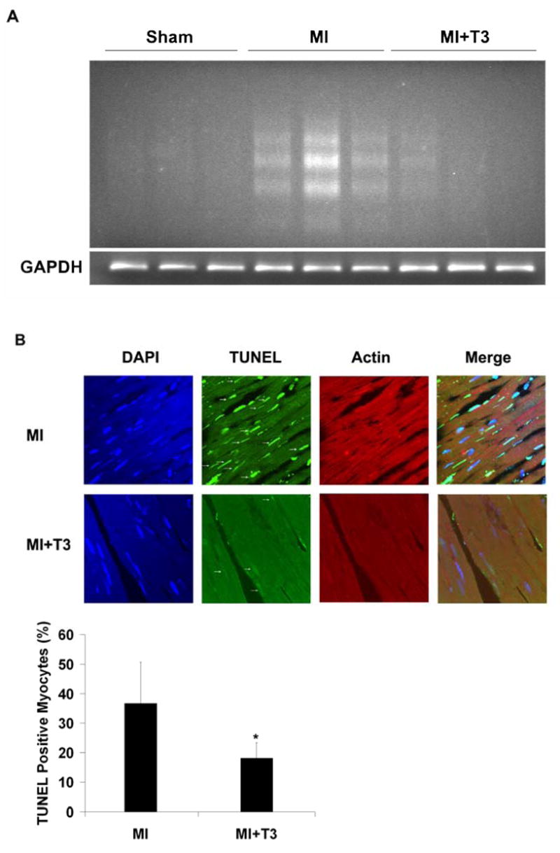
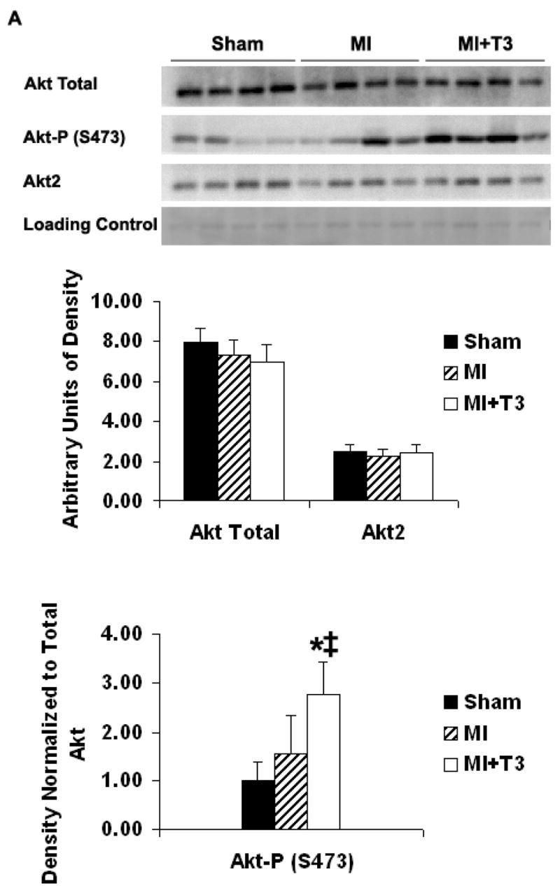
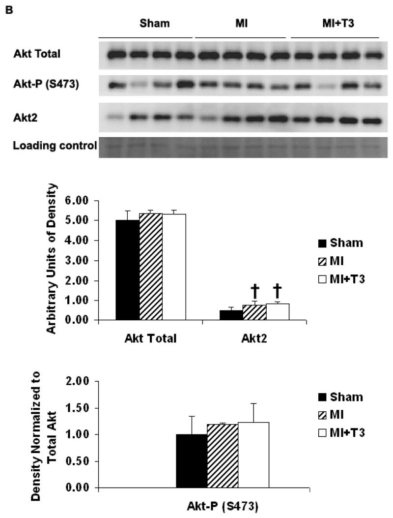
Thyroid hormone (TH) levels decline after a myocardial infarction (MI). Treatment with TH has been shown to improve left ventricular (LV) function in MI and other cardiovascular diseases, but the mechanisms are not clear. We have previously shown that TH can prevent myocyte apoptosis via Akt signaling in cultured neonatal rat cardiomyocytes. In this study, the effects of triiodo-L-thyronine (T3) on LV function and myocyte apoptosis after MI was examined in rats. After surgery, MI rats were treated with T3 for 3 days. Compared with sham-operated rats, MI rats showed significantly increased LV chamber dimension during systole and decreased LV function. T3 treatment increased LV +/-dP/dt but did not change LV chamber dimensions. MI rats also showed significantly increased myocyte apoptosis in the border area as assessed by DNA laddering and TUNEL assay. T3 treatment decreased the amount of DNA laddering, and reduced TUNEL positive myocytes in the border area, which was associated with phosphorylation of Akt at serine 473. These results suggest that T3 can protect myocytes against ischemia-induced apoptosis, which may be mediated by Akt signaling.
Figures
References
-
- Sam F, Sawyer DB, Chang DL, Eberli FR, Ngoy S, Jain M, et al. Progressive left ventricular remodeling and apoptosis late after myocardial infarction in mouse heart. Am J Physiol Heart Circ Physiol. 2000;279:H422–428. - PubMed
-
- Palojoki E, Saraste A, Eriksson A, Pulkki K, Kallajoki M, Voipio-Pulkki LM, et al. Cardiomyocyte apoptosis and ventricular remodeling after myocardial infarction in rats. Am J Physiol Heart Circ Physiol. 2001;280:H2726–2731. - PubMed
-
- Cheng W, Kajstura J, Nitahara JA, Li B, Reiss K, Liu Y, et al. Programmed myocyte cell death affects the viable myocardium after infarction in rats. Exp Cell Res. 1996;226:316–327. - PubMed
-
- Olivetti G, Quaini F, Sala R, Lagrasta C, Corradi D, Bonacina E, et al. Acute myocardial infarction in humans is associated with activation of programmed myocyte cell death in the surviving portion of the heart. J Mol Cell Cardiol. 1996;28:2005–2016. - PubMed
Publication types
MeSH terms
Substances
Grants and funding
LinkOut - more resources
Full Text Sources
Medical
Molecular Biology Databases
