. 2008 Feb;29(4):487-96.
doi: 10.1016/j.biomaterials.2007.08.050. Epub 2007 Oct 26.

Iron oxide nanoparticles as a drug delivery vehicle for MRI monitored magnetic targeting of brain tumors

Affiliations

Iron oxide nanoparticles as a drug delivery vehicle for MRI monitored magnetic targeting of brain tumors

Beata Chertok et al. Biomaterials. 2008 Feb.

Abstract

This study explored the possibility of utilizing iron oxide nanoparticles as a drug delivery vehicle for minimally invasive, MRI-monitored magnetic targeting of brain tumors. In vitro determined hydrodynamic diameter of approximately 100 nm, saturation magnetization of 94 emicro/g Fe and T2 relaxivity of 43 s(-1)mm(-)(1) of the nanoparticles suggested their applicability for this purpose. In vivo effect of magnetic targeting on the extent and selectivity of nanoparticle accumulation in tumors of rats harboring orthotopic 9L-gliosarcomas was quantified with MRI. Animals were intravenously injected with nanoparticles (12 mg Fe/kg) under a magnetic field density of 0 T (control) or 0.4 T (experimental) applied for 30 min. MR images were acquired prior to administration of nanoparticles and immediately after magnetic targeting at 1h intervals for 4h. Image analysis revealed that magnetic targeting induced a 5-fold increase in the total glioma exposure to magnetic nanoparticles over non-targeted tumors (p=0.005) and a 3.6-fold enhancement in the target selectivity index of nanoparticle accumulation in glioma over the normal brain (p=0.025). In conclusion, accumulation of iron oxide nanoparticles in gliosarcomas can be significantly enhanced by magnetic targeting and successfully quantified by MR imaging. Hence, these nanoparticles appear to be a promising vehicle for glioma-targeted drug delivery.


Figures

Figure 1
Figure 1
In Vitro characterization of magnetic nanoparticles. (A) Diagram of a magnetic nanoparticle consisting of magnetite (Fe3O4) core and a starch shell. (B) Intensity –weighted NICOMP particle size distribution of G100 colloid measured by Dynamic Light Scattering. (C) Typical TEM image of a single nanoparticle composed of multiple magnetite cores. (D) Magnetization curve of solid G100 at 293 K measured by SQUID exhibiting magnetic saturation (i.e. plateau at high magnetic field). The inset demonstrates negligible remanent magnetization indicative of superparamagnetic behavior.
Figure 2
Figure 2
Representative subset of kinetic series of MRI scans demonstrating nanoparticle accumulation in 9L gliosarcoma (A) with and (B) without magnetic targeting. The spin echo T2-weighted baseline images illustrate the tumor location clearly observable as a hyper-intense lesion. GE baseline images were acquired before the nanoparticle injection, while 1-hr and 3-hr images were acquired 1 and 3 hours after nanoparticle administration, respectively.
Figure 3
Figure 3
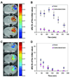
Kinetics of nanoparticle accumulation monitored by MRI in (A, B) targeted and (C, D) non-targeted groups of animals. (A, C) Representative series of R2 maps (msec-1) of the tumor tissue (color) superimposed onto the corresponding T2-weighted images acquired before (baseline) and 1–3 hours after nanoparticle administration in (A) targeted and (C) control rats. (B, D) Mean kinetic profiles of tumor and contra-lateral brain nanoparticle accumulation in (B) targeted and (D) control rats. Data expressed as MEAN±SE, n=5.
Figure 4
Figure 4
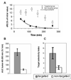
Analysis of nanoparticle distribution in the brains of 9L gliosarcoma-bearing rats. (A) Mean kinetic profiles of nanoparticle accumulation in the tumor of the targeted and control rats. Data expressed as MEAN ± SE, n=5. Exponential fit for the non-targeted group was calculated using nonlinear least squares regression method (R2= 0.97, RMSE=0.58). (B) Area under the dR2-time curve of the tumor ROI over the interval of observation. The plot shows statistically significant difference between the targeted and the control groups (p=0.005). (C) Target selectivity index of nanoparticle accumulation in tumor versus contra-lateral brain (p=0.025).
Figure 5
Figure 5
Analysis of nanoparticle concentration in excised animal tissues (glioma / contra-lateral brain) with ESR spectroscopy. (A) Typical ESR spectra of : #1- a standard solution of G100 magnetic nanoparticles; #2 – glioma of a rat injected with magnetic nanoparticles and subjected to magnetic targeting; #3 – glioma of a control rat not exposed to magnetic nanoparticles. (B) Nanoparticle concentration in excised tumor and contra-lateral brain tissues quantified by ESR spectroscopy. Data expressed as MEAN±SE, n=6. (C) A plot demonstrating linear correlation between the MRI-derived dR2 parameter and the ESR-determined nanoparticle concentration in excised tissue samples. (R2=0.88, slope=0.57, p=0.0001)

References

    1. Fisher PG, Buffler PA. Malignant Gliomas in 2005: Where to GO From Here? JAMA. 2005;293(5):615–617. - PubMed
    1. Newton HB. Primary brain tumors: review of etiology, diagnosis and treatment. American Family Physician. 1994;49(4):787–797. - PubMed
    1. Zhou R, Mazurchuk R, Straubinger RM. Antivasculature effects of doxorubicin-containing liposomes in an intracranial rat brain tumor model. Cancer Res. 2002;62(9):2561–2566. - PubMed
    1. Gutman RL, Peacock G, Lu DR. Targeted drug delivery for brain cancer treatment. Journal of Controlled Release. 2000;65:31–41. - PubMed
    1. Motl S, Zhuang Y, Waters CM, Stewart CF. Pharmacokinetic considerations in the treatment of CNS tumours. Clin Pharmacokinet. 2006;45(9):871–903. - PubMed

Publication types