. 2007 Dec;192(1-2):124-33.
doi: 10.1016/j.jneuroim.2007.09.024. Epub 2007 Oct 26.

Myelin oligodendrocyte glycoprotein peptide-induced experimental allergic encephalomyelitis and T cell responses are unaffected by immunoproteasome deficiency

Myelin oligodendrocyte glycoprotein peptide-induced experimental allergic encephalomyelitis and T cell responses are unaffected by immunoproteasome deficiency

Ricardo F Frausto et al. J Neuroimmunol. 2007 Dec.

Abstract

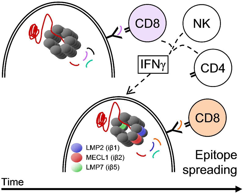
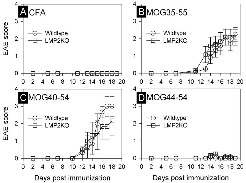
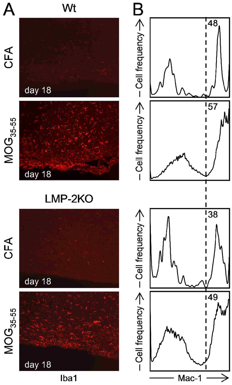
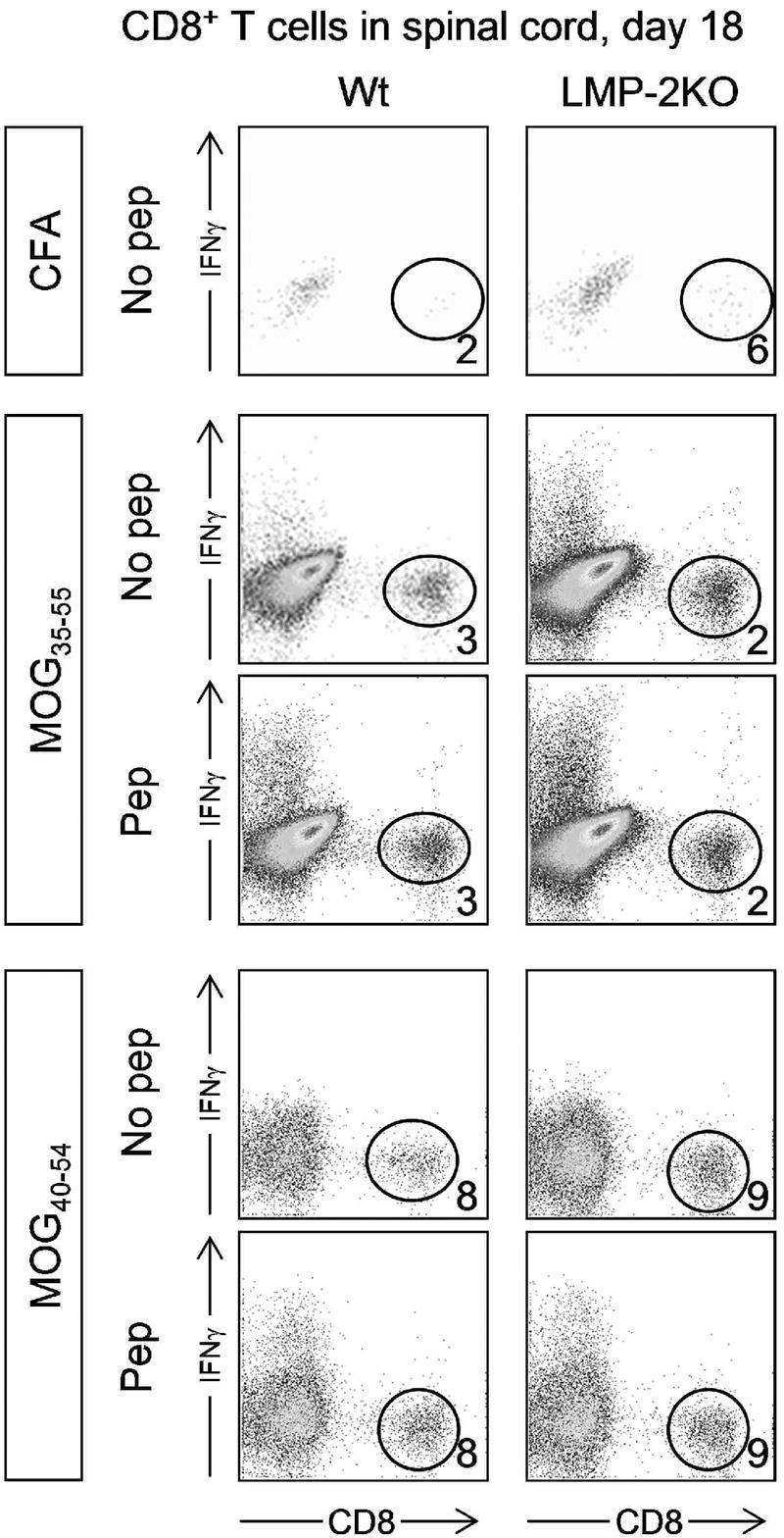
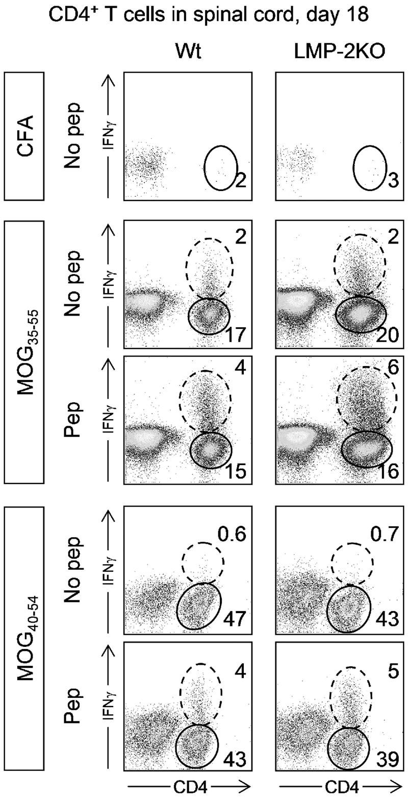
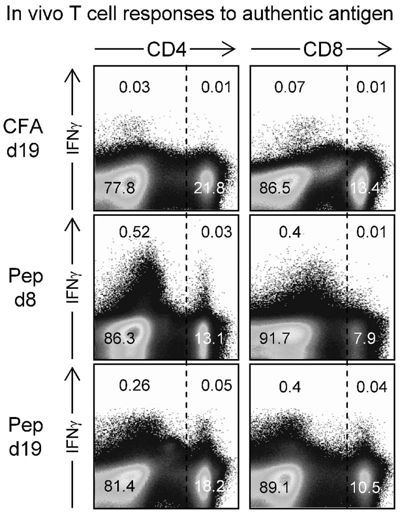
The inoculation of MOG peptides into C57BL/6 mice induces CD4(+) and CD8(+) T cells, and recent work has shown that adoptive transfer of the latter population, after extensive in vitro stimulation, can cause EAE in naïve recipient mice. Herein, we have evaluated the incidence and severity of EAE, and the induction of CD4(+) and CD8(+) T cells, following MOG peptide inoculation of wt mice and of LMP-2KO mice that lack an intact immunoproteasome, a cytoplasmic organelle that is induced by chronic inflammation and that may be important for the presentation of MHC class I epitopes to CD8(+) T cells. We report that EAE, evaluated by both clinical and histological criteria, is similar in LMP-2KO mice and wildtype C57B/6 mice (wt) in response to immunization with MOG peptides MOG(35-55) and MOG(40-54), suggesting that the immunoproteasome does not play a key role in the development of demyelinating disease. Furthermore, and consistent with previous reports, peptide-specific CD8(+) T cells were barely detectable in the CNS of peptide-immunized mice, although peptide-specific CD4(+) T cells were abundant. Therefore, we used a new technique to look for autoreactive CD8(+) T cells in MOG peptide-immunized mice, and we report the identification of CD4(+) and CD8(+) T cells that, as late as 19 days after peptide injection, are actively producing IFNgamma in vivo, in response to in vivo antigen contact.


Figures

Figure 1
Figure 1. Hypothesis: the immunoproteasome may be central to CD8+ T cell epitope spreading
The hypothesis described in the text is presented in diagrammatic form. NK = natural killer. The double line represents the plasma membrane of the cell, enclosing the multimeric proteasome (upper cell) or immunoproteasome (lower cell); the “Y” motif represents an MHC class I molecule.
Figure 2
Figure 2. Induction of EAE by MOG peptides, and the effect of immunoproteasome deficiency
The development of clinical signs of EAE in wildtype C57B/6 or LMP-2KO/B6 mice immunized with either emulsified Complete Freund’s Adjuvant (CFA, panel A), or CFA with one of the three indicated myelin oligodendrocyte glycoprotein (MOG) peptides (panels B-D). Data points represent the EAE score (mean ± SEM) for each genotype and treatment group (n=3-6/group/time point).
Figure 3
Figure 3. Similar changes in microglia/macrophage activation and recruitment in wt and LMP-2KO mice
Wt and LMP-2KO mice were inoculated with either CFA alone, or CFA with MOG35-55 peptide and, 18 days later, were sacrificed, and their CNS was harvested. (A) Immunohistochemical detection of Iba-1, a marker of macrophage/microglial activation, revealed significant activation of these phagocytes during the acute phase of EAE. (B) Mononuclear cells were isolated from the CNS tissues, and the presence of Mac1+ cells was determined by flow cytometry. Mac1 is a marker for macrophages/microglia. The relative abundance of Mac1+ cells increases during the acute phase of EAE in both wt mice and LMP-2KO mice.
Figure 4
Figure 4. Very few peptide-specific CD8+ T cells in the spinal cords of mice inoculated with MOG peptides
Wt C57BL/6 mice (left column) and LMP-2KO mice (right column) were immunized with either CFA, MOG35-55, or MOG40-54 (immunogen is indicated in boxes to the left of the dotplots). 18 days later, leukocytes were isolated from the spinal cords, and were analyzed for CD8+ T cell abundance (“No pep” rows) and peptide-responsiveness (“Pep” rows). Numbers represent the proportion of CD8+ T cells in each isolate (expressed as a percentage of all isolated leukocytes).
Figure 5
Figure 5. Peptide-specific CD4+ T cells are readily detectable in the spinal cords of peptide-inoculated mice
Wt C57BL/6 mice (left column) and LMP-2KO mice (right column) were immunized with either CFA, MOG35-55, or MOG40-54 (immunogen is indicated in boxes to the left of the dotplots). 18 days later, leukocytes were isolated from the spinal cords, and were analyzed for CD4+ T cell abundance (“No pep” rows) and peptide-responsiveness (“Pep” rows). Dashed ovals identify CD4+ T cells that are producing IFNγ, and solid ovals identify CD4+ T cells that do not produce this cytokine, even in response to the immunizing peptide. The numbers represent the proportion of CD4+ T cells in the adjacent oval (expressed as a percentage of all isolated leukocytes).
Figure 6
Figure 6. At the height of clinical EAE, both CD4+ and CD8+ T cells are actively producing IFNγ, indicating recent in vivo antigen contact
C57BL/6 mice received either CFA alone (CFA), or CFA with MOG35-55 peptide (rows labeled “Pep”). 8 or 19 days later (as indicated) the mice were injected with BFA (see Materials and Methods) and six hours later were sacrificed. The spleens were harvested, reduced to single cell suspension, and immediately stained (without ex vivo exposure to peptide) for CD4 or CD8, and for intracellular IFNγ. The numbers indicate the proportion of cells in each quadrant, as a percentage of total splenocytes.

References

    1. Abdul-Majid KB, Wefer J, Stadelmann C, Stefferl A, Lassmann H, Olsson T, Harris RA. Comparing the pathogenesis of experimental autoimmune encephalomyelitis in CD4-/- and CD8-/-DBA/1 mice defines qualitative roles of different T cell subsets. J Neuroimmunol. 2003;141:10–19. - PubMed
    1. Babbe H, Roers A, Waisman A, Lassmann H, Goebels N, Hohlfeld R, Friese M, Schroder R, Deckert M, Schmidt S, Ravid R, Rajewsky K. Clonal expansions of CD8+ T cells dominate the T cell infiltrate in active multiple sclerosis lesions as shown by micromanipulation and single cell polymerase chain reaction. J Exp Med. 2000;192:393–404. - PMC - PubMed
    1. Barnett LA, Whitton JL, Wada Y, Fujinami RS. Enhancement of autoimmune disease using recombinant vaccinia virus encoding myelin proteolipid protein. J Neuroimmunol. 1993;44:15–25. - PubMed
    1. Barnett LA, Whitton JL, Wang LY, Fujinami RS. Virus encoding an encephalitogenic peptide protects mice from experimental allergic encephalomyelitis. J Neuroimmunol. 1996;64:163–173. - PubMed
    1. Barry MA, Lai WC, Johnston SA. Protection against mycoplasma infection using expression-library immunization. Nature. 1995;377:632–635. - PubMed

Publication types

MeSH terms