

doi: 10.1016/j.compmedimag.2007.08.013.
Epub 2007 Oct 26.
Segmentation of arteries in MPRAGE images of the ventral medial prefrontal cortex
Affiliations
- PMID: 17964757
- PMCID: PMC2873191
- DOI: 10.1016/j.compmedimag.2007.08.013
Item in Clipboard
Segmentation of arteries in MPRAGE images of the ventral medial prefrontal cortex
Comput Med Imaging Graph.
2008 Jan.
Abstract
A method for removing arteries that appear bright with intensities similar to white matter in Magnetized Prepared Rapid Gradient Echo images of the ventral medial prefrontal cortex is described. The Fast Marching method is used to generate a curve within the artery. Then, the largest connected component is selected to segment the artery which is used to mask the image. The surface reconstructed from the masked image yielded cortical thickness maps similar to those generated by manually pruning the arteries from surfaces reconstructed from the original image. The method may be useful in masking vasculature in other cortical regions.
Figures
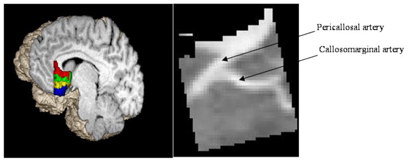
Left: Location of the ventral medial prefrontal cortex with colored gyri. Right: Sagittal MR slice of the VMPFC with callosomarginal and pericallosal arteries.
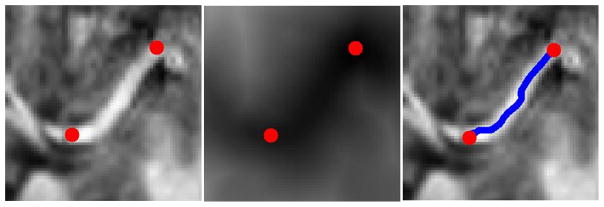
The steps of tracing a curve: a 2D slice with a vessel (left), the distance map calculated from the slice with lower values represented by darker intensities (center), and the path traced within the blood vessel (right); end points are marked in red. Lines thickened for emphasis.
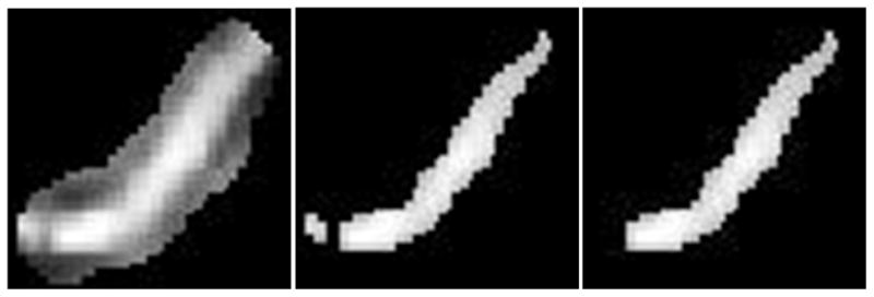
The steps of segmenting a vessel in a 2D slice: a tube is generated around the vessel (left), the tube is thresholded (center), and finally the largest connected component is selected (right).

Histogram of the distance of each automatically segmented voxel from the closest manually segmented one.

Top shows a VMPFC gray/white surface reconstructed with the automated method (green) with the blood vessels reconstructed from the original volume (blue). Bottom row shows the axial, coronal and sagittal views of the original and reconstructed surfaces indicating the presence of the blood vessels. The red ball on the surfaces (top row) corresponds to the cross-hair in the images (bottom row).
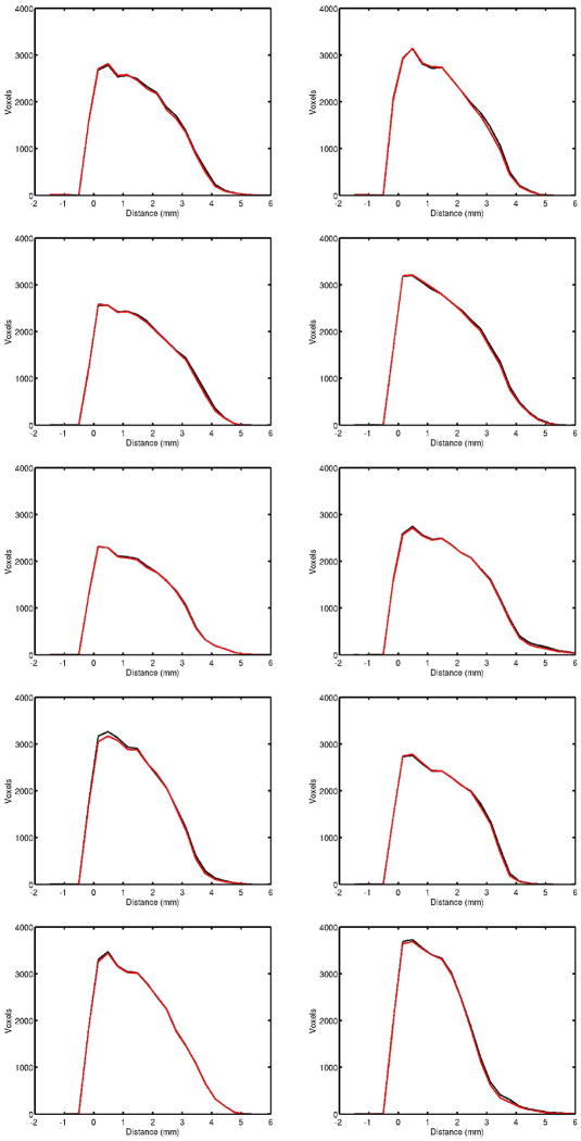
LCDMs comparing manual (black) and automatic removal (red) of blood vessels for subjects 1 to 10 (top-to-bottom, left-to-right).
Similar articles
-
Visualization of intra-thalamic nuclei with optimized white-matter-nulled MPRAGE at 7T.Neuroimage. 2014 Jan 1;84:534-45. doi: 10.1016/j.neuroimage.2013.08.069. Epub 2013 Sep 7. Neuroimage. 2014. PMID: 24018302 Free PMC article.
-
Statistical and topological atlas based brain image segmentation.Med Image Comput Comput Assist Interv. 2007;10(Pt 1):94-101. doi: 10.1007/978-3-540-75757-3_12. Med Image Comput Comput Assist Interv. 2007. PMID: 18051048
-
Flattening maps for the visualization of multibranched vessels.IEEE Trans Med Imaging. 2005 Feb;24(2):191-8. doi: 10.1109/tmi.2004.839368. IEEE Trans Med Imaging. 2005. PMID: 15707245 Free PMC article.
-
Quantitative comparison of two cortical surface extraction methods using MRI phantoms.Med Image Comput Comput Assist Interv. 2007;10(Pt 1):409-16. Med Image Comput Comput Assist Interv. 2007. PMID: 18051085
-
Adaptive averaging for improved SNR in real-time coronary artery MRI.IEEE Trans Med Imaging. 2004 Aug;23(8):1034-45. doi: 10.1109/TMI.2004.828677. IEEE Trans Med Imaging. 2004. PMID: 15338736 Clinical Trial.
Cited by
-
Cerebral blood flow with [15O]water PET studies using an image-derived input function and MR-defined carotid centerlines.Phys Med Biol. 2013 Mar 21;58(6):1903-23. doi: 10.1088/0031-9155/58/6/1903. Epub 2013 Feb 27. Phys Med Biol. 2013. PMID: 23442733 Free PMC article.
-
Feasibility of geometric-intensity-based semi-automated delineation of the tentorium cerebelli from MRI scans.J Neuroimaging. 2011 Apr;21(2):e148-55. doi: 10.1111/j.1552-6569.2009.00405.x. J Neuroimaging. 2011. PMID: 19659568 Free PMC article.
References
-
- Fischl B, Salat DH, van der Kouwe AJW, Makris N, Segonne F, Quinn BT, Dale AM. Sequence-independent segmentation of magnetic resonance images. NeuroImage. 2004;23(Supplement 1):S69–S84. - PubMed
-
- Duvernoy HM. The Human Brain: Surface, Three-Dimensional Sectional Anatomy with MRI and Blood Supply. Wien, Austria: Springer-Verlag; 1991.
-
- Botteron KN, Raichle ME, Drevets WC, Heath AC, Todd RD. Volumetric reduction in the left subgenual prefrontal cortex in early onset depression. Biol Psych. 2002;51:342–344. - PubMed
-
- Drevets WC, Price JL, Simpson JR, Todd RD, Reich T, Vannier M, Raichle ME. Subgenual prefontal cortex abnormalities in mood disorders. Nature. 1997;386:824–827. - PubMed
Publication types
MeSH terms
Grants and funding
- P50-MH071616/MH/NIMH NIH HHS/United States
- R01 MH062626/MH/NIMH NIH HHS/United States
- R01 MH056584/MH/NIMH NIH HHS/United States
- P01 AG003991/AG/NIA NIH HHS/United States
- P01-AG003991-21/AG/NIA NIH HHS/United States
- R01-MH56584/MH/NIMH NIH HHS/United States
- R01 EB000975/EB/NIBIB NIH HHS/United States
- P41 RR015241/RR/NCRR NIH HHS/United States
- R01-EB000975/EB/NIBIB NIH HHS/United States
- P41-RR15241/RR/NCRR NIH HHS/United States
- R01-MH626266/MH/NIMH NIH HHS/United States
- P01 AG026276/AG/NIA NIH HHS/United States
- P01-AG026276-01/AG/NIA NIH HHS/United States
- P50 MH071616/MH/NIMH NIH HHS/United States
- P41 EB015909/EB/NIBIB NIH HHS/United States
LinkOut - more resources
Full Text Sources
Medical
