. 2007 Dec 21;282(51):37064-73.
doi: 10.1074/jbc.M705015200. Epub 2007 Oct 26.

Calcineurin promotes hypoxia-inducible factor 1alpha expression by dephosphorylating RACK1 and blocking RACK1 dimerization

Affiliations

Calcineurin promotes hypoxia-inducible factor 1alpha expression by dephosphorylating RACK1 and blocking RACK1 dimerization

Ye V Liu et al. J Biol Chem. .

Abstract

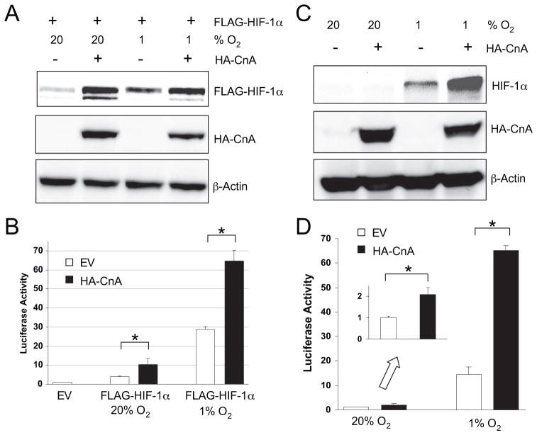
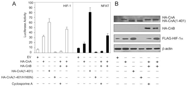
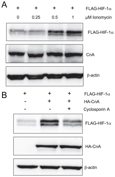
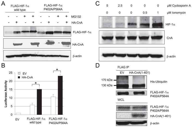
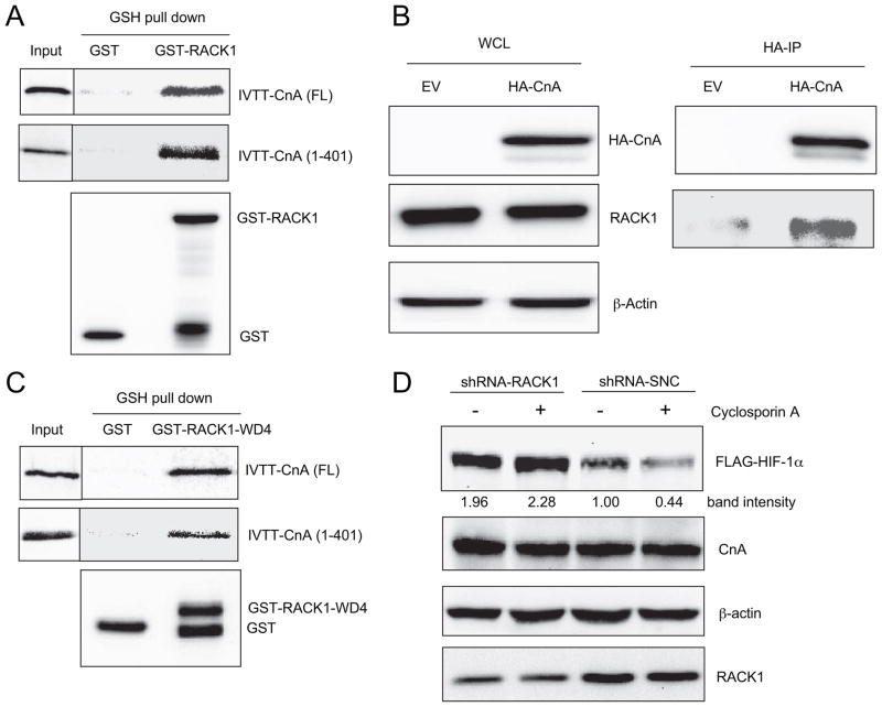
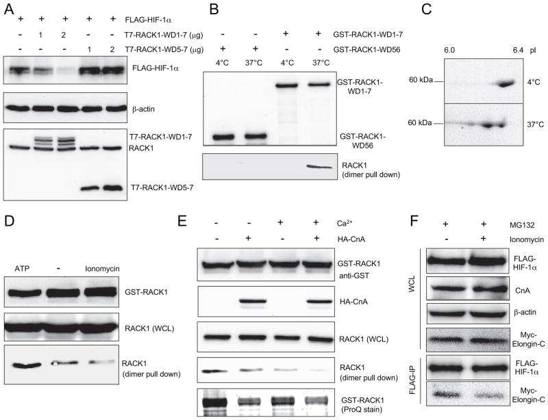
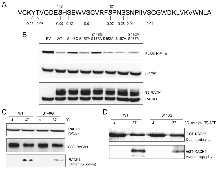
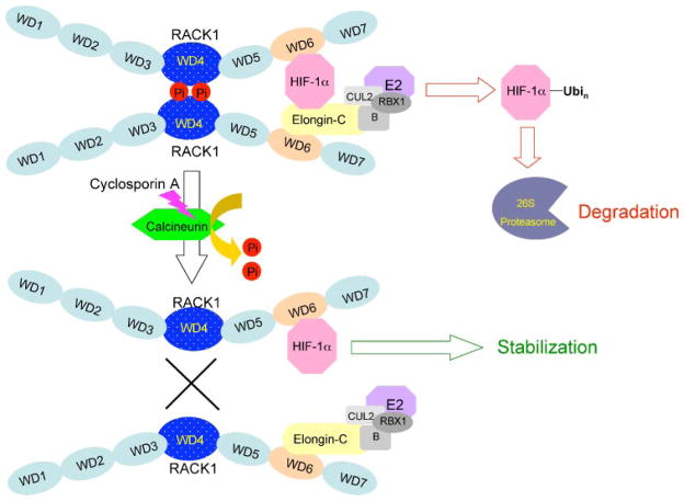
Oxygen homeostasis represents an essential organizing principle of metazoan evolution and biology. Hypoxia-inducible factor 1 (HIF-1) is a master regulator of transcriptional responses to changes in O2 concentration. HIF-1 is a heterodimer of HIF-1alpha and HIF-1beta subunits. O2-dependent degradation of the HIF-1alpha subunit is mediated by prolyl hydroxylase, von Hippel-Lindau protein (VHL)/Elongin-C E3 ubiquitin ligase, and the proteasome. O2-independent degradation of HIF-1alpha is regulated by the competition of RACK1 and HSP90 for binding to HIF-1alpha. RACK1 binding results in the recruitment of the Elongin-C E3 ubiquitin ligase, leading to VHL-independent ubiquitination and degradation of HIF-1alpha. In this report, we show that calcineurin inhibits the ubiquitination and proteasomal degradation of HIF-1alpha. Calcineurin is a serine/threonine phosphatase that is activated by calcium and calmodulin. The phosphatase activity of calcineurin is required for its regulation of HIF-1alpha. RACK1 binds to the catalytic domain of calcineurin and is required for HIF-1alpha degradation induced by the calcineurin inhibitor cyclosporine A. Elongin-C and HIF-1alpha each bind to RACK1 and dimerization of RACK1 is required to recruit Elongin-C to HIF-1alpha. Phosphorylation of RACK1 promotes its dimerization and dephosphorylation by calcineurin inhibits dimerization. Serine 146 within the dimerization domain is phosphorylated and mutation of serine 146 impairs RACK1 dimerization and HIF-1alpha degradation. These results indicate that intracellular calcium levels can regulate HIF-1alpha expression by modulating calcineurin activity and RACK1 dimerization.


Figures

FIGURE 1
FIGURE 1. Calcineurin A increases HIF-1αprotein levels under both hypoxic and non-hypoxic conditions
A, CnA increases HIF-1α protein levels in co-transfected cells. HEK293T cells were co-transfected with EV or expression vector encoding FLAG-HIF-1α or HA-CnA. The cells were exposed to 20 or 1% O2 for 4 h. WCL were subjected to IB assay to detect FLAG-HIF-1α, HA-CnA, and β-actin. B, calcineurin A increases transcription mediated by cotransfected HIF-1α. HEK293T cells were cotransfected with pSV-Renilla, in which Renilla luciferase is expressed from an SV40 promoter; p2.1, which contains a hypoxia response element upstream of an SV40 promoter and firefly luciferase coding sequences; and EV or vector encoding FLAG-HIF-1α or HA-CnA. The cells were exposed to 20 or 1% O2 for 24 h. Cells were lysed and the ratio of firefly: Renilla luciferase activity was determined. The results were normalized to those from cells transfected with EV and incubated at 20% O2 (mean ±S.D. shown). *, p <0.05 for indicated comparison. C, calcineurin A increases endogenous HIF-1α protein levels. HEK293T cells were transfected with EV or vector encoding HA-CnA. The cells were exposed to 20 or 1% O2 for 4 h. WCL were subjected to IB assay to detect HIF-1α, HA-CnA, and β-actin. D, calcineurin A increases transcription mediated by endogenous HIF-1α. HEK293T cells were co-transfected with pSV-Renilla, p2.1, and EV or HA-CnA. The cells were exposed to 20 or 1% O2 for 24 h. Cells were lysed and the ratio of firefly: Renilla luciferase activity was determined. The results were normalized to those from cells transfected with EV and incubated at 20% O2 (mean ± S.D. shown). *, p <0.05 for indicated comparison. Inset, luciferase activity at 20% O2.
FIGURE 2
FIGURE 2. Calcineurin has similar effects on HIF-1 and NFAT-dependent transcription
A, effect of calcineurin expression vectors on HIF-1 and NFAT-dependent transcription. HEK293T cells were co-transfected with control reporter plasmid pSV-Renilla and either HIF-1-dependent reporter plasmid p2.1 (left panel) or NFAT-dependent reporter plasmid pNFAT-Luc (right panel). Cells were co-transfected with FLAG-HIF-1α vector +additional expression vector(s) +treatment with 2.5 μM cyclosporine A as indicated. B, effect of HA-tagged calcineurin subunit expression on the levels of FLAG-HIF-1α and β-actin protein. The cells were transfected as in panel A except for the absence of reporter plasmids. IB assays were performed using aliquots of WCL prepared 24 h after transfection.
FIGURE 3
FIGURE 3. Calcineurin phosphatase activity regulates HIF-1α
A, ionomycin increases HIF-1α protein levels. HEK293T cells were transfected with FLAG-HIF-1α expression vector. The cells were treated with vehicle or ionomycin at the indicated concentration for 6 h. WCL were subjected to IB assay to detect FLAG-HIF-1α, CnA, and β-actin. B, cyclosporine A decreases HIF-1α protein expression. HEK293T cells were co-transfected with FLAG-HIF-1α and EV or HA-CnA expression vector. The cells were treated with 2.5 μM cyclosporine A for 6 h. WCL were subjected to IB assay to detect FLAG-HIF-1α, HA-CnA, and β-actin.
FIGURE 4
FIGURE 4. Calcineurin A regulates HIF-1α in a PHD/VHL-independent, ubiquitin/proteasome-dependent manner
A, calcineurin A increases the levels of both wild type and hydroxylation resistant mutant forms of HIF-1α protein by inhibiting their degradation in a proteasome dependent manner. HEK293T cells were co-transfected with EV, wild-type FLAG-HIF-1α, FLAG-HIF-1α-(P420A/P564A), or HA-CnA. The cells were treated with vehicle or 10 μM MG132 for 4 h. WCL were subjected to IB assay to detect FLAG-HIF-1α, HA-CnA, and β-actin. B, calcineurin A increases transcription mediated by both wild type and hydroxylation resistant mutant forms of HIF-1α. HEK293T cells were co-transfected with pSV-Renilla, p2.1, wild-type FLAG-HIF-1α or FLAG-HIF-1α(P420A/P564A), and EV or HA-CnA. Cells were lysed and the ratio of firefly: Renilla luciferase activity was determined. The results were normalized to those from cells transfected with EV and incubated at 20% O2 (mean ± S.D. shown). *, p < 0.05 for indicated comparison. C, calcineurin increases HIF-1α protein expression in a VHL independent manner. VHL-deficient RCC4 cells were treated with vehicle or the indicated concentration of ionomycin or cyclosporine A for 16 h. WCL were subjected to IB assay to detect HIF-1α, CnA, and β-actin. D, calcineurin A decreases the ubiquitination of HIF-1α. Aliquots of WCL containing FLAG-HIF-1α-(P402A/P564A) were incubated with WCL containing His-tagged ubiquitin with or without HA-CnA. IP of WCL was performed with anti-FLAG antibody. Anti-His and anti-FLAG antibodies were used to detect ubiquitinated and total FLAG-HIF-1α after FLAG IP.
FIGURE 5
FIGURE 5. RACK1 binds to calcineurin A and is required for cyclosporine A-induced HIF-1α degradation
A, RACK1 binds to both full-length calcineurin A and the isolated catalytic domain of calcineurin A in vitro. Purified GST or GST-RACK1 was incubated with in vitro transcribed, translated, and 35S-labeled full-length or the amino-terminal 401 amino acids of calcineurin A (IVTT-CnA(FL) or IVTT-CnA(1– 401), respectively); captured on glutathione (GSH)-Sepharose beads; and analyzed by SDS-PAGE followed by autoradiography (top and middle panels) and IB assay with anti-GST (bottom panel). An aliquot of the IVTT product was also applied directly to the gel (Input). B, co-IP of endogenous RACK1 and transfected HA-CnA. WCL were prepared from HEK293T cells transfected with EV or HA-CnA. IP was performed using anti-HA antibody. WCL and IP products were subject to IB assays to detect HA-CnA, RACK1, and β-actin. C, RACK1 WD4 domain binds to full-length and the amino-terminal catalytic domain of calcineurin A in vitro. Purified GST or GST-RACK1-WD4 were incubated with IVTT-CnA(FL) or IVTT-CnA(1– 401), captured on GSH-Sepharose beads, and analyzed by SDS-PAGE followed by autoradiography (top and middle panels) and IB assay with anti-GST (bottom panel). An aliquot of the IVTT product was also applied directly to the gel (Input). D, RACK1 knockdown blocks HIF-1α degradation induced by cyclosporine A. HEK293T cells were co-transfected with FLAG-HIF-1α and short hairpin RNA directed against RACK1 mRNA (shRNA-RACK1) or a scrambled negative control (shRNA-SNC). Cells were treated with vehicle or 2.5 μM cyclosporine A for 6 h. WCL were subjected to IB assays to detect FLAG-HIF-1α, CnA, β-actin, and RACK1. FLAG-HIF-1α levels were quantified by densitometric analysis (band intensity).
FIGURE 6
FIGURE 6. Dephosphorylation of RACK1 by calcineurin A blocks dimerization and HIF-1α degradation
A, RACK1 carboxyl-terminal domain is not sufficient to degrade HIF-1α. HEK293T cells were cotransfected with EV, FLAG-HIF-1α, full-length T7-RACK1-WD1–7, or carboxyl-terminal T7-RACK1-WD5–7. WCL were subjected to IB assay to detect FLAG-HIF-1α, β-actin, and RACK1. B, full-length GST-RACK1-WD1–7, but not GST-RACK1-WD56, can pull down endogenous RACK1 through dimerization only when incubated at 37 °C. GST-RACK1-WD1–7 or GST-RACK1-WD56 were incubated with WCL from HEK293T cells at 4 or 37 °C for 30 min. GST fusion proteins were captured on GSH-Sepharose beads and analyzed by SDS-PAGE followed by IB assays to detect GST-RACK1 with anti-GST antibody (top panel) and endogenous RACK1 with anti-RACK1 antibody (bottom panel). C, pI of GST-RACK1 was reduced upon incubation with WCL at 37 °C in the presence of ATP. GST-RACK1 was incubated with WCL from HEK293T cells at 4 or 37 °C with 1 mM ATP. GST-RACK1 was captured on GSH-Sepharose beads and analyzed by two-dimensional PAGE followed by Coomassie Blue staining. D, regulation of RACK1 dimerization by ATP and calcium. GST-RACK1 was incubated with WCL from HEK293T cells at 37 °C with 1 mM ATP or 10 μM ionomycin. GST-RACK1 was captured on GSH-Sepharose beads. The pull-down products or WCL were analyzed by IB assays using anti-GST or anti-RACK1 antibody. E, calcineurin A and calcium decrease RACK1 phosphorylation and inhibit RACK1 dimerization. GST-RACK1 was incubated with WCL prepared from HEK293T cells transfected with EV or HA-CnA expression vector, at 37 °C with or without 1 mM CaCl2. GST-RACK1 was captured on GSH-Sepharose beads. The pull-down products were analyzed by IB assays to detect GST-RACK1 with anti-GST (top panel) or RACK1 with anti-RACK1 (second panel from bottom) antibody and by SDS-PAGE followed by ProQ-Diamond phosphoprotein stain (bottom panel). WCL were subject to IB assay to detect HA-CnA (second panel from top) and RACK1 (middle panel). F, HEK293T cells were co-transfected with FLAG-HIF-1α and Myc-Elongin-C. Cells were treated with vehicle or 1 μM ionomycin in the presence of 10 μM MG132 for 6 h before WCL were collected. IP was performed with anti-FLAG antibody. The WCL and IP products were subjected to IB assays to detect FLAG-HIF-1α, CnA, β-actin, and Myc-Elongin-C.
FIGURE 7
FIGURE 7. Serine 146 is a target for phosphorylation and is required for efficient RACK1 dimerization and HIF-1α degradation
A, identification of candidate phosphorylation sites in the WD4 domain of RACK1 based on scores assigned by NetPhos. B, serine 146 is critical for RACK1-mediated HIF-1α degradation. HEK293T cells were co-transfected with vectors encoding FLAG-HIF-1α and wild type or the indicated mutant form of T7-RACK1. WCL were subjected to IB assays to detect FLAG-HIF-1α, RACK1, and β-actin. C, effect of serine 146 mutation on RACK1 dimerization. The wild type (WT) or S146G mutant form of GST-RACK1 was incubated with WCL at 4 or 37 °C. GST fusion proteins were captured on GSH-Sepharose beads. The pull-down products were analyzed by IB assays using anti-GST (middle panel) or anti-RACK1 (bottom panel) antibody. Endogenous RACK1 in WCL was also analyzed by IB assays (top panel). D, effect of serine 146 mutation on RACK1 phosphorylation. The WT or S146G mutant form of GST-RACK1 was incubated with WCL at 4 or 37 °C in the presence of 0.3 mCi of [γ-32P]ATP. GST fusion proteins were captured on GSH-Sepharose beads. The pull-down products were subjected to SDS-PAGE followed by Coomassie Blue staining and autoradiography.
FIGURE 8
FIGURE 8. Calcineurin dephosphorylates RACK1, inhibits RACK1 dimerization, and blocks RACK1-mediated ubiquitination and degradation of HIF-1α
Phosphorylation of the WD4 domain promotes RACK1 dimerization. The WD6 domain of each monomer binds to either HIF-1α or Elongin-C. HIF-1α is ubiquitinated by the E3 ubiquitin ligase complex (composed of Elongin-B (B), Cullin-2 (CUL2), ring box protein 1 (RBX1), and an E2 ubiquitin-conjugating enzyme (E2), which is associated with Elongin-C. Ubiquitination (Ubi) of HIF-1α targets the protein for degradation by the 26 S proteasome. The calcium-dependent phosphatase calcineurin dephosphorylates RACK1, inhibits RACK1 dimerization, and thereby reduces the association of HIF-1α with the Elongin-C E3 ubiquitin ligase complex. As a result, HIF-1α is protected from ubiquitination and proteasomal degradation. Cyclosporin A inhibits calcineurin activity, thereby increasing RACK1 dimerization and HIF-1α degradation.

Similar articles

Cited by

References

    1. Hu CJ, Wang LY, Chodosh LA, Keith B, Simon MC. Mol Cell Biol. 2003;23:9361–9374. - PMC - PubMed
    1. Manalo DJ, Rowan A, Lavoie T, Natarajan L, Kelly BD, Ye SQ, Garcia JG, Semenza GL. Blood. 2005;105:659–669. - PubMed
    1. Elvidge GP, Glenny L, Appelhoff RJ, Ratcliffe PJ, Ragoussis J, Gleadle JM. J Biol Chem. 2006;281:15215–15226. - PubMed
    1. Iyer NV, Kotch LE, Agani F, Leung SW, Laughner E, Wenger RH, Gassmann M, Gearhart JD, Lawler AM, Yu AY, Semenza GL. Genes Dev. 1998;12:149–162. - PMC - PubMed
    1. Ryan HE, Lo J, Johnson RS. EMBO J. 1998;17:3005–3015. - PMC - PubMed

Publication types

MeSH terms

Substances

LinkOut - more resources