. 2008 Jul;18(7):1536-48.
doi: 10.1093/cercor/bhm184. Epub 2007 Oct 26.

A molecular neuroanatomical study of the developing human neocortex from 8 to 17 postconceptional weeks revealing the early differentiation of the subplate and subventricular zone

Affiliations

A molecular neuroanatomical study of the developing human neocortex from 8 to 17 postconceptional weeks revealing the early differentiation of the subplate and subventricular zone

Nadhim Bayatti et al. Cereb Cortex. 2008 Jul.

Abstract

We have employed immunohistochemistry for multiple markers to investigate the structure and possible function of the different compartments of human cerebral wall from the formation of cortical plate at 8 postconceptional weeks (PCW) to the arrival of thalamocortical afferents at 17 PCW. New observations include the subplate emerging as a discrete differentiated layer by 10 PCW, characterized by synaptophysin and vesicular gamma-aminobutyric acid transporter expression also seen in the marginal zone, suggesting that these compartments may maintain a spontaneously active synaptic network even before the arrival of thalamocortical afferents. The subplate expanded from 13 to 17 PCW, becoming the largest compartment and differentiated further, with NPY neurons located in the outer subplate and KCC2 neurons in the inner subplate. Glutamate decarboxylase and calretinin-positive inhibitory neurons migrated tangentially and radially from 11.5 PCW, appearing in larger numbers toward the rostral pole. The proliferative zones, marked by Ki67 expression, developed a complicated structure by 12.5 PCW reflected in transcription factor expression patterns, including TBR2 confined to the inner subventricular and outer ventricular zones and TBR1 weakly expressed in the subventricular zone (SVZ). PAX6 was extensively expressed in the proliferative zones such that the human outer SVZ contained a large reservoir of PAX6-positive potential progenitor cells.

Keywords: cell migration; cortical development; immunohistochemistry; synaptogenesis.


Figures

Figure 1.
Figure 1.
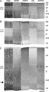
This figure shows cross-sections of the lateral wall of the cortex at 8 PCW near to its junction with the internal capsule and lateral ganglionic eminence. Adjacent paraffin sections have been immunostained for MAP2, GAP43, synaptophysin, and CR. The marginal zone expresses high levels of MAP2 and GAP43, but low levels of synaptophysin. Occasional CR-positive neurons are also seen. The cortical plate is clearly visible as a band of densely packed cells, which are strongly immunoreactive for MAP2. Some synaptophysin immunoreactivity is also observed and many CR-positive neurons are also present. GAP43 is most highly expressed in fibers in an intermediate zone, which contains few immunoreactive cell bodies. The SVZ is high in MAP2 expression and contains some GAP43-positive fibers. The VZ is characterized by a lack of synaptophysin immunoreactivity and little MAP2 immunostaining, although background staining reveals a high density of cell bodies. The occasional MAP2-positive neuron, can however, be seen in this region (asterisk) and may represent a tangentially migrating neuron. Scale bar = 50 μm.
Figure 2.
Figure 2.
This figure compares nuclear expression patterns of PAX6, TBR2, TBR1, and Ki67 immunoreactivity in paraffin-embedded sections demonstrating the differentiation of the human SVZ occurring between 9 and 12.5 PCW. At 9–11 PCW, PAX6 immunoreactive cells are observed at very high density within the VZ, with weak expression in the SVZ at 9 PCW that increases by 11 PCW. TBR2 was expressed in a layer of cells traversing the VZ/SVZ border, whereas TBR1-expressing cells were present predominantly in the cortical plate (CP), although expression throughout the SVZ, subplate/intermediate zone (SP/IZ), and marginal zone (MZ) was also noted. Ki67-positive nuclei, a marker for dividing cells, were present throughout the VZ at 9 PCW, but exhibited a discrete banding pattern at the ventricular surface of the VZ at 11 GW. Strong but less dense Ki67 immunoreactivity was observed throughout the SVZ at 9–11 GW. By 12 GW, the SVZ has differentiated into three discernible layers, an ISVZ and an OSVZ with a cell sparse-layer in between (marked by arrow, see also Fig. 3). PAX6-positive cells were expressed throughout the proliferative zones with some also observed in the IZ. A number of strongly TBR2-expressing cells were present in the VZ, whereas a more dense expression pattern of this protein was observed in the ISVZ. TBR2-immunoreactive nuclei were also present in the OSVZ to a lesser extent. Outside of the CP and SP, TBR1 was found to be present in a few cells within the IZ, and throughout the SVZ but with weaker expression. Ki67 expression at the ventricular surface of the VZ was detected at 12 GW, with the majority of staining within the SVZ being located in the OSVZ. Scale bars = 200 μm.
Figure 3.
Figure 3.
These montages are made from paraffin sections taken from the central/dorsolateral region of the cerebrum and illustrate the rapid growth in thickness of the cortex between 10 and 13 PCW, and in particular, the expansion of the cortical plate (CP) which is most clearly seen with Nissl staining. CR-positive neurons are found in the cortical plate around the border with the subplate (SP). The marginal zone (MZ) grows slowly in thickness. By 12.5 PCW (C) CR neurons form the very outermost layer of the marginal zone (the subpial granular layer) with strong GAP43 and MAP2 immunoreactivity sandwiched between this layer and the cortical plate. The SP and intermediate zone (IZ) appears as a homogenous cell poor zone in the Nissl-stained sections, although GAP43 immunostaining reveals a band particularly rich in growing axons in the outer part of this zone. GFAP immunoreactivity appears in these two compartments from 11.5 PCW (B). The SVZ appears as a homogenous band up to 11.5 PCW (B) but by 12.5 PCW (C) cell rich regions are divided by a cell poor band (arrow, see Fig. 2). The cell poor band and inner parts of the SVZ also contain numerous CR-positive neurons. The VZ is characterized by dense Nissl staining and an absence of MAP2 or GAP43 immunoreactivity. GFAP immunoreactivity is present at the ventricular surface only at 10.5 PCW (A) and but labels fibers throughout the VZ and SVZ at later stages of development. Scale bar = 200 μm.
Figure 4.
Figure 4.
These montages are made from paraffin sections taken from the parietal region, adjacent to those used in Fig. 3. A thin band of very low cellular density (arrow) can be clearly seen at the proximal border of the cortical plate, and this has been interpreted as representing the nascent subplate in a previous study (Kostović and Rakic 1990). However, GAP43, synaptophysin, and vGABA immunostains reveal a wider band displaying intense immunoreactivity for all three, similar to the marginal zone. We interpret this wider band as marking the full extent of the subplate (see text for further discussion). GFAP immunostaining also shows a subtle transition between the intermediate zone, where glial fibers are relatively disordered, and our definition of the subplate, where glial fibers adopt a parallel orientation, which is also seen in the cortical plate. The boundary between the intermediate zone and the SVZ is most clearly seen in the PAX6 immunostained section where immunoreactivity is almost completely confined to VZ and SVZ. Scale bar = 200 μm.
Figure 5.
Figure 5.
(A–D) Paraffin sections taken from the parietal region at 11.5 and 12.5 PCW, immunostained for GAD (11.5 PCW only) and CR. At 11.5 PCW, GAD- and CR-positive cells are found in similar locations in the marginal zone (MZ) and subplate (SP, arrows) however, a higher density of CR neurons is seen in the marginal zone and on the border between the cortical plate (CP) and subplate. By 12.5 PCW many more CR-positive cells appear, particularly in the SVZ. Many have the bipolar morphology characteristic of migrating neurons. Many are aligned for radial migration, but in addition tangentially migrating neurons can be observed in all zones including the SP and VZ (asterisks). IZ is the intermediate zone. At 12.5 PCW, CR expression was markedly different in the frontal (E, Fro) and occipital (F, Occ) lobes of the cortex with many more CR-positive neurons visible in the frontal section in all compartments except for the marginal zone. In addition, CR-positive fibers banded together in parallel bundles in the intermediate zone (IZ) of the occipital lobe only. (G) A series of bar charts summarizing counts of CR neurons (see text for details) in the different compartments of the cerebral wall of the frontal (F), parietal (P), and occipital (O) lobes at 12.5 PCW. It confirms there is a higher density of CR neurons is found in the frontal lobe compared with the occipital lobe in particular at this stage. Scale bar = 100 μm.
Figure 6.
Figure 6.
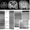
(A) (0.15 × 0.15 × 0.5 mm/voxel), (B) (0.25 × 0.25 × 1.0 mm/voxel), and (C) (0.25 × 0.30 × 1.0 mm/voxel) are from MRI scans of fixed brains at different developmental stages showing the parietal lobe in a coronal section. Higher signal intensities represent greater density of cellular material. They illustrate both the rapid growth and differentiation of the cortex. At 11.5 PCW (A) three layers can be identified in the cerebral wall; the proliferative layer (inner, white) and cortical plate (outer, white) separated by the intermediate/subplate zone. At 14.5 (B) and 17 PCW (C) four layers can be seen. Layer1 is very thin and not discernible everywhere and is probably the VZ. Layer 2 corresponds to the subventricular/intermediate zones and layer 3 to the subplate (as observed at 18 PCW by Radoš et al. 2006) and 4 is the cortical plate. (D and E) The entire cross-section of the cortex at 16 PCW. Nissl staining shows the different zones in the cerebral wall. The cell rich and cell poor layers in the SVZ first seen at 12.5 PCW are more pronounced by 16 PCW. Er81 immunostaining is found in cell nuclei the VZ and SVZ (H), and at high density in a layer in the middle of the cortical plate likely to be layer V (see text). This is shown in close-up in (F). CR immunoreactivity has a different location, being intense in the marginal zone (cell bodies and neurites) with CR-positive cell bodies in the cortical plate located mainly in a putative layer IV (G). In addition, lower power images (I, J) show dense PAX6 immunoreactivity in cell nuclei throughout the VZ and SVZ at this stage of development which coincides with GFAP immunoreactivity visible in cell bodies in the VZ and ISVZ but mostly localized to radial fibers in the rest of the SVZ. IZ intermediate zone; IFL, intermediate fiber layer. Scale bar = 5 mm in A–C, 500 μm in D, E, I, J and 200 μm in F, G, H.
Figure 7.
Figure 7.
This figure is taken from frozen sections from parietal cortex at 16 PCW. Synaptophysin immunoreactivity (A) shows clearly different regions in the cerebral wall. The marginal zone is intensely immunoreactive. The cortical plate is less so but immunoreactivity appears densest in layer IV. The subplate is found between the two small arrows and shows moderately dense immunoreactivity. In the intermediate zone, immunoreactivity appears to be expressed by fibers which are radial in the deeper parts but tangential near to the border with the subplate. (B) Synaptohysin immunoreactivity at a higher magnification at a border between the subplate and the cortical plate. Distinct punctuate staining which may correspond to synaptic terminals is seen in the neuropil sometimes appearing to surround a putative cell body (asterisk). (C and D) NPY and KCC2 immunoreactive neurons in the subplate, the large arrows show where in the subplate they are found. NPY neurons can be bipolar, orientated radially or tangentially, or multipolar, with varicose processes. KCC2 neurons are often pyramidal in shape. Scale bars = 100 μm in A, and 20 μm in B, C, D.
Figure 8.
Figure 8.
A qualitative summary diagram that shows the ontogenetic expression of several proteins found in all cortical regions and the average growth of the cerebral wall also across regions. Generalized immunostaining of many cells and/or their processes in a given layer is represented by blocks of color, whereas immunoreactivity observable in individual cells is represented circles of different colors. It can be seen that, initially, the cortical plate grows rapidly in thickness, but during the second half of the period studied it is the subplate and SVZ that undergo the greatest degree of expansion and differentiation. The SVZ divides into OSV and ISV cell rich regions that express PAX6 indicating that both compartments contain neural progenitor cells. This figure shows that CR-positive neurons are amongst the first differentiated neuronal cell types observed seen particularly in the marginal zone, but that a second cohort appears around 11–13 weeks, followed by migration to a final resting place in the cortical plate. NPY and KCC2 neurons appear at different locations in the subplate. Er81 positive neurons are born in the VZ but settle in layer V of the cortex. The scale bar should be considered as only giving approximate values for thickness of the cerebral wall and each layer, as this diagram is derived from sections that have undergone different methods of tissue processing and have different angles of cut.

References

    1. Aguado F, Carmona MA, Pozas E, Aguilo A, Martinez-Guijarro FJ, Alcantara S, Borrell V, Yuste R, Ibanez CF, Soriano E. BDNF regulates spontaneous correlated activity in early developmental stages by increasing synaptogenesis and expression of the K+/Cl- co-transporter KCC2. Development. 2003;130:1267–1280. - PubMed
    1. Altman J, Bayer SA. Regional differences in the stratified transitional field and honeycomb matrix of the developing human cerebral cortex. J Neurocytol. 2002;31:613–622. - PubMed
    1. Bacci A, Huguenard JR, Prince DA. Differential modulation of synaptic transmission by neuropeptide Y in rat neocortical neurons. Proc Natl Acad Sci USA. 2002;99:17125–17130. - PMC - PubMed
    1. Ben-Ari Y, Khalilov I, Represa A, Gozlan H. Interneurons set the tune of developing networks. Trends Neurosci. 2004;27:422–427. - PubMed
    1. Benowitz LI, Routtenberg A. GAP43; an intrinsic determinant of neuronal development and plasticity. Trends Neurosci. 1997;20:84–91. - PubMed

Publication types