. 2007 Nov 6;104(45):17783-8.
doi: 10.1073/pnas.0706406104. Epub 2007 Oct 26.

Bone marrow cells adopt the cardiomyogenic fate in vivo

Affiliations

Bone marrow cells adopt the cardiomyogenic fate in vivo

Marcello Rota et al. Proc Natl Acad Sci U S A. .

Abstract

The possibility that adult bone marrow cells (BMCs) retain a remarkable degree of developmental plasticity and acquire the cardiomyocyte lineage after infarction has been challenged, and the notion of BMC transdifferentiation has been questioned. The center of the controversy is the lack of unequivocal evidence in favor of myocardial regeneration by the injection of BMCs in the infarcted heart. Because of the interest in cell-based therapy for heart failure, several approaches including gene reporter assay, genetic tagging, cell genotyping, PCR-based detection of donor genes, and direct immunofluorescence with quantum dots were used to prove or disprove BMC transdifferentiation. Our results indicate that BMCs engraft, survive, and grow within the spared myocardium after infarction by forming junctional complexes with resident myocytes. BMCs and myocytes express at their interface connexin 43 and N-cadherin, and this interaction may be critical for BMCs to adopt the cardiomyogenic fate. With time, a large number of myocytes and coronary vessels are generated. Myocytes show a diploid DNA content and carry, at most, two sex chromosomes. Old and new myocytes show synchronicity in calcium transients, providing strong evidence in favor of the functional coupling of these two cell populations. Thus, BMCs transdifferentiate and acquire the cardiomyogenic and vascular phenotypes restoring the infarcted heart. Together, our studies reveal that locally delivered BMCs generate de novo myocardium composed of integrated cardiomyocytes and coronary vessels. This process occurs independently of cell fusion and ameliorates structurally and functionally the outcome of the heart after infarction.


Conflict of interest statement

The authors declare no conflict of interest.

Figures
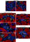
Fig. 1.
Fig. 1.
BMCs engraft and divide. (A and B) Clusters of BMCs within the recipient myocardium after infarction. BMCs are c-kit-positive (green) and carry the Y chromosome (Y-chr, white dots). Connexin 43 (A, Cnx-43, yellow dots) and N-cadherin (B, N-cadh, yellow dots) are present between male BMCs (arrows) and between male BMCs and female myocytes (α-sarcomeric-actin, α-SA, red; arrows) and fibroblasts (procollagen, col, magenta; arrows). (C–E) BrdU (C, yellow), Ki67 (D, yellow), and phospho-H3 (E, yellow) are detected in dividing BMCs (c-kit, green; (Y-chr, red dots). Inset in E shows metaphase chromosomes. (F and G) Apoptosis (TdT, magenta) of BMCs (c-kit, F, green; G, white; Y-chr, white dots). EGFP (G, green). Cnx-43 (G, yellow) is absent in apoptotic BMCs.
Fig. 2.
Fig. 2.
BMCs acquire the cardiogenic fate. (A and B) BMCs are mostly CD45-positive at 12 h (white) and mostly CD45 negative at 48 h. (C) At 48 h, BMCs (c-kit, green; Y-chr:, white dots) express Cnx-43 (yellow) and are CD45 negative. (D) Values are mean ± SD. *, P < 0.05 vs. 12 h; **, P < 0.05 vs. 24–36 h. (E and F) BMCs (EGFP, green; Y-chr, white dots) express Nkx2.5 (magenta, arrowheads) and Cnx-43 (yellow). (G–J) BMCs (G, EGFP, green) show von Willebrand factor (H, vWF, white, arrows) and α-SA (I, red, arrowheads). J shows a merge.
Fig. 3.
Fig. 3.
BMCs interact with cardiomyocytes. (A–C) DiI-labeled BMCs (A, red) were cocultured with calcein-labeled myocytes (B, green). Calcein transferred to BMCs (C, yellow-green, arrows). (D–O) EGFP-positive BMCs (D and J, green) cocultured with myocytes (E and K, dotted lines) were injected with cascade blue (F and L, arrows). Cascade blue transferred to myocytes adjacent to BMCs (H, I, and N). Rhodamine-labeled dextran (O, red) did not transfer from BMC to the adjacent myocyte. (P–U) Cocultured BMCs differentiated into myocytes that contracted spontaneously (R) or after stimulation (U). (P) Myocyte derived from a DiI-labeled BMC (Q, red). (S) BMC-derived mononucleated myocytes express connexin 43 (T, white dots). Neonatal myocytes were labeled by DiI (T, red). (V–X) BMC from α-MHC-EGFP mouse differentiated into a myocyte that expressed EGFP (V, green), α-SA (W, red), and Cnx-43 (X, white). The new myocyte possesses 2n DNA content. (W) Values of DNA content.
Fig. 4.
Fig. 4.
BMCs and myocardial regeneration. (A and B) BMCs from α-MHC-EGFP (A) and α-MHC-c-myc-tagged-nuc-Akt (B) mice regenerated myocardial infarcts. Formed myocytes (arrowheads) express α-SA (red), EGFP (A, green) and c-myc (B, green). (C and D) Infarcts treated with BMCs from β-actin-EGFP mice. EP, epicardium; EN, endocardium. (C) Left, Center, and Right show EGFP (green), regenerated myocytes (MHC, red), and their merge. Arrows, nonregenerated infarct. (D) Y-chr localization across the infarct. Left, Center, and Right illustrate regenerated myocytes (MHC, red), distribution of Y-chr (white dots), and their merge. Arrows, nonregenerated infarct. (E and F) Arterioles formed by BMCs from α-MHC-c-myc-tagged-nuc-Akt mice. SMCs (α-smooth muscle actin, α-SMA, red) and ECs (vWF, yellow) carry the Y-chr (white dots). Vessel wall is c-myc-negative; myocytes (Lower) are c-myc-positive (green), carry the Y-chr, and are α-SA-positive (magenta).
Fig. 5.
Fig. 5.
Myocyte differentiation and functional competence. (A) DNA sequences of EGFP and c-myc-tag by PCR. DNA from the tail of donor transgenic (TG) and wild-type (WT) mice was used as positive and negative control. (B) Transcripts for EGFP and c-myc-tag by real-time RT-PCR in infarcted treated hearts (+). Samples in the absence of RT reaction (−). RNA from hearts of TG and WT was used as positive and negative control. (C) EGFP and c-myc-tag protein by Western blotting. Protein lysates from hearts of TG were used as positive control, and protein lysates from untreated infarcted hearts were used as negative control. (D) At 2–3 days, EGFP-positive cells lacked electrical activity. (E) Electrical properties of BMC-derived and spared myocytes. (F) At 30 days, newly formed EGFP-positive myocytes were electrically excitable. (G) Spared myocytes had depressed fractional shortening. Values are mean ± SD. *, P < 0.05 vs. new myocytes. (H) EGFP-myocytes derived from BMC differentiation used for the evaluation of cell mechanics. Cell volume is indicated (μm3). (I) Mouse heart at 30 days after coronary artery ligation and implantation of BMCs from α-MHC-EGFP mouse. Calcium transient was detected in EGFP-positive-myocytes and EGFP-negative-myocytes.

References

    1. Tosh D, Slack JM. Nat Rev Mol Cell Biol. 2002;3:187–194. - PubMed
    1. Orlic D, Kajstura J, Chimenti S, Jakoniuk I, Anderson SM, Li B, Pickel J, McKay R, Nadal-Ginard B, Bodine DM, et al. Nature. 2001;410:701–705. - PubMed
    1. Yoon YS, Wecker A, Heyd L, Park JS, Tkebuchava T, Kusano K, Hanley A, Scadova H, Qin G, Cha DH, et al. J Clin Invest. 2005;115:326–338. - PMC - PubMed
    1. Murry CE, Soonpaa MH, Reinecke H, Nakajima H, Nakajima HO, Rubart M, Pasumarthi KB, Virag JI, Bartelmez SH, Poppa V, et al. Nature. 2004;428:664–668. - PubMed
    1. Balsam LB, Wagers AJ, Christensen JL, Kofidis T, Weissman IL, Robbins RC. Nature. 2004;428:668–673. - PubMed