. 2007 Oct;19(10):3146-56.
doi: 10.1105/tpc.107.053017. Epub 2007 Oct 26.

Arabidopsis cryptochrome 2 completes its posttranslational life cycle in the nucleus

Affiliations

Arabidopsis cryptochrome 2 completes its posttranslational life cycle in the nucleus

Xuhong Yu et al. Plant Cell. 2007 Oct.

Abstract

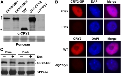
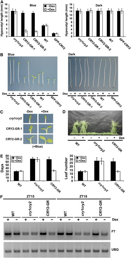
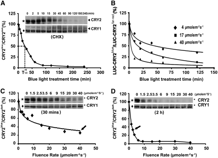
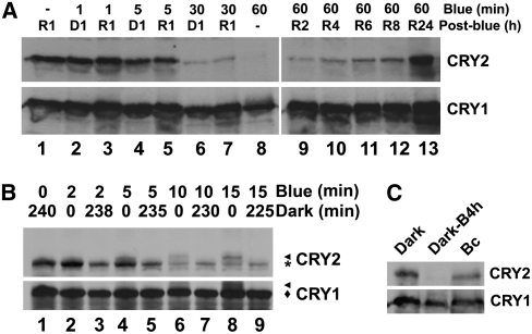
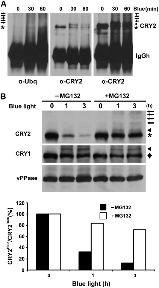
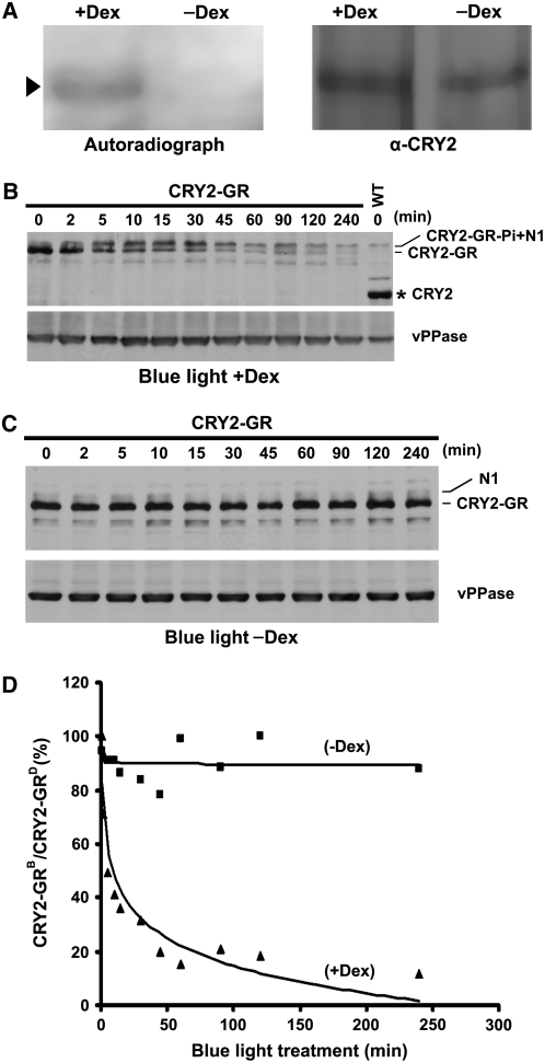
CRY2 is a blue light receptor regulating light inhibition of hypocotyl elongation and photoperiodic flowering in Arabidopsis thaliana. The CRY2 protein is found primarily in the nucleus, and it is known to undergo blue light-dependent phosphorylation and degradation. However, the subcellular location where CRY2 exerts its function or undergoes blue light-dependent phosphorylation and degradation remains unclear. In this study, we analyzed the function and regulation of conditionally nuclear-localized CRY2. Our results show that CRY2 mediates blue light inhibition of hypocotyl elongation and photoperiodic promotion of floral initiation in the nucleus. Consistent with this result and a hypothesis that blue light-dependent phosphorylation is associated with CRY2 function, we demonstrate that CRY2 undergoes blue light-dependent phosphorylation in the nucleus. CRY2 phosphorylation is required for blue light-dependent CRY2 degradation, but only a limited quantity of CRY2 is phosphorylated at any given moment in seedlings exposed to blue light, which explains why continuous blue light illumination is required for CRY2 degradation. Finally, we showed that CRY2 is ubiquitinated in response to blue light and that ubiquitinated CRY2 is degraded by the 26S proteasome in the nucleus. These findings demonstrate that a photoreceptor can complete its posttranslational life cycle (from protein modification, to function, to degradation) inside the nucleus.


Figures

Figure 1.
Figure 1.
Expression and Dex-Dependent Nuclear Localization of CRY2-GR. (A) Immunoblot showing relative levels of expression of CRY2-GR in two independent lines (CRY2-GR-1 and CRY2-GR-2), the control GFP-CRY2 in the respective transgenic lines, and endogenous CRY2 in wild-type plants. Samples were prepared from etiolated seedlings of indicated genotypes, fractioned by a 10% SDS-PAGE gel, blotted, stained with Ponceau S (Ponceau), and probed with the anti-CRY2 antibody (α-CRY2). The bracket indicates CRY2 fusion proteins, and the asterisk indicates the endogenous CRY2. (B) Immunostaining (red) showing accumulation of CRY2-GR (line CRY2-GR-1) in the nucleus (4′,6-diamidino-2-phenylindole stain, blue) in the presence of Dex. Top panel: nuclei were isolated from CRY2-GR/cry1 cry2 seedlings grown in Murashige and Skoog (MS) medium containing 30 μM Dex (+Dex) or mock solution (−Dex) in the dark. Bottom panel: CRY2 immunostaining of the nuclei isolated from wild-type and cry1 cry2 mutant plants. The immunostaining was probed with anti-CRY2 (α-CRY2) antibody and then the second antibody conjugated with the fluorescent dye rhodamine red-x. (C) Immunoblot comparing the level of CRY2-GR (line CRY2-GR-1) in 7-d-old seedlings grown in MS medium containing 30 μM Dex (+Dex) or mock solution (−Dex) in the dark or continuous blue light. The blot was probed with anti-CRY2, stripped, and reprobed with anti-vPPase (vacuolar pyrophosphatase) to show the relative loading. The arrowhead indicates phosphorylated CRY2-GR, and the bracket indicates unphosphorylated CRY2-GR.
Figure 2.
Figure 2.
Only Nuclear-Localized CRY2-GR Can Rescue the Long-Hypocotyl and Late-Flowering Phenotypes of the cry1 cry2 Mutant. (A) Hypocotyl lengths of 5-d-old seedlings with indicated genotypes grown on soil watered with 30 μM Dex (+Dex) or mock solution (−Dex) in continuous blue light (16 μmol m−2 s−1) or in the dark. The means of hypocotyl lengths (n ≥ 20) and standard deviations of two independent lines (CRY2-GR-1 and CRY2-GR-2) are shown. (B) Representative seedlings from samples shown in (A). (C) A close-up cotyledon image of the seedling shown in the left panel of (B), with the indicated genotype and treatment shown. (D) Representative 50-d-old plants of indicated genotypes (CRY2-GR/cry1 cry2 is line CRY2-GR-1). Plants were grown in long-day photoperiods (16 h light/8 h dark) and sprayed daily (from days 10 to 30) with 30 μM Dex (+Dex) or mock solution (−Dex). (E) The flowering times of plants, for which the representatives are shown in (D). The flowering times are measured as days to flower (days) or number of rosette leaves (leaf number) at flowering, and the means and standard deviations (n ≥ 20) are shown. (F) FT mRNA expression in the wild type, cry1 cry2, and CRY2-GR/cry1 cry2 (line CRY2-GR-1). Plants were grown on MS medium containing 30 μM Dex (+Dex) or mock solution (−Dex) in long-day photoperiods (16 h light/8 h dark). Samples were harvested at 15 (ZT15) and 18 (ZT18) h after light on, and the RT-PCR results are shown.
Figure 3.
Figure 3.
Kinetics of Blue Light–Dependent Degradation of Endogenous CRY2 in Wild-Type Seedlings or LUC-CRY2 in Transgenic Seedlings. (A) Immunoblot showing endogenous CRY2 in etiolated wild-type seedlings exposed to blue light (16 μmol m−2 s−1) for the indicated time. Five-day-old etiolated seedlings were treated with cycloheximide (CHX) for 4 h before transferring to blue light for the indicated duration. Protein samples were extracted, fractioned by 10% SDS-PAGE, and blotted. The immunoblot was probed with anti-CRY2, stripped, and reprobed with anti-CRY1 antibodies. The relative level of CRY2 (including both phosphorylated upper bands and unphopsphorylated lower band) is calculated as described (see Methods) and presented as CRY2blue (level of CRY2 after blue light treatment)/CRY2dark (level of CRY2 before blue light treatment). (B) Change in luminescence of LUC-CRY2 seedlings in response to blue light. LUC-CRY2 seedlings were grown on MS medium (∼150 seedlings per Petri dish) in far-red light for 5 d and exposed to blue light of the fluence rates and durations indicated. The luminescence of untreated and blue light–treated seedlings was measured using a CCD camera, and the relative luminescence per seedling is presented as the percentage of LUC-CRY2blue (luminescence after blue light treatment)/LUC-CRY2far-red (luminescence before blue light treatment). (C) and (D) Immunoblot showing endogenous CRY2 in etiolated wild-type seedlings exposed to blue light with the fluence rates indicated for 30 min (C) or 2 h (D). The relative level of CRY2 is calculated as in (A). Arrows indicate the fluence rates by which ∼70% of CRY2 was degraded.
Figure 4.
Figure 4.
Continuous Blue Light and CRY2 Phosphorylation Is Required for CRY2 Degradation. (A) Immunoblots showing the level of CRY2 in etiolated seedlings exposed to blue light for different durations and then transferred to red light or dark (post-blue) for 1 h (lanes 1 to 8) or the level of CRY2 in etiolated seedlings exposed to blue light for 60 min (blue) and then transferred to red light for different durations (lanes 9 to 13). (B) Immunoblot showing changes in the levels of phosphorylated CRY2 and unphosphorylated CRY2 in etiolated seedlings exposed to blue light followed by a posttreatment in the dark. Five-day-old etiolated wild-type seedlings were exposed to blue light (15 μmol m−2 s−1) for the durations indicated (blue). Samples were either analyzed immediately after blue light exposure (lanes 2, 4, 6, and 8) or analyzed after a dark treatment for the durations indicated (lanes 3, 5, 7, and 9). Arrowhead and asterisk (or diamond) indicate phosphorylated and unphosphorylated CRY2 (or CRY1), respectively. (C) Immunoblot showing CRY2 in 5-d-old etiolated seedlings (dark) exposed to blue light for 4 h (20 μmol m−2 s−1; Dark-B4 h) or 5-d-old seedlings germinated and grown in continuous blue light (20 μmol m−2 s−1; Bc). Similar amounts of total protein from different samples were loaded as shown by the similar level of CRY1 (bottom).
Figure 5.
Figure 5.
CRY2 Is Ubiquitinated in Response to Blue Light and Degraded by the 26S Proteasome. (A) Immunoblots showing ubiquitin conjugates of the CRY2-IP products. Five-day-old etiolated wild-type seedlings were exposed to blue light (15 μmol m−2 s−1) for 30 or 60 min. CRY2 was immunoprecipitated with the anti-CRY2 antibody, fractionated by 10% SDS-PAGE, and blotted. Immunoblots were probed by the anti-ubiquitin antibody (left panel) or anti-CRY2 antibody (middle and right panels). The middle and right panels are different exposures of the same blot. Arrows, arrowhead, and asterisk indicate ubiquitinated CRY2, phosphorylated CRY2, and unphosphorylated CRY2, respectively. (B) Inhibition of CRY2 degradation by the proteasome inhibitor MG132. Five-day-old wild-type seedlings grown in the dark were excised and incubated with MG132 or mock solution and exposed to blue light (12 μmol m−2 s−1) for 1 or 3 h. Top: the immunoblot was probed with anti-CRY2 antibody (CRY2; arrows, arrowhead, and asterisk indicate ubiquitinated CRY2, phosphorylated CRY2, and unphosphorylated CRY2, respectively), stripped, and reprobed with the anti-CRY1 antibody (CRY1; arrowhead and diamond indicate phosphorylated and unphosphorylated CRY1, respectively) and then with the antivacuolar pyrophosphatase antibody (vPPase). Bottom: the relative level of CRY2 was calculated as described (see Methods) and presented as the percentage of CRY2blue (level of CRY2 after blue light treatment)/CRY2Dark (level of CRY2 before blue light treatment).
Figure 6.
Figure 6.
Blue Light–Dependent Phosphorylation and Degradation of CRY2-GR Occur in the Nucleus. (A) Autoradiograph showing CRY2-GR was phosphorylated only in the presence of Dex. CRY2-GR/cry1 cry2 seedlings were grown on MS medium with (+Dex) or without (−Dex) Dex, preincubated with 32P for 2 h, and exposed to blue light (18 μmol m−2 s−1) for 15 min. CRY2 was isolated by IP, fractionated, and blotted. After autoradiography (left), the blot was probed with anti-CRY2 antibody (right) to show the level of CRY2. Arrowhead indicates phosphorylated CRY2-GR. (B) and (C) Immunoblots showing that CRY2-GR underwent blue light–dependent degradation only in the presence of Dex. Five-day-old etiolated CRY2-GR/cry1 cry2 seedlings were treated with Dex (B) or mock solution (C) for 2 h and then exposed to blue light (15 μmol m−2 s−1) for the durations indicated. Protein extracts were fractioned by 10% SDS-PAGE gels, and the immunoblots were probed using anti-CRY2 and then anti-vPPase antibodies. CRY2-GR and CRY2-GR-Pi indicate unphosphorylated or phosphorylated CRY2-GR, respectively, N1 indicates the position of a band nonspecifically recognized by the anti-CRY2 antibody, and the asterisk indicates endogenous CRY2. (D) The CRY2-GR signals were digitized and calculated as described (see Methods) and presented as the percentage of CRY2-GRB (level of CRY2-GR after blue light treatment)/CRY2-GRD (level of CRY2-GR before blue light treatment).

References

    1. Ahmad, M., and Cashmore, A.R. (1993). HY4 gene of A. thaliana encodes a protein with characteristics of a blue-light photoreceptor. Nature 366 162–166. - PubMed
    1. Ahmad, M., Jarillo, J.A., and Cashmore, A.R. (1998). Chimeric proteins between cry1 and cry2 Arabidopsis blue light photoreceptors indicate overlapping functions and varying protein stability. Plant Cell 10 197–208. - PMC - PubMed
    1. Banerjee, R., Schleicher, E., Meier, S., Viana, R.M., Pokorny, R., Ahmad, M., Bittl, R., and Batschauer, A. (2007). The signaling state of Arabidopsis cryptochrome 2 contains flavin semiquinone. J. Biol. Chem. 282 14916–14922. - PubMed
    1. Bouly, J.P., Giovani, B., Djamei, A., Mueller, M., Zeugner, A., Dudkin, E.A., Batschauer, A., and Ahmad, M. (2003). Novel ATP-binding and autophosphorylation activity associated with Arabidopsis and human cryptochrome-1. Eur. J. Biochem. 270 2921–2928. - PubMed
    1. Bouly, J.P., Schleicher, E., Dionisio-Sese, M., Vandenbussche, F., Van Der Straeten, D., Bakrim, N., Meier, S., Batschauer, A., Galland, P., Bittl, R., and Ahmad, M. (2007). Cryptochrome blue light photoreceptors are activated through interconversion of flavin redox states. J. Biol. Chem. 282 9383–9391. - PubMed

Publication types

MeSH terms