. 2008 Feb 1;111(3):1240-7.
doi: 10.1182/blood-2007-08-109918. Epub 2007 Oct 26.

The tertiary structure and domain organization of coagulation factor VIII

Affiliations

The tertiary structure and domain organization of coagulation factor VIII

Betty W Shen et al. Blood. .

Abstract

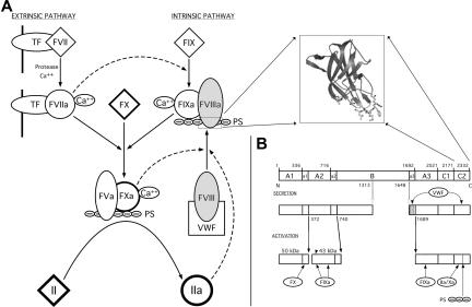
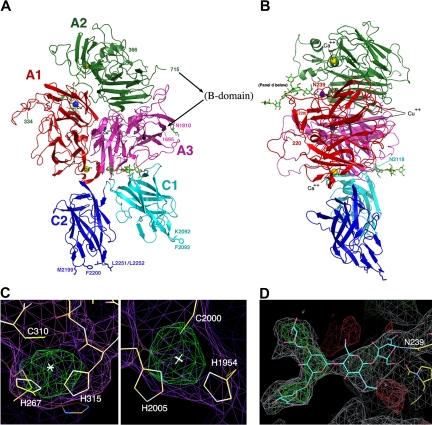
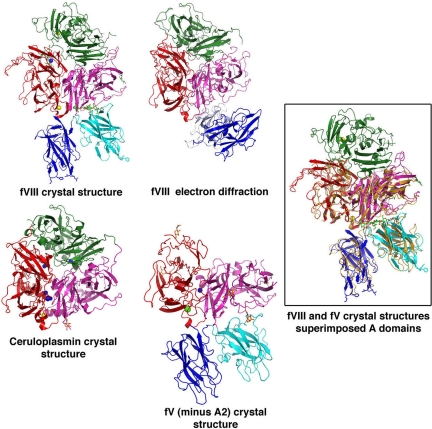
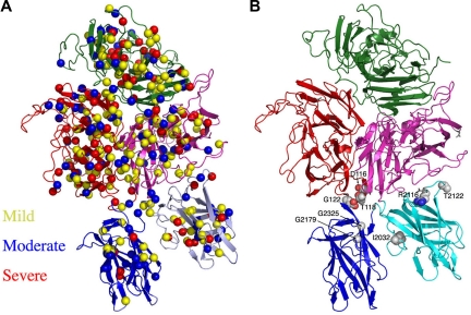
Factor VIII (fVIII) is a serum protein in the coagulation cascade that nucleates the assembly of a membrane-bound protease complex on the surface of activated platelets at the site of a vascular injury. Hemophilia A is caused by a variety of mutations in the factor VIII gene and typically requires replacement therapy with purified protein. We have determined the structure of a fully active, recombinant form of factor VIII (r-fVIII), which consists of a heterodimer of peptides, respectively containing the A1-A2 and A3-C1-C2 domains. The structure permits unambiguous modeling of the relative orientations of the 5 domains of r-fVIII. Comparison of the structures of fVIII, fV, and ceruloplasmin indicates that the location of bound metal ions and of glycosylation, both of which are critical for domain stabilization and association, overlap at some positions but have diverged at others.


Figures

Figure 1
Figure 1
Factor VIII and the coagulation cascade. (A) The blood coagulation cascade consists of 2 pathways (extrinsic and intrinsic) that are initiated by the exposure of tissue factor (TF) or phosphatidylserine groups (PS) of activated platelet membranes to circulating protein factors, respectively. fVIII is a plasma glycoprotein that acts as an initiator and regulator of the intrinsic pathway. Upon proteolytic activation by either fXa or thrombin, fVIIIa dissociates from VWF, associates with the fIXa serine protease, and directs the localization of the resulting complex to the membrane surface of activated platelets via an interaction with its C-terminal C2 domain (structure in inset). The membrane-bound complex between fVIIIa and fIXa complex functions to proteolytically activate fX, which then activates thrombin (fII). (B) Domain structure of fVIII. fVIII is synthesized as a single polypeptide chain of 2332 residues. Based on sequence homology, fVIII has the domain structure A1-A2-B-A3-C1-C2. Linker regions between domains are denoted with lowercase letters (“a1,” etc). The location of domain boundaries and primary sites of proteolytic processing during secretion and activation are denoted by residue numbers. The circulating fVIII heterodimer is associated with VWF primarily through interactions with the “a3” acidic region at the light chain N-terminus and with the C2 domain. Various proteases interact with the activated heterotrimer at positions denoted at the bottom panel. Membrane association is primarily accomplished through the C2 domain; its deletion completely abrogates binding of fVIII to platelet surfaces.
Figure 2
Figure 2
The structure of the B domain deleted fVIII heterodimer. (A,B) The fVIII domains are individually labeled and colored. The C-terminal end of the heavy chain and the N-terminal end of the light chain are indicated with residue numbers (715 and 1695). Residues 1563 to 1694 (the N-terminal 80 residues of the light chain) are present in the construct but are poorly ordered. The N- and C-termini of 2 additional disordered regions are also indicated with residue numbers: residues 220 and 228 that flank a surface loop in the A1 domain (right panel) and residues 334 and 366 that flank the linker region between the A1 and A2 domains (left panel). Shown are 2 bound calcium ions, 2 bound copper ions, and 3 well-ordered and visible N-linked oligosaccharide structures. Shown and labeled for reference are 4 residues on the C2 domain that are thought to be involved in membrane binding and a similarly positioned pair of residues on the C1 domain. (C) Anomalous difference peaks at the sites of bound copper ions buried in the A1 and A3 domains (contoured at 5σ; both are approximately 9σ peak height overall). The residues involved in metal binding at this site are conserved in the analogous copper-binding site in ceruloplasmin, but are diverged from fV. (D) Difference density for 1 of 3 N-linked oligosaccharide structures, which modifies Asn239 in the A1 domain. The density is readily apparent for the entire pentameric polysacharide mannose core of an N-linked sugar, and represents unbiased density prior to any modeling of the covalent modification.
Figure 3
Figure 3
Comparisons of fV and fVIII structures. The relative domain orientations of r-fVIII with a previously reported model generated from electron diffraction studies, and with 2.8-Å resoloution crystal structures of ceruloplasmin and of an inactive (A2-deleted) fV construct are shown. The structures are all oriented similarly with respect to the A domains. Metals ions modeled in the various structures as calcium and copper are indicated as yellow and blue spheres, respectively. The docked orientations of the C domains of r-fVIII, and their interactions with each other and with the A3 domain of the light chain, are both rotated by approximately 90° with respect to the electron diffraction model, but in excellent agreement with their homologous domains in fV. The box shows superposition of full-length fVIII (colored by domains) versus inactive (A2-deleted) fV (shown in orange). The RMSD for aligned α-carbons is approximately 2 Å.
Figure 4
Figure 4
Interface dimensions and packing in the r-fVIII molecule. (A) The light chain and packing of the C domains. The C1 domain (cyan ribbon and pink side chains) is engaged in an extensive docked interaction against the A3 domain (light magenta ribbon and yellow side chains) that involves multiple aromatic, aliphatic, and hydrophilic side chains, several of which are sites of missense mutations associated with protein dysfunction and hemophilia A. An N-linked glycosyl modification (red) is also involved in the C1-A3 interface. In contrast, the C2 domain (blue ribbon and side chains) is loosely tethered to the r-fVIII molecule, displaying small, entirely hydrophilic interfaces with the A3 and C1 domains. Interestingly, a number of residues in these interfaces are also associated with hemophilia. (B) Packing of the trimer of A domains and close up of the packing of the A2 domain (green) against the A3 domain, with residues that have been previously mutated to create engineered disulfide cross-links indicated. These residues are located on 2 adjacent loops in the A domains, which appear to display sufficient flexibility to permit covalent disulfide formation while maintaining r-fVIII activity in vivo.
Figure 5
Figure 5
Location of hemophilia A–associated missense mutations across the r-fVIII structure. (A) The disease phenotype/symptom designations of “mild,” “moderate,” and “severe” are taken from the HamSters database and correspond to plasma fVIIIa activity levels of 6% to 30%, 1% to 5% and less than 1%, respectively. (B) The presence of mutations in the interfaces of the C domains with each other and with the A3 domain.

References

    1. Davie EW, Fujikawa K, Kisiel W. The coagulation cascade: initiation, maintenance, and regulation. Biochemistry. 1991;30:10363–10370. - PubMed
    1. Davie EW, Ratnoff OD. Waterfall sequence for intrinsic blood clotting. Science. 1964;145:1310–1312. - PubMed
    1. Macfarlane RG. An enzyme cascade in the blood clotting mechanism, and its function as a biochemical amplifier. Nature. 1964;202:498–499. - PubMed
    1. Mann KG. Biochemistry and physiology of blood coagulation. Thromb Haemost. 1999;82:165–174. - PubMed
    1. Osterud B, Rapaport SI. Activation of factor IX by the reaction product of tissue factor and factor VII: additional pathway for initiating blood coagulation. Proc Natl Acad Sci U S A. 1977;74:5260–5264. - PMC - PubMed

Publication types