Review
. 2007 Nov;10(11):1377-86.
doi: 10.1038/nn2004.

Astrocyte metabolism and signaling during brain ischemia

Affiliations
Review

Astrocyte metabolism and signaling during brain ischemia

David J Rossi et al. Nat Neurosci. 2007 Nov.

Abstract

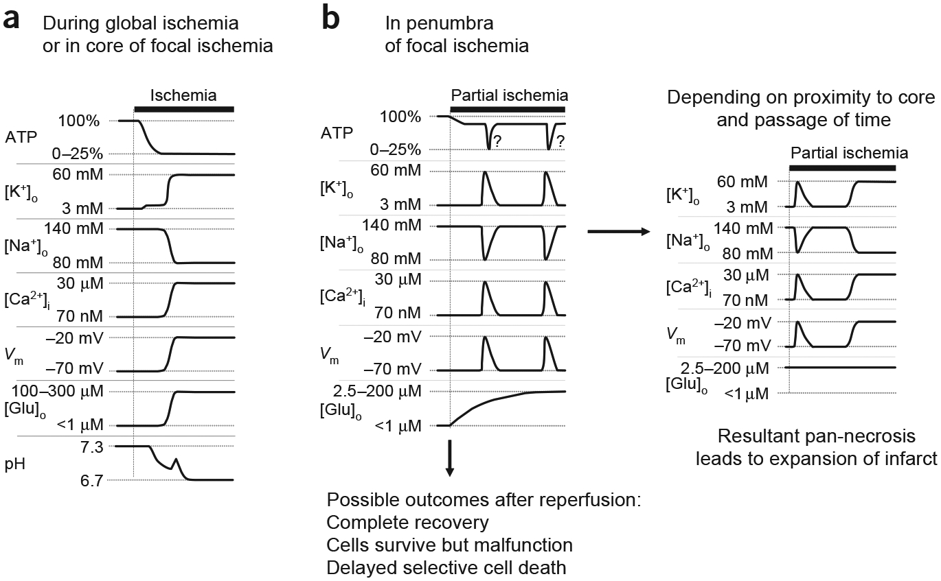
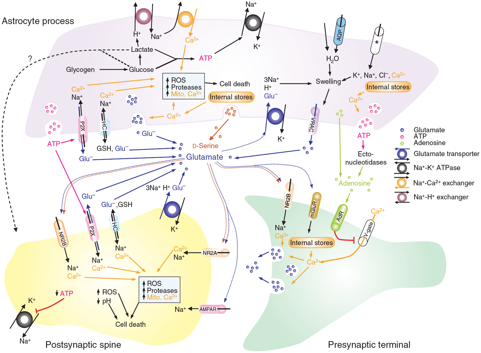
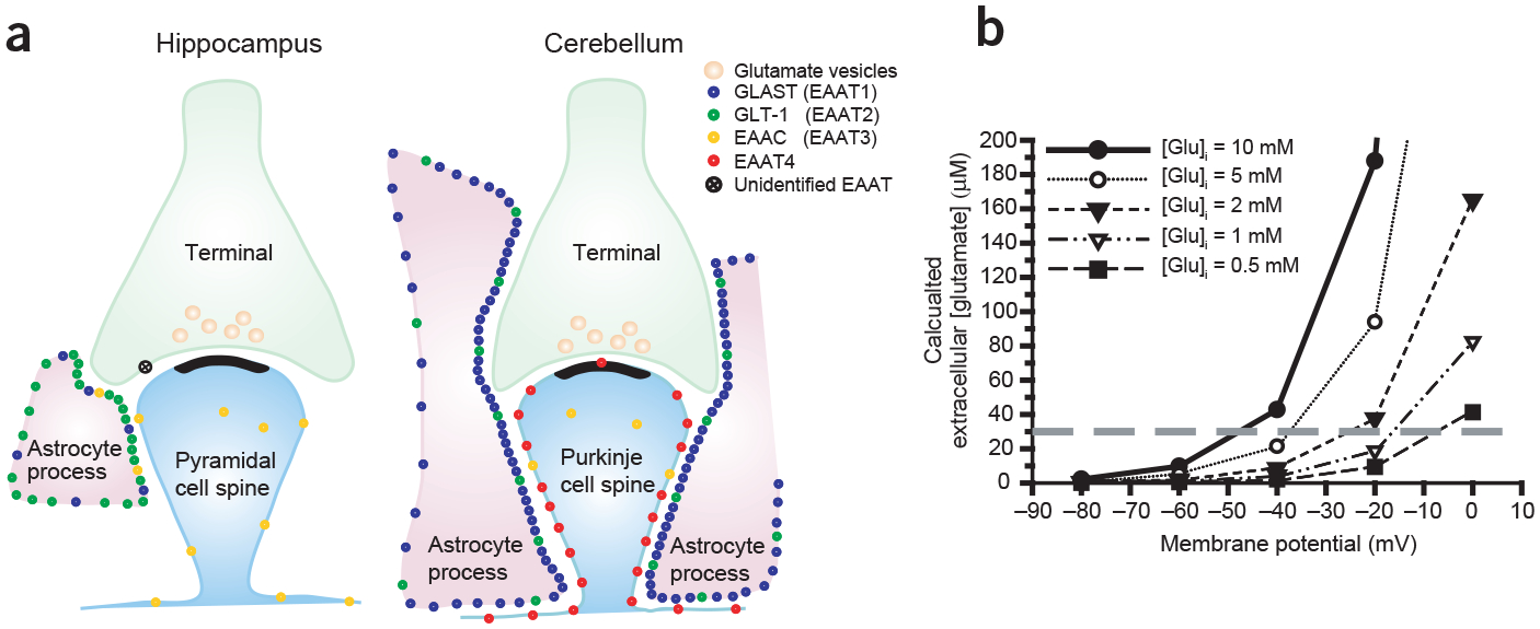
Brain ischemia results from cardiac arrest, stroke or head trauma. These conditions can cause severe brain damage and are a leading cause of death and long-term disability. Neurons are far more susceptible to ischemic damage than neighboring astrocytes, but astrocytes have diverse and important functions in many aspects of ischemic brain damage. Here we review three main roles of astrocytes in ischemic brain damage. First, we consider astrocyte glycogen stores, which can defend the brain against hypoglycemic brain damage but may aggravate brain damage during ischemia due to enhanced lactic acidosis. Second, we review recent breakthroughs in understanding astrocytic mechanisms of transmitter release, particularly for those transmitters with known roles in ischemic brain damage: glutamate, D-serine, ATP and adenosine. Third, we discuss the role of gap-junctionally connected networks of astrocytes in mediating the spread of damaging molecules to healthy 'bystanders' during infarct expansion in stroke.


Figures

Figure 1
Figure 1
Events in brain ischemia. (a) Schematic illustration of events that occur during severe brain ischemia, as occurs in global ischemia and in the core of focal ischemia. Interruption of ATP production leads to inhibition of the Na+-K+ ATPase and to a consequent decrease of the transmembrane ion gradients. The disruption of ion homeostasis depolarizes cells and causes a large release of glutamate into the extracellular space. Ischemia also leads to extracellular acidification. (b) Events in the penumbra of focal ischemia. Initially, the drop in ATP is less severe, but triggers repeated transient depolarizations and associated ion shifts, while [Glu]o rises slowly but steadily (left panel). If reperfusion is initiated soon enough, complete recovery or selective damage may occur (bottom). With increasing duration of ischemia and proximity to the core, the transient ischemic depolarizations may evolve into terminal depolarization and ionic disruption, which leads to pan-necrosis and expansion of the infarct (right panel). Question marks next to transient ATP dips indicate our speculation that ischemic transient depolarizations may induce hypoxia, similarly to spreading depression in nonischemic brain tissue, and resultant transient enhanced ATP loss. Such an effect may act synergistically with ongoing partial ischemia to promote terminal disregulation and exacerbate damage.
Figure 2
Figure 2
Simulated ischemia affects three aspects of glutamatergic signaling. (a) Voltage-clamp recording (holding voltage, Vh = −30 mV) of current in a CA1 pyramidal cell in a hippocampal slice during simulated ischemia. Blockade of large inward current by NMDA receptor antagonist AP5 (50 μM) and non-NMDA receptor antagonist NBQX (25 μM) indicates that it is generated by glutamate receptors. Inset shows typical examples of changes (*before ischemia compared with **after ischemia) in miniature EPSC (mEPSC) frequency (top) and suppression of evoked EPSC (eEPSC) amplitude (bottom) from different cells. Traces are adapted from data gathered in ref. . (b) Plot shows average (n = 3 or 4) membrane potential of Purkinje cells in cerebellar slices during simulated ischemia under control conditions or in the presence of glutamate receptor antagonists (as in a). Data from ref. .
Figure 3
Figure 3
Summary diagram of the processes in neurons and astrocytes which have been shown to or could in principle contribute to the rise in [Glu]o and [Ca2+]i. AdR, adenosine receptor; AMPAR, AMPA receptor; AQP, aquaporin channel; ASIC, acid-sensing ion channels; GSH, glutathione; HC, connexin hemichannel; mGluR, metabotropic glutamate receptor; mito. Ca2+, mitochondrial calcium; NR2A/B, NMDA receptor with subunit 2A or B, respectively; P2XR, P2X receptor; V-gate, voltage-gated calcium channel; *ion flux through various unidentified ion channels. Details of evidence and our speculations in the text.
Figure 4
Figure 4
Expression and operation of Na+-dependent plasma-membrane glutamate transporters. (a) Schematic representation of distribution and relative densities of different glutamate transporter subtypes (based on quantitative immunocytochemistry, adapted from ref. 61). EAAT1–4 are members of the family of five high-affinity Na+-dependent plasma-membrane glutamate transporters. (EAAT1 is the human homolog of GLAST, and EAAT2 is the human homolog of GLT-1.) (b) Plot showing predicted (from equation (1)) extracellular glutamate concentration at equilibrium for ionic gradients during ischemia, given different membrane potentials and different intracellular concentrations of glutamate. The concentrations of co- and countertransported ions used were (in mM) [Na+]o = 87, [Na+]i = 39, [K+]o = 60, [K+]i = 131, [H+]o = 2 and [H+]i = 4. Gray dashed line shows concentration below which cultured neurons can tolerate 5 min exposure without detectable damage.
Figure 5
Figure 5
Schematic illustrations of possible interactions between healthy and dying cells through gap junctions. (a) Toxic substances from dying cells in a focal ischemic core can diffuse through gap junctions to healthy cells in the penumbra, thereby spreading damage. Simultaneously, protective substances from healthy cells in the penumbra can diffuse into and protect damaged cells in the core. (b) The number of gap junctions determines the rate and extent of equilibration of substances between gap-junctionally coupled cells. (c,d) The ratio of toxic cells to healthy ‘bystanders’ determines whether toxic substances are safely diluted (c) or spread damage (d).

References

    1. Choi DW & Rothman SM The role of glutamate neurotoxicity in hypoxic-ischemic neuronal death. Annu. Rev. Neurosci 13, 171–182 (1990). - PubMed
    1. Lipton P Ischemic cell death in brain neurons. Physiol. Rev 79, 1431–1568 (1999). - PubMed
    1. Silver IA, Deas J & Erecinska M Ion homeostasis in brain cells: differences in intracellular ion responses to energy limitation between cultured neurons and glial cells. Neuroscience 78, 589–601 (1997). - PubMed
    1. Abramov AY, Scorziello A & Duchen MR Three distinct mechanisms generate oxygen free radicals in neurons and contribute to cell death during anoxia and reoxygenation. J. Neurosci 27, 1129–1138 (2007). - PMC - PubMed
    1. Lowry OH, Passonneau JV, Hasselberger FX & Schulz DW Effect of ischemia on known substrates and cofactors of the glycolytic pathway in brain. J. Biol. Chem 239, 18–30 (1964). - PubMed

Publication types