. 2007 Dec;8(12):1303-12.
doi: 10.1038/ni1525. Epub 2007 Oct 28.

'Role reversal' for the receptor PAR1 in sepsis-induced vascular damage

Affiliations

'Role reversal' for the receptor PAR1 in sepsis-induced vascular damage

Nicole C Kaneider et al. Nat Immunol. 2007 Dec.

Abstract

Sepsis is a deadly disease characterized by considerable derangement of the proinflammatory, anti-inflammatory and coagulation responses. Protease-activated receptor 1 (PAR1), an important regulator of endothelial barrier function and blood coagulation, has been proposed to be involved in the lethal sequelae of sepsis, but it is unknown whether activation of PAR1 is beneficial or harmful. Using a cell-penetrating peptide (pepducin) approach, we provide evidence that PAR1 switched from being a vascular-disruptive receptor to a vascular-protective receptor during the progression of sepsis in mice. Unexpectedly, we found that the protective effects of PAR1 required transactivation of PAR2 signaling pathways. Our results suggest therapeutics that selectively activate PAR1-PAR2 complexes may be beneficial in the treatment of sepsis.


Figures
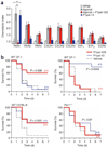
Figure 1
Figure 1
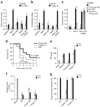
Effects of PAR1 agonist and antagonist pepducins on the survival of septic mice. (a) Migration of HEK cells transiently transfected with cDNA encoding human G protein–coupled receptors (horizontal axis) toward RPMI medium plus 0.5% BSA alone or containing the following agonists: TFLLRN (10 µM), for PAR1; SLIGKV (10 µM), for PAR2; AYPGKF (100 µM), for PAR4; interleukin 8 (50 nM), for CXCR1 and CXCR2; SDF-1α (100 nM), for CXCR4; S1P (100 nM), for S1P1 and S1P3; or RANTES (20 ng/ml), for CCR5 (ref. 4). P1pa1-12S (2 µM) or P1pal-13 (1 µM) was added at time 0 and migration in the Transwell apparatus (8-µm pore) was stopped after 20 h. The chemotactic index (mean ± two s.e.m.) is the ratio of directed migration to random migration (RPMI); n = 5–7 wells per data point. *, P < 0.05. (b) Survival of wild-type CF-1 mice (n = 10–15), wild-type C57BL/6 mice (n = 12) or Par1−/− C57BL/6 mice (n = 8–12) subjected to CLP, then given vehicle, P1pal-12S or P1pal-13 (2.5 mg/kg) subcutaneously immediately (0 h) or 4 h later; mice then received a maintenance dose of pepducins (1 mg/kg) subcutaneously every 24 h until day 6. P values, pepducin versus vehicle (Kaplan-Meier comparisons made with the nonparametric log-rank test). Data are representative of three experiments.
Figure 2
Figure 2
Effect of activation or inhibition of PAR1 on lung vascular permeability in septic mice. (a) Lung vascular permeability after injection of CF-1 mice (n = 5) with peptide (5 mg/kg) or vehicle (Veh; 100 µl of 80% PBS and 20% DMSO) over 1 min, quantified at 45 min as the accumulation of Evans blue dye in lung interstitium over 30 min and presented as peptide treatment relative to vehicle treatment (set as 1). (b) Lung vascular permeability after intraperitoneal injection of LPS (10 mg/kg; nonlethal dose) or buffer (PBS), measured as described in a and presented as LPS treatment relative to buffer treatment (n = 6 mice). (c) Lung vascular permeability after injection of LPS (10 mg/kg, intraperitoneally), then pepducins (5 mg/kg, subcutaneously) or hirudin (10 mg/kg, intraperitoneally) either immediately (0 h) or 2 h later, measured as described in a at 4 h (n = 3 mice). (d) Lung vascular permeability after the induction of sepsis by CLP at time 0, assessed as described in a from 2 h to 48 h. (e) Lung vascular permeability in mice subjected to CLP and injected with P1pal-12S or P1pal-13 (each 2.5 mg/kg, subcutaneously), assessed as described in a at 24 h. *, P < 0.05 (a,c,e). Data are representative of three experiments.
Figure 3
Figure 3
Treatment of septic mice with PAR1-based pepducins inhibits DIC. (a) Time course of thrombocytopenia after CLP of CF-1 mice (n = 6) at time 0; platelets were counted in platelet-rich plasma. (b) ELISA of the time course of the changes in plasma concentrations of D-dimer after CLP of CF-1 mice (n = 6). (c) Thrombocytopenia in mice (n = 5) injected once with pepducins (2.5 mg/kg, subcutaneously) at 0, 2, 4 or 8 h after CLP; platelets were counted 24 h after CLP. *, P < 0.05. (d) Formation of D-dimers in CF-1 mice (n = 6) treated with P1pal-12S (2.5 mg/kg) or P1pal-13 (2.5 mg/kg) either immediately (0 h) or 8 h after CLP and assessed 48 h later. Sham, laparotomy only. *, P < 0.05. Data are representative of three experiments.
Figure 4
Figure 4
The beneficial effects of PAR1 agonists on endothelial barrier function require PAR2. (a) Permeability of quiescent EA.hy926 monolayers grown to confluence on Transwell membranes (3 µm pore), then stimulated with LPS (1 µg/ml), TFLLRN (10 µM), SFLLRN (100 µM), SLIGKV (100 µM), AYPGKF (100 µM), P1pal-13 (0.3 µM) or P1pal-12S (0.3 µM); after 30–240 min of incubation, Evans blue (30 mg/ml in DMEM) was added to the upper well and leakage (15 min) into the bottom well was measured as absorbance at 650 nm. Results are presented as stimulated relative to unstimulated control. (b) Immunoblot analysis of the time course of LPS activation of Rho in EA.hy926 monolayers grown to confluence and treated with LPS (time, above lanes); cell lysates were analyzed for active Rho-GTP and total Rho. (c) Permeability of confluent endothelial monolayers stimulated with LPS (1 µg/ml) and then treated with peptide agonists (concentrations as in a) or 0.2% DMSO vehicle immediately after LPS challenge (0 h) or 2 h later; permeability was measured at 4 h (0% is the permeability of quiescent cells; 100% is the permeability of LPS-stimulated cells). *, P < 0.05. (d,e) Permeability of EA.hy926 cells (d) or HPAECs (e) grown on membranes (3-µm pore) and transfected with siRNA specific for Par1, Par2 or luciferase (Luci) control; after 48 h, cells were stimulated with LPS and then peptide agonists (100 µM) were added 2 h later and permeability was measured at 4 h. *, P < 0.05. (f) Intracellular calcium mobilization in EA.hy926 cells transfected with siRNA specific for Par1, Par2 or luciferase control; after 48 h, cells were ‘lifted’ with 1 mM EDTA in PBS and loaded with the dye Fura-2AM, and calcium mobilization (Ca2+) was monitored as the ratio of fluorescence excitation intensity at 340 to that at 380 nm, as described. AU, arbitrary units. (g) Immunoblot (IB) of lysates of siRNA-transfected cells (48 h), analyzed with monoclonal antibodies ATAP2 (anti-PAR1) and SLIGK (anti-PAR2). Data are representative of three (a,cf), or four (b,g) independent experiments.
Figure 5
Figure 5
The protective effects of the PAR1-agonist pepducin on the survival, DIC and vascular permeability of septic mice require PAR2. (a–c) Migration of HEK cells left untransfected (–) or transiently transfected with pcDEF3 vector, PAR1 (wild-type), PAR1-RD or PAR2 (wild-type) toward chemotactic gradients of thrombin (T; 0.3 nM), P1pal-13 (1 µM), SLIGKV (10 µM), P1pal-12S (3 µM) or RPMI, for 24 h in a Transwell microchemotaxis apparatus (8-µm pore), presented as described in Figure 1a. * P < 0.01. (d) Survival of Par2−/− C57BL/6 mice (n = 17–20) subjected to CLP and then injected subcutaneously with vehicle, P1pal-12S (2.5 mg/kg) or P1pal-13 immediately (vehicle or P1pal-12S) or 4 h later (P1pal-13); mice then received pepducin (1 mg/kg) subcutaneously every 24 h until day 6. (e) ELISA of TAT complex concentrations in the blood of Par2−/− mice subjected to CLP, then injected subcutaneously with P1pal-12S or P1pal-13 (2.5 mg/kg) either immediately or 4 h later, and assessed 48 h after CLP. (f) Thrombocytopenia in Par2−/− mice subjected to CLP, then injected subcutaneously with pepducins (2.5 mg/kg) or vehicle immediately or 4 h later; platelets were counted 24 h after CLP. (g) Lung vascular permeability in Par2−/− mice subjected to CLP, then injected subcutaneously with P1pal-12S or P1pal-13 (2.5 mg/kg) immediately or 4 h later; permeability was assessed at 24 h and is presented relative to that of vehicle-treated mice (set as 100%). *, P < 0.05. Data are representative of six (ac) or three (dg) experiments.
Figure 6
Figure 6
The endothelial barrier–restoring activity of thrombin and APC requires PAR1 and PAR2. (a) Barrier integrity of EA.hy926 cells grown to confluence on Transwell membranes (3-µm pore), then exposed to LPS (1 µg/ml) or 0.2% DMSO vehicle (−) at time 0 (right margin), and then treated with vehicle, thrombin (0.3 nM), APC (180 nM) or P1pal-13 (0.3 µM) either at the time of LPS challenge or 2 h later (left margin); at 4 h, all cells were stained with fluorescein isothiocyanate–phalloidin and analyzed for retraction or spreading. Original magnification, ×160. (b) Permeability of confluent EA.hy926 cells stimulated with LPS (1 µg/ml) and then treated with thrombin (Thr) or APC immediately (0 h) or 2 h later and assessed at 4 h. Results are presented relative to unstimulated control. (c,d) Permeability of EA.hy926 cells (c) or HPAECs (d) transfected with siRNA specific for Par1, Par2 or luciferase; 2 d later, cells were stimulated with LPS (1 µg/ml), treated with thrombin or APC 2 h later, and assessed at 4 h. *, P < 0.05. Data are representative of three independent experiments.
Figure 7
Figure 7
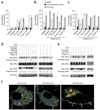
PAR1–PAR2 switching of Rac and Rho signaling in endotoxin-stimulated endothelium. (a) Permeability of quiescent confluent HPAECs treated overnight with DMEM containing buffer (PBS), C3 transferase (100 µg/ml) or pertussis toxin (PTX; 100 ng/ml), then washed and exposed to thrombin (0.3 nM), TFLLRN (10 µM), SLIGKV (100 µM), P1pal-12S (0.3 µM) or P1pal-13 (0.3 µM); permeability was assessed as the leakage of Evans blue into the lower wells. (b,c) Permeability of HPAECs (b) or EA.hy926 cells (c) pretreated with C3 transferase or PTX and stimulated with LPS (1 µg/ml); PAR agonists or antagonists were added 2 h later and endothelial permeability was assessed at 4 h. (d,e) Immunoblot of EA.hy926 cells transfected with luciferase-specific siRNA (d) or Par2-specific siRNA (e); 2 d later, cells were exposed for 5 min to LPS or buffer (−), then thrombin (0.3 nM), SLIGKV (100 µM), TFLLRN (10 µM), P1pal-12S (0.3 µM) or P1pal-13 (0.3 µM) was added and, at 2 h, cells were lysed and activated Rho-GTP was precipitated with GST-Rhotekin beads and Rho-GTP was quantified by analysis with RhoA-Ab. For the corresponding Rac assays, EA.hy926 cells were pretransfected with Myc-tagged Rac; activated Rac was precipitated with GST-PAK beads and was quantified by immunoblot analysis with antibody to Myc. Total Rho and Rac, immunoblot of whole-cell lysates (loading control). Intensity (below lanes) is relative to negative control, set as 1. (f) Confocal photobleaching FRET microscopy of EA.hy926 endothelial cells transfected with PAR2Δ372-CFP (donor) and PAR1Δ377-YFP (acceptor). Blue indicates low FRET efficiency; red (arrowheads) indicates high FRET efficiency. Original magnification, ×400. Data are representative of three experiments (ac); four to five experiments with similar results (d,e); or 10–20 cells per condition in three independent experiments (f).

Similar articles

Cited by

References

    1. Hotchkiss RS, Karl IE. The pathophysiology and treatment of sepsis. N. Engl. J. Med. 2003;348:138–150. - PubMed
    1. Riedemann NC, Guo RF, Ward PA. Novel strategies for the treatment of sepsis. Nat. Med. 2003;9:517–524. - PubMed
    1. Parrillo JE. Pathogenetic mechanisms of septic shock. N. Engl. J. Med. 1993;328:1471–1477. - PubMed
    1. Kaneider NC, Agarwal A, Leger AJ, Kuliopulos A. Reversing systemic inflammatory response syndrome with chemokine receptor pepducins. Nat. Med. 2005;11:661–665. - PubMed
    1. Xiao H, Siddiqui J, Remick DG. Mechanisms of mortality in early and late sepsis. Infect. Immun. 2006;74:5227–5235. - PMC - PubMed

Publication types