. 2008 Feb;153(3):557-67.
doi: 10.1038/sj.bjp.0707542. Epub 2007 Oct 29.

Curcumin inhibits connective tissue growth factor gene expression in activated hepatic stellate cells in vitro by blocking NF-kappaB and ERK signalling

Affiliations

Curcumin inhibits connective tissue growth factor gene expression in activated hepatic stellate cells in vitro by blocking NF-kappaB and ERK signalling

A Chen et al. Br J Pharmacol. 2008 Feb.

Abstract

Background and purpose: Gene expression of connective tissue growth factor (CTGF) is induced in activated hepatic stellate cells (HSC), the major effectors in hepatic fibrosis, and production of extracellular matrix (ECM) is consequently increased. We previously reported that curcumin, the yellow pigment in curry, suppressed ctgf expression, leading to decreased production of ECM by HSC. The purpose of this study is to evaluate signal transduction pathways involved in the curcumin suppression of ctgf expression in HSC.

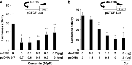
Experimental approaches: Transient transfection assays were performed to evaluate effects of activation of signalling pathways on the ctgf promoter activity. Real-time PCR and Western blotting analyses were conducted to determine expression of genes.

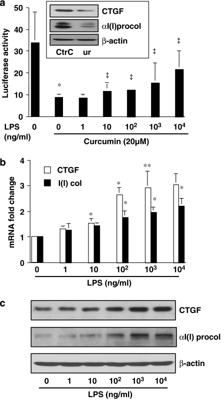
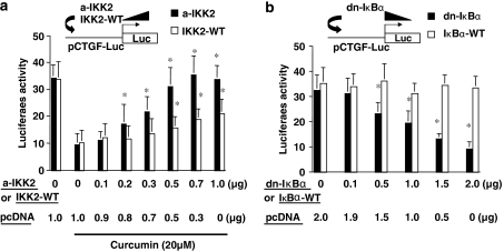
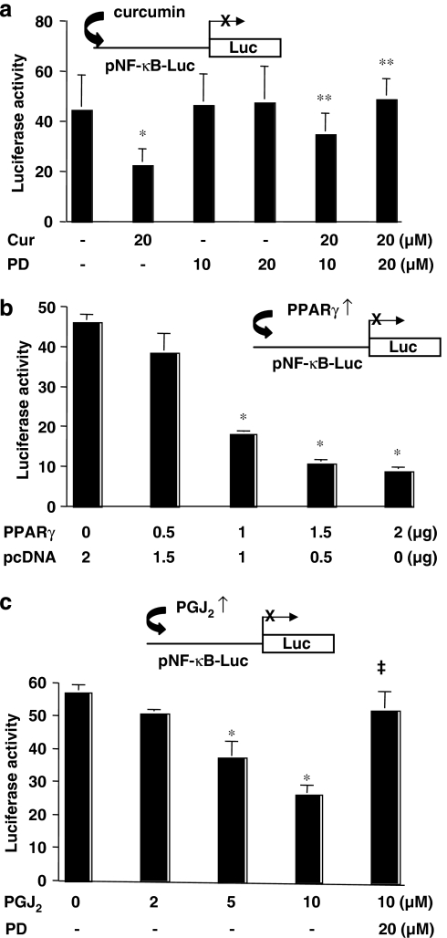
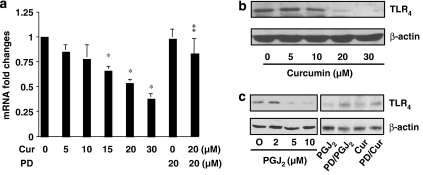
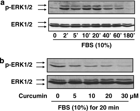
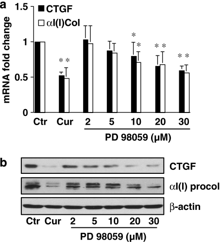
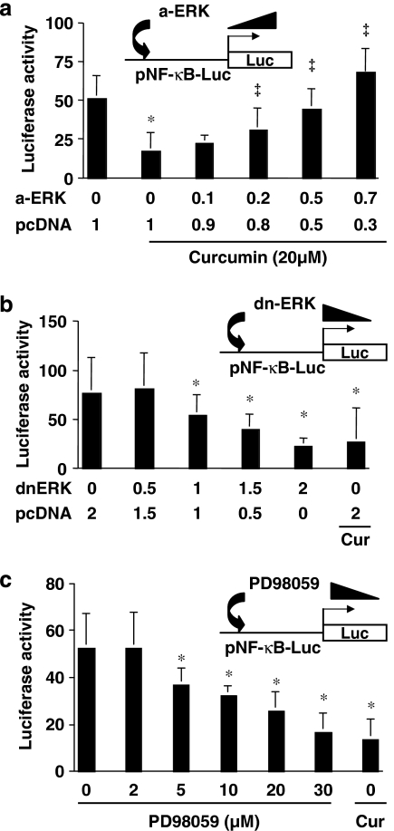
Results: Suppression of ctgf expression by curcumin was dose-dependently reversed by lipopolysaccharide (LPS), an NF-kappaB activator. LPS increased the abundance of CTGF and type I collagen in HSC in vitro. Activation of NF-kappaB by dominant active IkappaB kinase (IKK), or inhibition of NF-kappaB by dominant negative IkappaBalpha, caused the stimulation, or suppression of the ctgf promoter activity, respectively. Curcumin suppressed gene expression of Toll-like receptor-4, leading to the inhibition of NF-kappaB. On the other hand, interruption of ERK signalling by inhibitors or dominant negative ERK, like curcumin, reduced NF-kappaB activity and in ctgf expression. In contrast, the stimulation of ERK signalling by constitutively active ERK prevented the inhibitory effects of curcumin.

Conclusions and implications: These results demonstrate that the interruption of NF-kappaB and ERK signalling by curcumin results in the suppression of ctgf expression in activated HSC in vitro.


Figures

Figure 1
Figure 1
Lipopolysaccharide (LPS) dose-dependently abolishes the inhibitory effect of curcumin on the connective tissue growth factor (CTGF) gene promoter and induces gene expression of CTGF and αI(I) collagen in activated hepatic stellate cell (HSC) in vitro. Passaged HSCs were treated with LPS at indicated concentrations for 24 h in the presence and absence of curcumin (20 μM). (a) Luciferase assays of cells transfected with the plasmid pCTGF-Luc. Luciferase activities were expressed as relative units after β-galactosidase normalization (n⩾6). *P<0.05 versus cells with no treatment (the first column on the left). P<0.05 versus cells with curcumin only (the second column on the left). The inset demonstrates that curcumin (Cur) reduced the protein abundance of CTGF and αI(I) procollagen (αI(I) procol) analysed by western blotting analyses. (b) Real-time PCR analyses of the steady-state mRNA levels of CTGF and αI(I)collagen (αI(I) col). Glyceraldehyde-3-phosphate dehydrogenase (GAPDH) was used as an invariant control for calculating mRNA fold changes. Values are expressed as means±s.d. (n=3). *P<0.05, versus the untreated control (the corresponding first column on the left). (c) Western blotting analyses of the abundance of CTGF and αI(I)procollagen (αI(I) procol). β-Actin was used as an invariant control for equal loading. Representative blots from three independent experiments are shown.
Figure 2
Figure 2
Nuclear factor kappa B (NF-κB) activity plays a critical role in regulating the promoter activity of connective tissue growth factor (CTGF) gene in activated hepatic stellate cell (HSC) in vitro. Passaged HSCs were co-transfected with the ctgf promoter luciferase reporter plasmid pCTGF-Luc plus a cDNA expression plasmid. A total of 3.5 μg (a) or 4.5 μg (b) of plasmid DNA per well was used for co-transfection of HSC in six-well culture plates. It included 2 μg of pCTGF-Luc, 0.5 μg of pSV-β-gal and 1.0 μg (a) or 2 μg (b) of the cDNA expression plasmid at indicated doses plus the empty vector pcDNA. The latter was used to ensure an equal amount of total DNA in transfection assays. After recovery, cells were treated with or without curcumin for 24 h. Luciferase assays were performed. Luciferase activities were expressed as relative units after β-galactosidase normalization (means±s.d.; n⩾6). (a) Luciferase assays of cells co-transfected with pCMV-IKK-2 S177E/S181E (a-IKK2), encoding constitutively active form of IKK2, or with pCMV-IKK-2-WT (IKK2-WT), expressing wild-type IKK2. *P<0.05 versus cells transfected with no pa-IKK2 or pIKK2-WT, but treated with curcumin (the second column on the left). (b) Luciferase assays of cells co-transfected with pCMV-IκBα-M, encoding dominant-negative form of IκBα (dn-IκBα) or with pCMV-IκBα-WT encoding wild-type IκBα (IκBα-WT). *P<0.05 versus cells transfected with no dn-IκBα-M or IκBα-WT (the first column on the left).
Figure 3
Figure 3
Activation of peroxisome proliferator-activated receptor gamma (PPARγ) reduces nuclear factor kappa B (NF-κB) activity in activated hepatic stellate cell (HSC) in vitro. Semiconfluent HSCs were transiently transfected with the plasmid pNF-κB-Luc. After overnight recovery, cells were pretreated with or without the specific PPARγ inhibitor PD68235 (10 or 20 μM) for 30 min prior to the treatment as indicated in the following for additional 24 h. Luciferase assays were performed. Luciferase activities were expressed as relative units after β-galactosidase normalization (means±s.d.; n⩾6). *P<0.05 versus cells with no treatment (the first column on the left). **P<0.05 versus cells with curcumin only (the second column on the left). The insets denote the pNF-κB-Luc construct in use and the application of a treatment, or a co-transfected plasmid, to the system. (a) Cells were treated with curcumin (Cur; 20 μM) with or without PD68235 (PD; 10 or 20 μM). (b) Cells were co-transfected with the PPARγ cDNA expression plasmid pPPARγ at indicated doses. A total of 4.5 μg of plasmid DNA per well was transfected to HSC in six-well culture plates. It included 2 μg of pNF-κB-Luc, 0.5 μg of pSV-β-gal and 2 μg of pPPARγ at indicated doses plus the empty vector pcDNA. The latter was used to ensure an equal amount of total DNA in transfection assays. After overnight recovery, cells were cultured in Dulbecco's modified Eagle's medium (DMEM) with fetal bovine serum (FBS) (10%) for 24 h with no additional treatment. (c) Cells were treated with the natural PPARγ agonist 15-deoxy-Δ12,14-prostaglandin J2 (PGJ2) at the indicated concentrations with or without PD68235 (20 μM).
Figure 4
Figure 4
Activation of peroxisome proliferator-activated receptor-gamma (PPARγ) suppresses gene expression of TLR4 in activated hepatic stellate cell (HSC) in vitro. Semiconfluent HSCs were pretreated with or without the specific PPARγ inhibitor PD68235 (20 μM) for 30 min prior to the treatment as indicated, for an additional 24 h. Total RNA or whole-cell extracts were prepared from these cells. (a) Real-time PCR analyses of the steady-state levels of TLR4 in cells treated with curcumin at indicated concentrations. Glyceraldehyde-3-phosphate dehydrogenase (GAPDH) was used as an invariant control for calculating mRNA fold changes. Values are expressed as means±s.d. (n=3). *P<0.05 versus the untreated control (the first column on the left). (b) Western blotting analyses of the abundance of TLR4 in cells treated with curcumin at indicated concentrations. β-Actin was used as an invariant control for equal loading. Representative blots from three independent experiments are shown. (c) Western blotting analyses of the abundance of TLR4 in cells treated with 15-deoxy-Δ12,14-prostaglandin J2 (15d-PGJ2) (PGJ2) at indicated concentrations, or with 15d-PGJ2 (10 μM) or curcumin (Cur; 20 μM), with or without the pre-exposure to PD68235 (PD; 20 μM). β-Actin was used as an invariant control for equal loading. Representative blots from three independent experiments are shown.
Figure 5
Figure 5
Curcumin inhibits the activation of ERK in activated hepatic stellate cell (HSC) in vitro. After serum starvation for 48 h, HSCs were stimulated with fetal bovine serum (FBS) (10%). Whole-cell extracts were prepared for western blotting analyses of the level of phosphorylated ERK1/2 (p-ERK1/2). Total ERK1/2 was used as an internal invariant control for equal loading. Representative blots from three independent experiments are shown. (a) Serum-starved cells were stimulated with FBS (10%) for indicated minutes. (b) Serum-starved cells were pretreated with curcumin at indicated concentration for 30 min prior to the stimulation with FBS (10%) for additional 20 min.
Figure 6
Figure 6
The inhibition of ERK activity by PD98059 suppresses gene expression of connective tissue growth factor (CTGF) and αI(I) collagen in activated hepatic stellate cell (HSC) in vitro. Serum-starved cells were pretreated with curcumin (Cur) at 20 μM or with PD98059 at indicated concentrations for 30 min prior to the stimulation with fetal bovine serum (FBS) (10%) for additional 24 h. Total RNA or whole-cell extracts were prepared from these cells. (a) Real-time PCR analyses of the steady-state levels of CTGF and αI(I) procollagen (αI(I) col). Glyceraldehyde-3-phosphate dehydrogenase (GAPDH) was used as an invariant control for calculating mRNA fold changes. Values are expressed as means±s.d. (n=3). *P<0.05, versus the untreated control (the corresponding first column on the left). (b) Western blotting analyses of the abundance of CTGF and αI(I) procollagen (αI(I) procol). β-Actin was used as an invariant control for equal loading. Representative blots from three independent experiments are shown.
Figure 7
Figure 7
ERK activity plays a role in the regulation of the promoter activity of connective tissue growth factor (CTGF) in activated hepatic stellate cell (HSC) in vitro. HSCs were co-transfected with pCTGF-Luc (2 μg per well) plus a cDNA-expressing plasmid of pa-ERK (a-ERK) or pdn-ERK (dn-ERK) at indicated doses. The empty vector pcDNA was used to ensure an equal amount of total DNA in transfection assays. After overnight recovery, cells were serum-starved in Dulbecco's modified Eagle's medium (DMEM) for 24 h prior to the stimulation with fetal bovine serum (FBS) (10%) in the presence and absence of curcumin (20 μM) for additional 24 h. Luciferase assays were performed. Luciferase activities were expressed as relative units after β-galactosidase normalization (means±s.d.; n⩾6). *P<0.05 versus cells with no treatment (the first column on the left); **P<0.05 versus cells transfected with pcDNA only (the second column on the left). (a) Luciferase assays of cells co-transfected with pCTGF-Luc plus pa-ERK. (b) Luciferase assays of cells co-transfected with pCTGF-Luc plus pdn-ERK.
Figure 8
Figure 8
ERK activity affects the transactivation activity of nuclear factor kappa B (NF-κB) in activated hepatic stellate cell (HSC) in vitro. HSCs were co-transfected with the plasmid pNF-κB-Luc plus pa-ERK or pdn-ERK at indicated doses. After recovery, cells were serum-starved in Dulbecco's modified Eagle's medium (DMEM) for 24 h prior to the stimulation with fetal bovine serum (FBS) (10%) in the presence and absence of curcumin (20 μM) for additional 24 h. Luciferase assays were performed. Luciferase activities were expressed as relative units after β-galactosidase normalization (means±s.d.; n⩾6). *P<0.05 versus cells with no treatment (the first column on the left); P<0.05 versus cells transfected with pcDNA only (the second column on the left). (a) Luciferase assays of cells co-transfected with pNF-κB-Luc plus pa-ERK. (b) Luciferase assays of cells co-transfected with pNF-κB-Luc plus pdn-ERK. (c) Luciferase assays of cells transfected with pNF-κB-Luc only, and then treated with the ERK inhibitor PD98059 at indicated concentrations for 24 h.

Similar articles

Cited by

References

    1. Aga M, Watters JJ, Pfeiffer ZA, Wiepz GJ, Sommer JA, Bertics PJ. Evidence for nucleotide receptor modulation of cross talk between MAP kinase and NF-kappa B signaling pathways in murine RAW 264.7 macrophages. Am J Physiol Cell Physiol. 2004;286:C923–C930. - PubMed
    1. Ammon HP, Wahl MA. Pharmacology of Curcuma longa. Planta Med. 1991;57:1–7. - PubMed
    1. Blom IE, Goldschmeding R, Leask A. Gene regulation of connective tissue growth factor: new targets for antifibrotic therapy. Matrix Biol. 2002;21:473–482. - PubMed
    1. Bourgier C, Haydont V, Milliat F, Francois A, Holler V, Lasser P, et al. Inhibition of Rho kinase modulates radiation induced fibrogenic phenotype in intestinal smooth muscle cells through alteration of the cytoskeleton and connective tissue growth factor expression. Gut. 2005;54:336–343. - PMC - PubMed
    1. Brown K, Gerstberger S, Carlson L, Franzoso G, Siebenlist U. Control of I kappa B-alpha proteolysis by site-specific, signal-induced phosphorylation. Science. 1995;267:1485–1488. - PubMed

Publication types

MeSH terms