. 2008 Jan;153(1):110-23.
doi: 10.1038/sj.bjp.0707541. Epub 2007 Oct 29.

Electromechanical characterization of cinnamophilin, a natural thromboxane A2 receptor antagonist with anti-arrhythmic activity, in guinea-pig heart

Affiliations

Electromechanical characterization of cinnamophilin, a natural thromboxane A2 receptor antagonist with anti-arrhythmic activity, in guinea-pig heart

G-J Chang et al. Br J Pharmacol. 2008 Jan.

Abstract

Background and purpose: Cinnamophilin, a thromboxane A(2) receptor antagonist, has been identified as a prominent anti-arrhythmic agent in rat heart. This study aimed to determine its electromechanical and anti-arrhythmic effects in guinea-pig hearts.

Experimental approach: Microelectrodes were used to study action potentials in ventricular papillary muscles. Fluo-3 fluorimetric ratio and whole-cell voltage-clamp techniques were used to record calcium transients and membrane currents in single ventricular myocytes, respectively. Intracardiac electrocardiograms were obtained and the anti-arrhythmic efficacy was determined from isolated perfused hearts.

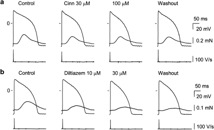
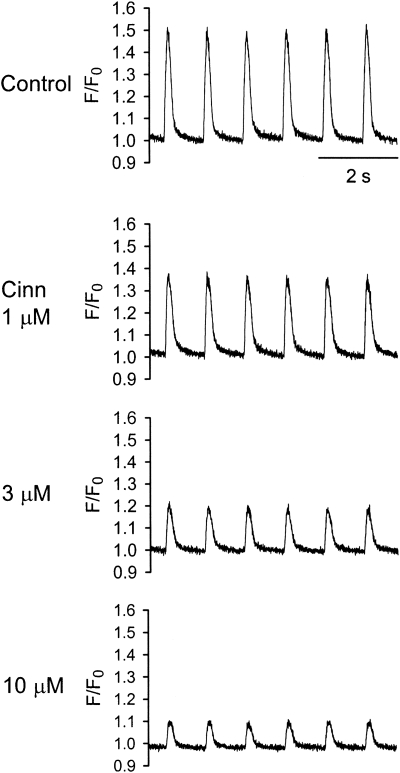
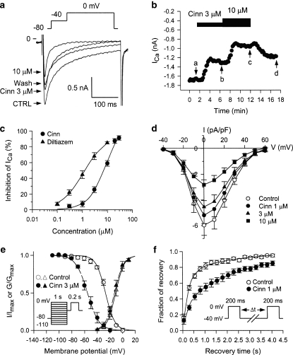
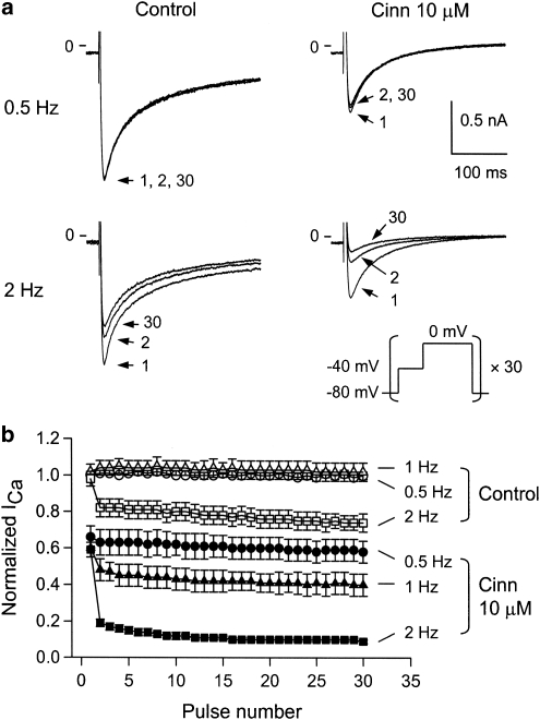
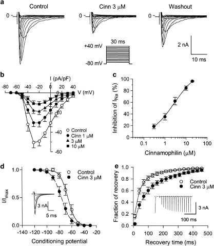
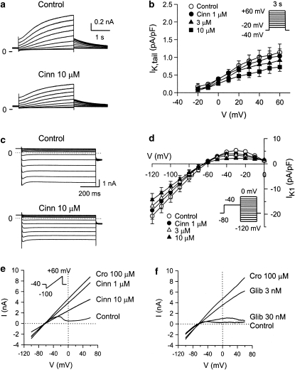
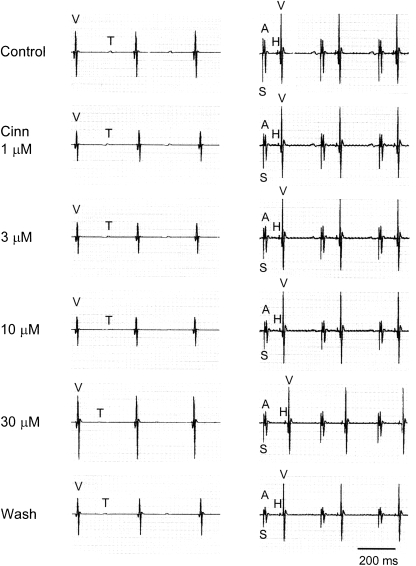
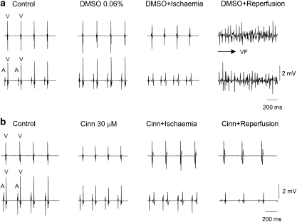
Key results: In papillary muscles, cinnamophilin decreased the maximal rate of upstroke (V(max)) and duration of action potential, and reduced the contractile force. In single ventricular myocytes, cinnamophilin reduced Ca(2+) transient amplitude. Cinnamophilin decreased the L-type Ca(2+) current (I(Ca,L))(IC(50)=7.5 microM) with use-dependency, induced a negative shift of the voltage-dependent inactivation and retarded recovery from inactivation. Cinnamophilin also decreased the Na(+) current (I(Na)) (IC(50)=2.7 microM) and to a lesser extent, the delayed outward (I(K)), inward rectifier (I(K1)), and ATP-sensitive (I(K,ATP)) K(+) currents. In isolated perfused hearts, cinnamophilin prolonged the AV nodal conduction interval and Wenckebach cycle length and the refractory periods of the AV node, His-Purkinje system and ventricle, while shortening the ventricular repolarization time. Additionally, cinnamophilin reduced the occurrence of reperfusion-induced ventricular fibrillation.

Conclusions and implications: These results suggest that the promising anti-arrhythmic effect and the changes in the electromechanical function induced by cinnamophilin in guinea-pig heart can be chiefly accounted for by inhibition of I(Ca,L) and I(Na).


Figures

Figure 1
Figure 1
Original records of transmembrane action potential and isometric contraction in guinea-pig papillary muscles driven at 1 Hz before and following cumulative exposures to 30 and 100 μM cinnamophilin (Cinn) (a) or 10 and 30 μM diltiazem (b), and after 60 min washout. Traces from top to bottom in each panel show contractile force, action potential and Vmax, respectively. Records were obtained at the end of a 20-min exposure time to each concentration of drug. Vmax, maximal upstroke velocity of action potential.
Figure 2
Figure 2
Concentration-dependent effects of cinnamophilin on intracellular Ca2+ transients in ventricular myocytes stimulated at 1 Hz. Original steady-state intracellular Ca2+ transients in the absence (control) and in the presence of 1, 3 and 10 μM cinnamophilin (Cinn) were measured using fluo-3 as relative fluorescence (F/F0).
Figure 3
Figure 3
Effects of cinnamophilin on ICa,L recorded in guinea-pig ventricular myocytes. (a) Individual sample traces recorded in one myocyte in response to 300 ms steps to 0 mV in the absence, in the presence of 3 and 10 μM cinnamophilin (Cinn), and after washout of drug. Each step to 0 mV was preceded by a 150-ms prestep to –40 mV (holding potential=−80 mV; pulse protocol shown at top). (b) Time courses of alteration of ICa,L amplitude. Step pulses were applied every 10 s. Letters on the curve correspond to traces in (a) (point a: control; point b: Cinn 3 μM; point c: Cinn 10 μM and point d: washout). (c) Concentration–response curves for the effect of cinnamophilin (n=12) and diltiazem (n=10) on ICa,L. Solid lines were drawn by fitting to the Hill equation. (d) I–V curves of ICa,L in the absence (control) and presence of different concentrations of cinnamophilin. Each data point represents means±s.e.mean from 10 cells. ICa,L was elicited by various depolarizing pulses (300 ms duration) ranging from −40 to +60 mV in 10 mV increments at 0.1 Hz. (e) Voltage dependence of steady-state ICa,L activation and inactivation in the absence and presence of 3 μM cinnamophilin. The activation curves were constructed using I–V relationships shown in (d). Normalized Ca2+ conductance is plotted as a function of the membrane potential (n=10). Smooth curves are Boltzmann fittings for control (solid line) and cinnamophilin-treated (dashed line) groups. Voltage-dependent steady-state inactivation was examined with a two-pulse protocol as shown in the inset. Preconditioning pulses of 1 s duration were applied in 10 mV steps between −110 and 0 mV from a holding potential of −80 mV, and then the test pulse of 200 ms duration was applied to 0 mV (interpulse duration was 30 ms). The inactivation curves were obtained by normalizing the current amplitudes (I) to the maximal value (Imax) and plotted as a function of the prepulse potential in the absence and presence of 3 μM cinnamophilin (n=7). (f) Effects of cinnamophilin on the recovery of ICa,L from inactivation. Recovery was measured by a double-pulse protocol (see inset). Both pre- and test-pulse were stepped to 0 mV with identical duration of 200 ms and the pulse interval was varied between 50 and 4050 ms. Each two-pulse sequence was separated by a 30 s interval. The ratio of ICa,L obtained by the test pulse to that elicited by the prepulse (fraction of recovery) is plotted as a function of the interpulse interval. Solid and dashed lines represent biexponential curves fitted to the data in control and cinnamophilin-treated groups (n=7), respectively. ICa,L, L-type Ca2+ inward current; IV, current–voltage.
Figure 4
Figure 4
Tonic block and use-dependent block of ICa,L. (a) Original recordings of ICa,L obtained under control conditions and in the presence of cinnamophilin (10 μM). ICa,L were elicited by 30 consecutive 300 ms depolarizing step pulses to 0 mV (each one preceded by a 150-ms prepulse to −40 mV from a holding potential of −80 mV) at 0.5 Hz (upper) or 2 Hz (lower). The pulse protocol is shown in the inset. For each condition, pulses 1, 2 and 30 are shown. (b) Peak amplitudes of ICa,L were normalized to that elicited by the first test pulse before drug application at 0.5 Hz and plotted against the number of pulses applied at different rates in the absence and presence of cinnamophilin (10 μM). Each data point represents means±s.e.mean (n=7). ICa,L, L-type Ca2+ inward current.
Figure 5
Figure 5
Effects of cinnamophilin on INa in guinea-pig ventricular myocytes. (a) Superimposed current traces elicited by 30 ms depolarizing test pulses (–70 to +40 mV in 10 mV increments) from a holding potential of –80 mV, before (control), 5 min after bath application of 3 μM cinnamophilin, and upon 5-min washout. The pulse protocol is shown in the inset. (b) I–V curves of INa in the absence and presence of 1, 3 and 10 μM cinnamophilin. Data are means±s.e.mean (n=7). (c) Concentration–response curve for cinnamophilin on INa is shown. Solid curve was drawn by fitting to the Hill equation. INa was elicited by a series of 30 ms test pulses to –20 mV from a holding potential of –80 mV at 0.2 Hz. (d) Voltage dependence of steady-state INa inactivation in the absence and presence of 3 μM cinnamophilin. Conditioning pulses (1 s long) to potentials ranging from –120 to –30 mV were applied before 50 ms depolarizing test pulses to –20 mV. The holding potential was –80 mV. The predrug superimposed current traces are shown in the inset. Solid and dashed lines drawn through the data points were the best fit to the Boltzmann equation before and after cinnamophilin (n=7), respectively. (e) Recovery of INa from inactivation in the absence and presence of 3 μM cinnamophilin. The twin-pulse protocol used consisted of a 50 ms prepulse from –80 to –20 mV was followed after various recovery times by a 20 ms test pulse to –20 mV. An example of control current traces is shown in the inset. The normalized current is plotted against the recovery time. Lines (control, solid line; cinnamophilin, dashed line) show best-fits of a double exponential function to the data (n=7). INa, Na+ inward current; IV, current–voltage.
Figure 6
Figure 6
(a) Effects of cinnamophilin on IK in guinea-pig ventricular myocytes. Superimposed current traces obtained during 3 s depolarizing pulses to potentials ranging from −20 to +60 mV in 10 mV steps applied from a holding potential of –40 mV before (control) and after 5 min exposure to 10 μM cinnamophilin. (b) I–V relationships for IK tail currents recorded under control conditions and during exposure to increasing concentrations of cinnamophilin. Data are means±s.e.mean (n=9). (c) Effects of cinnamophilin on IK1 in ventricular myocytes. Superimposed current traces in the absence and after 5 min of exposure to 10 μM cinnamophilin were serially elicited (from 0 to –120 mV for 800 ms in 10 mV steps) after a prepulse of –40 mV. (d) I–V curves of IK1 in the absence and presence of 1, 3, and 10 μM cinnamophilin. Data are means±s.e.mean (n=8). (e) and (f) Effects of cinnamophilin (Cinn) or glibenclamide (Glib) on whole-cell IK,ATP induced by 100 μM cromakalim (Cro) in ventricular myocytes, respectively. The quasi steady-state currents were evoked by a 9-s long voltage ramp from −100 to +60 mV (holding potential=–40 mV). The voltage protocol is shown in the inset of (e). IK, delayed rectifier K+ current; IK1, inward rectifier K+ current; IK,ATP, ATP-sensitive K+ current; IV, current–voltage.
Figure 7
Figure 7
Representative ventricular electrograms at spontaneous rhythm (left) and His bundle electrograms at paced rhythm (300 ms cycle length, right) after cinnamophilin (Cinn) of the guinea-pig heart. A, atrial depolarization; H, His bundle depolarization; S, stimulation artifact; T, ventricular repolarization; V, ventricular depolarization. The paper speed was 100 mm s−1.
Figure 8
Figure 8
Protection against globally ischaemia/reperfusion-induced arrhythmias by cinnamophilin. (a) Electrogram recorded from a DMSO-perfused control heart in which VF can be readily induced after ischaemia/reperfusion. (b) Electrogram recorded from a cinnamophilin (30 μM)-perfused heart which maintained sinus rhythm after ischaemia/reperfusion. Upper and lower traces in both (a) and (b) show the ventricular electrogram and the electrogram recorded at lower atrium, respectively, and show the atrial (A) and ventricular depolarization (V). DMSO, dimethylsulphoxide; VF, ventricular fibrillation.

Similar articles

Cited by

References

    1. Bean BP, Cohen AJ, Tsien RW. Lidocaine block of cardiac sodium channels. J Gen Physiol. 1983;81:613–642. - PMC - PubMed
    1. Bernauer W. Concerning the effect of the K+ channel blocking agent glibenclamide on ischaemic and reperfusion arrhythmias. Eur J Pharmacol. 1997;326:147–156. - PubMed
    1. Billman GE. Role of ATP sensitive potassium channel in extracellular potassium accumulation and cardiac arrhythmias during myocardial ischaemia. Cardiovasc Res. 1994;28:762–769. - PubMed
    1. Chang GJ, Su MJ, Hung LM, Lee SS. Cardiac electrophysiologic and antiarrhythmic actions of a pavine alkaloid derivative, O-methyl-neocaryachine, in rat heart. Br J Pharmacol. 2002;136:459–471. - PMC - PubMed
    1. Chang GJ, Su MJ, Kuo SC, Lin TP, Lee YS. Multiple cellular electrophysiological effects of a novel antiarrhythmic furoquinoline derivative HA-7 [N-benzyl-7-methoxy-2,3,4,9-tetrahydrofuro[2,3-b]quinoline-3,4-dione] in guinea pig cardiac preparations. J Pharmacol Exp Ther. 2006;316:380–391. - PubMed

Publication types

MeSH terms