. 2007 Nov;117(11):3350-8.
doi: 10.1172/JCI32727.

Rational strain selection and engineering creates a broad-spectrum, systemically effective oncolytic poxvirus, JX-963

Affiliations

Rational strain selection and engineering creates a broad-spectrum, systemically effective oncolytic poxvirus, JX-963

Steve H Thorne et al. J Clin Invest. 2007 Nov.

Abstract

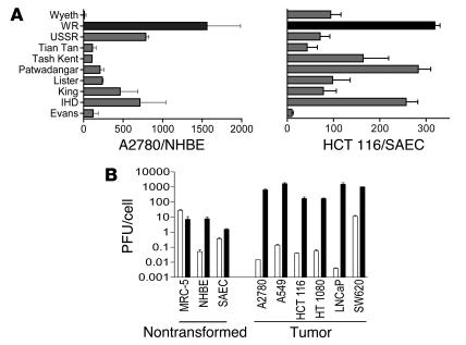
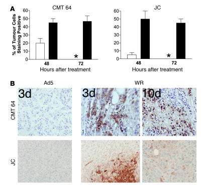
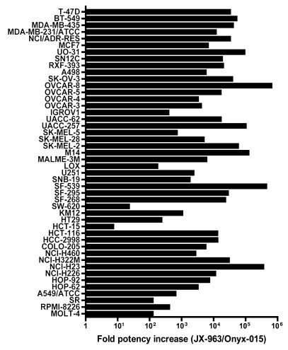
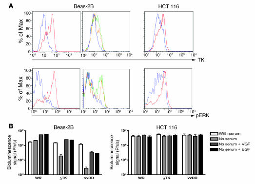
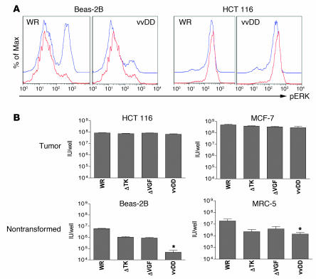
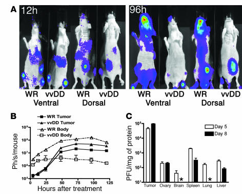
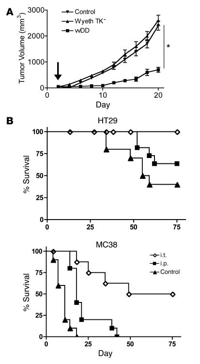
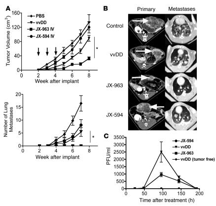
Replication-selective oncolytic viruses (virotherapeutics) are being developed as novel cancer therapies with unique mechanisms of action, but limitations in i.v. delivery to tumors and systemic efficacy have highlighted the need for improved agents for this therapeutic class to realize its potential. Here we describe the rational, stepwise design and evaluation of a systemically effective virotherapeutic (JX-963). We first identified a highly potent poxvirus strain that also trafficked efficiently to human tumors after i.v. administration. This strain was then engineered to target cancer cells with activation of the transcription factor E2F and the EGFR pathway by deletion of the thymidine kinase and vaccinia growth factor genes. For induction of tumor-specific cytotoxic T lymphocytes, we further engineered the virus to express human GM-CSF. JX-963 was more potent than the previously used virotherapeutic Onyx-015 adenovirus and as potent as wild-type vaccinia in all cancer cell lines tested. Significant cancer selectivity of JX-963 was demonstrated in vitro in human tumor cell lines, in vivo in tumor-bearing rabbits, and in primary human surgical samples ex vivo. Intravenous administration led to systemic efficacy against both primary carcinomas and widespread organ-based metastases in immunocompetent mice and rabbits. JX-963 therefore holds promise as a rationally designed, targeted virotherapeutic for the systemic treatment of cancer in humans and warrants clinical testing.


Figures

Figure 1
Figure 1. Virus species and strain selection.
(A) Burst ratio of vaccinia strains in tumor relative to normal cells. Different vaccinia strains were used to infect both primary normal cells (normal human bronchial epithelial [NHBE] or small airway bronchial epithelial [SAEC]) and tumor cell lines (A2780 or HCT 116) at an MOI of 1.0 PFU/cell. Virus collected 48 hours later was titered by plaque assay, and the ratio of virus produced (per cell) in tumor relative to normal cells is represented. (B) Different cell lines were infected with either WR (black bars) or Ad5 (white bars) at an MOI of 1.0 PFU/cell. Amounts of virus produced (infectious units/cell) 48 hours later were titered by plaque assay (left grouping: nontransformed and primary cells; right grouping: tumor cell lines).
Figure 2
Figure 2. Systemic delivery of viral strains to tumors.
1 × 10⁹ PFU of vaccinia strain WR or Ad5 were delivered i.v. to immuno­competent mice bearing subcutaneous CMT 64 or JC tumors (both lines susceptible to replication by both viruses after intratumoral injection). Mice were sacrificed after 48 or 72 hours or after 10 days and immunohistochemistry performed against viral coat proteins on paraffin-embedded sections of tumor tissue. (A) Graphs show scoring of positive cells in each tumor (asterisk indicates none detectable). For each condition, results are based on tumors from 3 mice, and for each tumor, 10 randomly chosen fields of view were scored. (B) Representative photographs show sections at 72 hours and 10 days after treatment (original magnification, ×100).
Figure 3
Figure 3. Cytopathic effect of JX-963 (vvDD expressing GM-CSF) versus Onyx-015 on a panel of human tumor cell lines.
EC50 values were determined 3 days following infection of tumor cell lines with JX-963 and 6 days after Onyx-015 infection. The ratio of the Onyx-015 EC50 to the JX-963 EC50 was plotted (a value greater than 1.0 indicates that JX-963 was more potent).
Figure 4
Figure 4. Mechanisms of selectivity of vvDD for tumor cells.
(A) Levels of pERK and TK within cells under different conditions. Cell lines (Beas-2B, nontransformed; and HCT 116, transformed) were grown overnight in media with serum (red); without serum (blue); without serum and with EGF added 30 minutes before sampling (green); or serum starved with VGF added 30 minutes before sampling (yellow). Cells were then fixed and permeabilized before staining for pERK or TK and analyzed by flow cytometry (y axes are percent of maximum). (B) Viral gene expression early after infection of cells under different conditions. Cells grown as above were infected with vaccinia strains (WR; WR with TK deletion [ΔTK]; vvDD) expressing luciferase at an MOI of 1.0. Luciferase levels were measured by bioluminescence imaging 4 hours after infection.
Figure 5
Figure 5. Selectivity and potency of vaccinia deletion mutants in vitro.
(A) Beas-2B (nontransformed) and HCT 116 (tumor) human cells were grown overnight in media without serum and infected with an MOI of 1.0 of viruses WR or vvDD. Cells were sampled 0 (red) or 4 hours (blue) after infection, stained for pERK, and assayed by flow cytometry. (B) Human tumor cell lines (HCT 116 and MCF-7) or human immortalized but nontransformed cell lines (Beas-2B and MRC-5), grown serum starved overnight, were treated with different strains of vaccinia at an MOI of 1.0 PFU/cell. Strains used were WR and WR containing deletions in the TK gene (ΔTK), the VGF gene (ΔVGF), or both of these genes (vvDD). Virus produced after 48 hours was titered by plaque assay. Asterisks indicate vvDD replication significantly reduced relative to WR, P < 0.05.
Figure 6
Figure 6. Intravenous delivery and tumor selectivity of vvDD versus wild-type vaccinia (WR).
(A) Biodistribution of WR and vvDD following systemic delivery to tumor-bearing mice. Athymic CD1 nu/nu mice bearing subcutaneous human HCT 116 tumors (arrows) were treated with 1 × 10⁷ PFU of vaccinia strains via tail vein injection. Viral strains (WR and vvDD) expressed luciferase, and the subsequent biodistribution of viral gene expression was detected by bioluminescence imaging in an IVIS 100 system (Xenogen; Caliper Life Sciences) following addition of the substrate luciferin at the times indicated after treatment. Representative mice from n = 5/group are shown; the remaining mice are shown in Supplemental Figure 6. (B) Viral gene expression, as quantified by light production, was plotted over time for the regions of interest covering the whole body (ventral image) (dashed line, open symbols) or from the tumor only (dorsal view, region of interest over tumor) (solid line, filled symbols) for BALB/c mice bearing subcutaneous JC tumors (n = 5 mice/group) and treated with 1 × 10⁷ PFU of either virus by tail vein injection. (C) Recovery of vvDD delivered systemically (i.p. injection of 1 × 10⁹ PFU) to C57BL/6 mice bearing subcutaneous MC38 tumors. Mice were sacrificed on days 5 or 8 after treatment (n = 8/group), different tissues recovered, and viral infectious units (PFU/mg tissue) titered by plaque assay (asterisk indicates below the limits of detection).
Figure 7
Figure 7. Efficacy of vvDD following delivery by different routes to tumor-bearing mouse models.
(A) Single i.v. injections of 1 × 10⁹ PFU of viral strain vvDD or vaccinia Wyeth strain bearing a TK deletion were delivered to immunocompetent mice bearing subcutaneous TIB-75 tumors (50–100 mm3) (3 days after implantation; arrow). Tumor volume was measured by calipers (n = 8/group). *P = 0.04 for vvDD relative to Wyeth TK. (B) 1 × 10⁹ PFU of vvDD was delivered intratumorally (i.t.) or i.p. to SCID mice bearing subcutaneous HT29 tumors or BALB/c mice bearing subcutaneous MC38 tumors. Kaplan-Meier survival curves were compared with those for the PBS-injected control group (n = 8/group). Mice were euthanized when tumors reached 1.4 cm3. P < 0.05 for all treatment groups relative to PBS.
Figure 8
Figure 8. Rabbits bearing VX2 tumors implanted into the liver were followed by CT imaging at the indicated times after tumor implantation.
(A) 1 × 10⁸ PFU of viruses JX-594 (Wyeth strain, TK-deleted, expressing human GM-CSF), vvDD, or JX-963 (vvDD–GM-CSF) were delivered by ear vein injection at 2, 3, and 4 weeks after tumor implantation (arrows), when tumors measured approximately 5 cm3. The primary tumor burden (top) and the number of detectable lung metastases (by CT scanning; bottom) were measured (n = 18 for control-treated animals; n = 6 for vvDD-treated; n = 6 for JX-963–treated) *P < 0.05. (B) Representative CT scans of the primary tumor in the liver (left panels; tumor indicated by arrows) and metastases in the lungs (right panels) at 6 weeks after implantation. (C) Animals were also bled at the indicated times after treatment, and the numbers of viral infectious units (PFU/ml) were titered in the blood .

References

    1. Thorne S.H., Hermiston T., Kirn D. Oncolytic virotherapy: approaches to tumor targeting and enhancing antitumor effects. Semin. Oncol. 2005;32:537–548. - PubMed
    1. Parato K.A., Senger D., Forsyth P.A., Bell J.C. Recent progress in the battle between oncolytic viruses and tumours. Nat. Rev. Cancer. 2005;5:965–976. - PubMed
    1. Smith G.L., Symons J.A., Khanna A., Vanderplasschen A., Alcami A. Vaccinia virus immune evasion. Immunol. Rev. 1997;159:137–154. - PubMed
    1. Buller R.M., Palumbo G.J. Poxvirus pathogenesis. Microbiol. Rev. 1991;55:80–122. - PMC - PubMed
    1. Hallden G., et al. Novel immunocompetent murine tumor models for the assessment of replication-competent oncolytic adenovirus efficacy. Mol. Ther. 2003;8:412–424. - PubMed

Publication types

Substances