. 2007 Oct 26;3(10):1502-10.
doi: 10.1371/journal.ppat.0030156.

Evidence for direct involvement of the capsid protein in HIV infection of nondividing cells

Affiliations

Evidence for direct involvement of the capsid protein in HIV infection of nondividing cells

Masahiro Yamashita et al. PLoS Pathog. .

Abstract

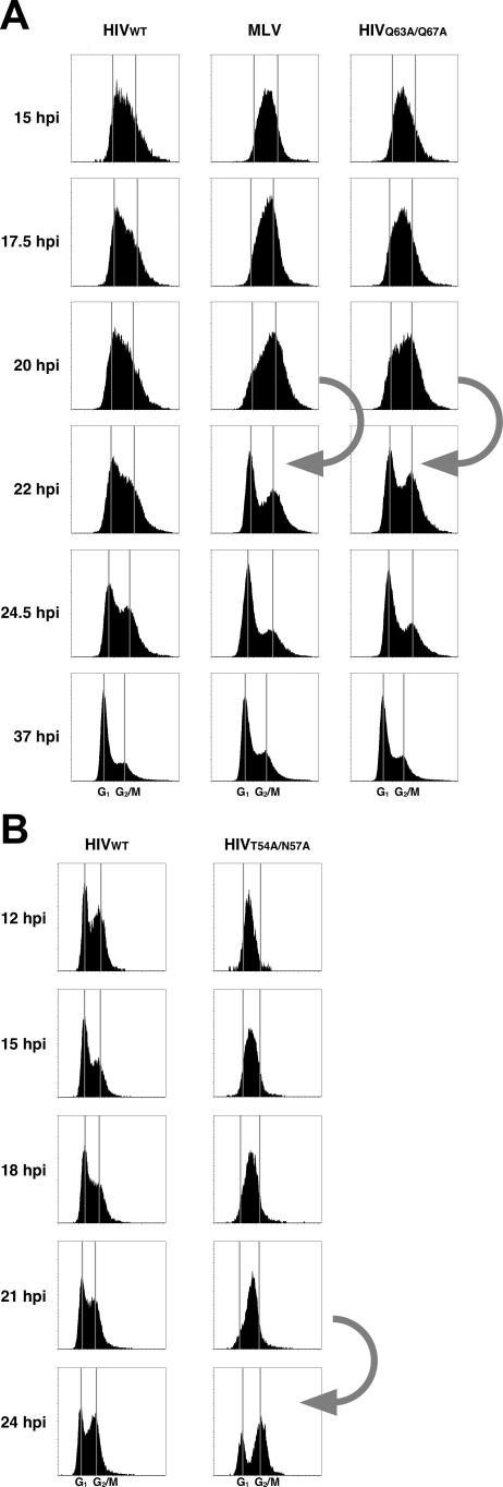
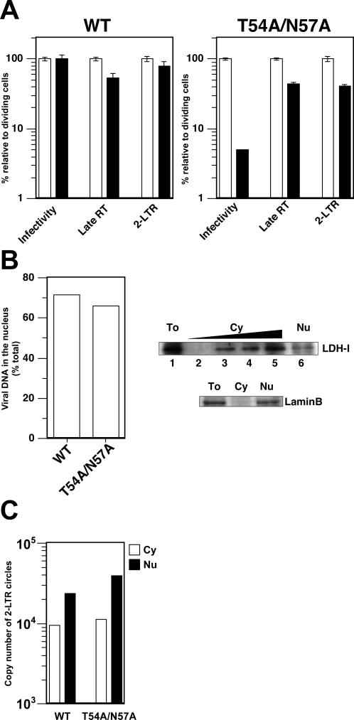
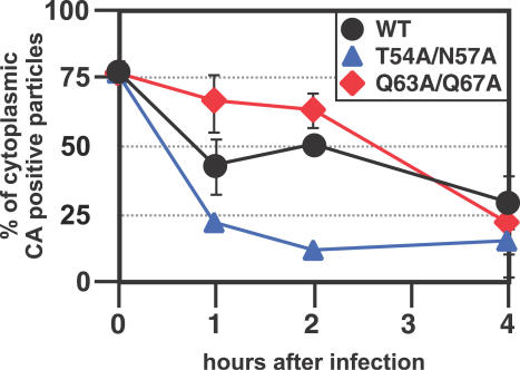
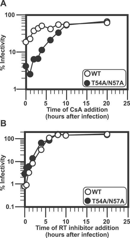
HIV and other lentiviruses can productively infect nondividing cells, whereas most other retroviruses, such as murine leukemia virus, require cell division for efficient infection. However, the determinants for this phenotype have been controversial. Here, we show that HIV-1 capsid (CA) is involved in facilitating HIV infection of nondividing cells because amino acid changes on CA severely disrupt the cell-cycle independence of HIV. One mutant in the N-terminal domain of CA in particular has lost the cell-cycle independence in all cells tested, including primary macrophages. The defect in this mutant appears to be at a stage past nuclear entry. We also find that the loss of cell-cycle independence can be cell-type specific, which suggests that a cellular factor affects the ability of HIV to infect nondividing cells. Our data suggest that CA is directly involved at some step in the viral life cycle that is important for infection of nondividing cells.


Conflict of interest statement

Competing interests. The authors have declared that no competing interests exist.

Figures

Figure 1
Figure 1. HIV CA Mutants Require Cell Division for Efficient Infection
(A) Dose-independent restriction of several CA mutants in nondividing cells. Nondividing cells were prepared by treatment of HeLa cells with aphidicolin and then infected with increasing amount of GFP-encoding viruses. Virus infectivity was measured by quantifying GFP-positive cells 2 d after infection. WT HIV-1 and MLV were used as controls. Open circles indicate infections with cycling cells and closed circles indicate infections with nondividing cells. (B) Infection of MDMs with HIV-1. The WT strains together with CA mutants T54A/N57A and Q63A/67A were used to infect a human T cell line (MT4: upper panels) or MDMs (lower panels). Ten times more virus was used in MDMs, which are not as permissive as MT4 cells to HIV-1 infection. Expression of GFP, which is encoded by the reporter virus constructs, was examined 2 or 3 d after infection. The % of GFP-positive cells is indicated in the upper half of each flow cytometry panel. The data are representative of three independent experiments using at least three different donor PBMCs. (C) Viral infectivity of CA mutants in HOS cells. Cell-cycle requirement for virus infection was examined by infectivity in dividing and nondividing HOS cells with WT or CA mutants encoding the GFP gene. Nondividing cells were prepared by treatment of aphidicolin (2 μg per ml) and the chemical was added throughout the experiment. Open circles indicate infections with cycling cells and closed circles indicate infections with nondividing cells. Shown here are the data that represent at least two independent experiments.
Figure 2
Figure 2. HIV-1 CA Mutants Resemble MLV in Their Cell-Cycle Requirements for Transduction
HeLa cells were infected with GFP-encoding viruses HIV-1, MLV, and HIV-1 CA mutants (A) Q63A/Q67A or (B) T54A/N57A. The cell-cycle profiles of the GFP-positive populations of cells are shown. The y-axis in each plot is the number of cells and the x-axis is the cell-cycle position and the approximate positions of cells in G1 (left vertical line) and G2/M (right vertical line) are indicated. HIV-1 in (A) carries the mutated vpr gene, whereas the virus in (B) contains the intact vpr gene. The arrow indicates the progression of the transduced populations infected with MLV or with the HIV-1 CA mutants from G2/M to G1. Note that WT HIV remains asynchronous in the cell cycle throughout the experiment.
Figure 3
Figure 3. Reverse Transcription and Nuclear Import of Viral DNA in Nondividing Cells
(A) Viral DNA synthesis and formation of 2-LTR circles. Dividing (shown in white bars) and nondividing HeLa cells (shown in black bars) prepared by aphidicolin treatment were infected with reporter virus constructs bearing the WT CA or T54A/N57A mutation. Virus infectivity was assessed by quantifying GFP-positive cells 2 d after infection. Relative infectivity is shown as percentage of infectivity in dividing cells. Total DNA was extracted 1 d after infection and used in real-time PCR to measure the copy number of late products of reverse transcription (RT) as well as that of 2-LTR circles. The data is shown as relative values where the amount in dividing cells for each different virus is set at 100%. Infections were done in triplicate and error bars indicate standard deviation. Two independent experiments were performed with similar results. One representative experiment is shown here. (B) Nuclear localization of viral DNA. Nondividing cells infected with either WT HIV-1 or the CA mutant T54A/N57A were separated into cytoplasmic and nuclear fractions (left panel). DNA extracted from each fraction was used as template for real-time PCR to measure newly synthesized viral DNA. The efficiency of nuclear migration of viral DNA was examined by dividing the copy number of viral DNA in nuclear fractions by that in both fractions (cytoplasmic plus nuclear fractions). The data shown here is a representative of two independent experiments; although the ratio of viral DNA associated with nuclear fractions differed between two experiments, the amount of viral DNA associated with nuclear fraction remains the same between WT and the CA mutant. Controls with reverse transcriptase inhibitors showed that contamination by plasmid DNA accounted for less than 1% of the values (not shown). Western blotting analysis was performed to ensure the integrity of subcellular fractionation (right panels). Contamination was checked by checking a cytoplasmic protein, LDH-I (upper lanes) and a nuclar protein, lamin B (lower lanes). To, total cell lysates; Cy, cytoplasmic extract; Nu, nuclear lysates. Twenty migrograms of proteins were loaded on the gel except for the following dilutions. Cytoplasmic extract was diluted by 5-fold; 5-, 25-, and 125-fold dilutions correspond to lanes 4, 3, and 2 (upper lanes). (C) Subcellular localization of 2-LTR circles. Copy numbers of 2-LTR circles in cytoplasmic (white boxes) and nuclear (black boxes) fractions were measured by real-time PCR. The same fractions as in the (B) were used in this PCR assay.
Figure 4
Figure 4. In Situ Analysis of Kinetics of p24 Loss from Cytoplasmic HIV
HeLa cells were spinoculated with VSV-g pseudotyped, S15-mCherry, GFP-Vpr labeled HIV-1ΔEnv virions for 2 h at 17 °C. Infection was synchronized by washing off innocula and replaced with 37 °C media. HeLa cells were then fixed and immunostained for p24CA (Cy-5) at the indicated time post infection and imaged. GFP (+) pucnta were then quantified and individually examined for the presence of mCherry and Cy-5 (p24CA) signal. The identity of the samples was blinded before the experiment. The percentage of the total number of virions that have stained for p24CA over time following fusion is shown. The 0-h time point represents total number of GFP (+) virions that stained positive for p24CA. HIVWT is represented by black lines/circles, the Q63/67A CA mutant by red line/diamonds, and the T54A/N57A CA mutant by blue line/triangles. The results shown are from three independently performed experiments and the standard deviation at each time point is shown.
Figure 5
Figure 5. Kinetics of HIV-1 Sensitivity to CsA
(A) CsA affects HIV-1 with the CA mutation T54A/N57A much longer than HIV-1 with WT CA. Human T cell Jurkat cells were infected with replication-competent HIV-1 virus encoding the WT envelope and the luciferase gene, which contains either the WT CA sequence or the T54A/N57A mutation. CsA was added to the well at indicated time points. Virus was added at 8 °C by spinolculation, and infections were synchronized by washing off the innocula and adding media at 37 °C. The relative percentage of infectivity was determined by assigning the luciferase value of the sample without CsA as 100% for each virus. White circles indicate WT, black circles the mutant (T54A/N57A). Similar results were observed in three independent experiments. (B) Reverse transcription (RT) of T54A/N57A progresses similarly to that of WT. Similar experiments were done as above (A), except that nevirapine (50 μM) was added instead of CsA.

Similar articles

Cited by

References

    1. Katz RA, Greger JG, Skalka AM. Effects of cell cycle status on early events in retroviral replication. J Cell Biochem. 2005;94:880–889. - PubMed
    1. Yamashita M, Emerman M. Retroviral infection of non-dividing cells: old and new perspectives. Virology. 2006;344:88–93. - PubMed
    1. Zhang Z, Schuler T, Zupancic M, Wietgrefe S, Staskus KA, et al. Sexual transmission and propagation of SIV and HIV in resting and activated CD4+ T cells. Science. 1999;286:1353–1357. - PubMed
    1. Weinberg JB, Matthews TJ, Cullen BR, Malim MH. Productive human immunodeficiency virus type 1 (HIV-1) infection of nonproliferating human monocytes. J Exp Med. 1991;174:1477–1482. - PMC - PubMed
    1. Lewis PF, Emerman M. Passage through mitosis is required for oncoretroviruses but not for the human immunodeficiency virus. J Virol. 1994;68:510–516. - PMC - PubMed

Publication types

Substances