. 2007 Oct;3(10):1991-2006.
doi: 10.1371/journal.pgen.0030182. Epub 2007 Sep 10.

The essential role of Drosophila HIRA for de novo assembly of paternal chromatin at fertilization

Affiliations

The essential role of Drosophila HIRA for de novo assembly of paternal chromatin at fertilization

Emilie Bonnefoy et al. PLoS Genet. 2007 Oct.

Abstract

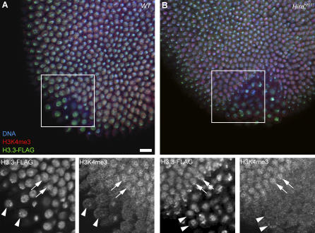
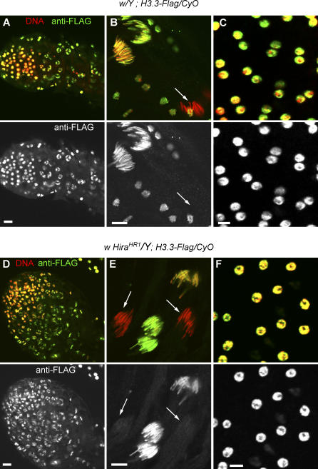
In many animal species, the sperm DNA is packaged with male germ line--specific chromosomal proteins, including protamines. At fertilization, these non-histone proteins are removed from the decondensing sperm nucleus and replaced with maternally provided histones to form the DNA replication competent male pronucleus. By studying a point mutant allele of the Drosophila Hira gene, we previously showed that HIRA, a conserved replication-independent chromatin assembly factor, was essential for the assembly of paternal chromatin at fertilization. HIRA permits the specific assembly of nucleosomes containing the histone H3.3 variant on the decondensing male pronucleus. We report here the analysis of a new mutant allele of Drosophila Hira that was generated by homologous recombination. Surprisingly, phenotypic analysis of this loss of function allele revealed that the only essential function of HIRA is the assembly of paternal chromatin during male pronucleus formation. This HIRA-dependent assembly of H3.3 nucleosomes on paternal DNA does not require the histone chaperone ASF1. Moreover, analysis of this mutant established that protamines are correctly removed at fertilization in the absence of HIRA, thus demonstrating that protamine removal and histone deposition are two functionally distinct processes. Finally, we showed that H3.3 deposition is apparently not affected in Hira mutant embryos and adults, suggesting that different chromatin assembly machineries could deposit this histone variant.


Conflict of interest statement

Competing interests. The authors have declared that no competing interests exist.

Figures

Figure 1
Figure 1. Targeting the Hira Gene by Homologous Recombination
(A) Schematic representation of the wild-type (WT) Hira locus, the HiraHR1 recombined allele, and the pW25-HiraHR1-Flag reporter transgene. The dotted lines indicate the region that is replaced by the pW25 vector sequence in HiraHR1. The gray and white boxes indicate the Hira and white exons, respectively, and the black box is the 3X-Flag tag at the 3' end of the pW25-HiraHR1-Flag transgene. The dark gray hexagons represent termination codons in the six reading frames. The positions of the primer pairs used in (B) are shown (arrows). (B) Example of a genomic PCR with the primer pairs shown in (A). Note that the primer pair #1 does not amplify the large pW25 insertion in the HiraHR1 allele. The tested male genotypes are indicated. (C) Anti-FLAG and anti-tubulin western blot analysis of embryo extracts from Hira-Flag and HiraHR1-Flag transgenic lines. The arrow indicates the HIRA-FLAG protein. Other smaller bands are interpreted as HIRA-FLAG degradation products.
Figure 2
Figure 2. HIRA Is Not Detected in HiraHR1 Eggs
Confocal sections of eggs or embryos stained for DNA (red) and anti-HIRA antibodies (green). (A) In wild-type (WT) fertilized eggs, HIRA is specifically detected in the male nucleus (arrowhead in the inset). (B) In eggs from HiraHR1 females, HIRA is not detected in the male nucleus (inset). Note that the HIRA antibody 830 non-specifically binds the sperm tail (elongated structure visible in the green channel) [17]. (C) A Cycle 5 haploid embryo from a Hirassm female stained with antibody 830. The only stained nucleus is the condensed male nucleus (arrowheads). (D) Apposed pronuclei in a Hirassm egg stained with HIRA antibody PG1 showing a strong signal in the male nucleus (arrowhead). (E) A HiraHR1 egg at the same stage stained with the same antibody. F: Female pronucleus. PB: Polar Bodies. Bars: 10 μm.
Figure 3
Figure 3. HIRA Accumulates in the Germinal Vesicle in Wild-Type but Not in HiraHR1 Oocytes
Stage 10 egg chambers stained for DNA (red) and anti-HIRA PG1 or anti-FLAG antibodies (green). (A) In wild-type egg chambers, HIRA is specifically detected in the germinal vesicle where it occupies the whole nuclear volume. The karyosome, the compact structure containing the maternal chromosomes, is visible in the DNA channel (arrow). (B) In HiraHR1 egg chambers, the antibody does not detect HIRA in the germinal vesicle (arrow). (C) In transgenic Hira-Flag egg chambers, HIRA-FLAG protein is found in the germline vesicle (arrows) like the endogenous protein. (D) No HIRA-FLAG protein is detected in the oocyte nucleus in HiraHR1-Flag transgenic egg chambers.
Figure 4
Figure 4. HiraHR1 Eggs Are Unable to Assemble Paternal Chromatin at Fertilization
Confocal sections of eggs and embryos stained for DNA (red) and anti-acetylated histone H4 antibody (green). (A) A wild-type egg in meiosis II with the elongated fertilizing male nucleus (M) that brightly stains for acetylated-H4 (arrow). (B) A HiraHR1 egg at the same stage with no acetylated-H4 detected in the male nucleus. (C) A cycle 3 haploid embryo from a HiraHR1 mother. The maternal nuclei, but not the male nucleus, stain for acetylated-H4. Bar: 10 μm.
Figure 5
Figure 5. HIRA Is Not Required for Protamine Removal from the Decondensing Sperm Nucleus
(A) Left panel: in a fixed ProtA-GFP transgenic testis, the GFP fluorescence is very strong in the most condensed spermatid nuclei (asterisk), whereas less condensed nuclei are much less bright (arrow). Middle panel: the same testis stained with an anti-GFP antibody considerably enhances the GFP detection in less condensed nuclei (arrow, compare with left panel), whereas highly condensed nuclei are comparatively less stained. Right panel: the same testis stained with the DNA dye TO-PRO3. (B) In wild-type (WT) eggs fertilized with sperm from snky1 ; ProtA-GFP males, the sperm nucleus is not activated (arrow), remains at the egg periphery, and its protamines are not removed. (C) In wild-type eggs fertilized with ProtA-GFP sperm and fixed before the end of meiosis II (MII), ProtA-GFP is never detected in the decondensing male nucleus (arrows). (D) The same result is obtained for HiraHR1 eggs. Eggs in (B–D) were stained with an anti-GFP antibody revealed with a green secondary antibody to cumulate the GFP and secondary antibody respective fluorescence in the green channel of the confocal microscope. Identical results were obtained with ProtB-GFP transgenic males (unpublished data). PB: Polar Body. Bar: 10 μm.
Figure 6
Figure 6. The Male Nucleus Does Not Recondense in Hira ; sra Double Mutant Eggs
(A) In sraA108/Df(3R)sbd45 mutant eggs, the female meiosis arrests in anaphase of the first meiotic division (MI). The male nucleus (arrowhead and bottom panels) appears condensed but irregular in shape and stains with anti-acetylated histone H4 antibodies (bottom right panel). Note that the DNA positive dots that are visible in this egg are Wolbachia bacteria that naturally infect the stock. (B) In Hirassm ; sraA108/Df(3R)sbd45 Double Mutant Eggs, the Male Nucleus Is Round and Does Not Stain with Anti-Histone Antibodies. Bars: 2 μm.
Figure 7
Figure 7. ASF1 Is Not Directly Involved in the RI Paternal Chromatin Assembly
Confocal sections of eggs stained for DNA (red) and anti-ASF1 antibody (green). (A) In wild-type fertilized eggs, ASF1 is not detected in the male nucleus or in maternal nuclei during the decondensation phase. (B) ASF1 is not detected in the male nucleus during pronuclear migration. (C) ASF1 stains both pronuclei in a wild-type egg during the first S phase. (D) ASF1 is not detected in the male nucleus in Hirassm eggs. (D) The same result was obtained with the HiraHR1 allele. F: Female pronucleus, M: Male pronucleus, PB: Polar Bodies. Bar: 10 μm.
Figure 8
Figure 8. Dynamics of H3.3 Deposition in Wild-Type and HiraHR1 Early Embryos
Confocal sections of eggs/embryos stained with propidium iodide (red) and anti-FLAG antibody (green). (A) In wild-type (WT) eggs, RI deposition of maternal H3.3-FLAG is observed in the decondensing male nucleus (M) before the first zygotic S phase. (B) H3.3-FLAG is not detected in the male nucleus in HiraHR1 eggs. (C) At pronuclear apposition, during the first S phase, limited RC deposition of H3.3-FLAG is detected in the female pronucleus (arrow) in WT eggs. (D). The same, faint H3.3-FLAG staining of the female pronucleus is observed in HiraHR1 eggs (arrow). (E) A WT embryo in anaphase of the third nuclear division. At this early stage, the stronger H3.3-FLAG staining of the paternally derived chromosomes (arrowheads) is still detectable (note that paternal and maternal chromosomes tend to remain separated during the early syncytial mitoses). (F) A HiraHR1 haploid embryo in its fourth mitosis showing a weak H3.3-FLAG staining on maternally derived chromosomes (arrows). (G) A wild-type, diploid blastoderm embryo in metaphase showing a strong H3.3-FLAG chromosomal staining on all nuclei. (H) H3.3-FLAG is also detected on the chromosomes of HiraHR1 haploid blastoderm embryos. (I) Embryos from wild-type mothers crossed with H3.3-Flag/CyO males showing no detection of zygotic H3.3-FLAG at this stage. (J) Zygotic H3.3-FLAG appears in the chromatin of gastrula embryos. (K) A wild-type, cycle 3 embryo in anaphase showing a strong H3-FLAG staining on all chromosomes. (L) A blastoderm embryo with a strong maternal H3-FLAG staining. Gray panels or insets show the H3.3-FLAG or H3-FLAG staining for a representative group of nuclei. Bar: 10 μm.
Figure 9
Figure 9. H3.3-FLAG Is Deposited in the Germ Line Chromatin in Blastoderm Embryos
Confocal sections of blastoderm embryos stained for DNA (blue), H3.3-FLAG (green), and H3K4me3 (red). (A) H3.3-FLAG (left inset) is deposited at equivalent levels in somatic (arrows) and germ line (arrowheads) nuclei in wild-type embryos. H3K4me3 is enriched in somatic nuclei (right inset). (B) An identical situation is observed in HiraHR1 embryos. Bar: 10 μm.
Figure 10
Figure 10. HiraHR1 Does Not Affect the Distribution of H3.3-FLAG in Adult Testis
Testis and accessory glands from H3.3-Flag/CyO transgenic adult males with a wild-type (A–C) or HiraHR1 (D–F) X chromosome, stained with anti-FLAG antibody and propidium iodide. (A) Apical tip of a wild-type testis. (B) A group of elongating spermatids in a wild-type testis showing a bright H3.3-FLAG nuclear staining that disappears in late condensing spermatid nuclei (arrow). (C) Nuclei from a wild-type accessory gland. (D) Apical tip of a HiraHR1 testis. (E) Spermatid nuclei in a HiraHR1 testis. H3.3-FLAG is not detected in late spermatid nuclei (arrows). (F) Nuclei from a HiraHR1 accessory gland. Bars: 10 μm.

Similar articles

Cited by

References

    1. Polo SE, Almouzni G. Chromatin assembly: a basic recipe with various flavours. Curr Opin Genet Dev. 2006;16(2):104–111. - PubMed
    1. Akey CW, Luger K. Histone chaperones and nucleosome assembly. Curr Opin Struct Biol. 2003;13(1):6–14. - PubMed
    1. Poccia D, Collas P. Transforming sperm nuclei into male pronuclei in vivo and in vitro. Curr Top Dev Biol. 1996;34:25–88. - PubMed
    1. McLay DW, Clarke HJ. Remodelling the paternal chromatin at fertilization in mammals. Reproduction. 2003;125(5):625–633. - PMC - PubMed
    1. Wright SJ. Sperm nuclear activation during fertilization. Curr Top Dev Biol. 1999;46:133–178. - PubMed

Publication types

MeSH terms

LinkOut - more resources