. 2007 Dec;133(6):1840-8.
doi: 10.1053/j.gastro.2007.08.009. Epub 2007 Aug 2.

Inactivation of the UNC5C Netrin-1 receptor is associated with tumor progression in colorectal malignancies

Affiliations

Inactivation of the UNC5C Netrin-1 receptor is associated with tumor progression in colorectal malignancies

Agnès Bernet et al. Gastroenterology. 2007 Dec.

Abstract

Background & aims: The UNC5H netrin-1 receptors (UNC5H1-3 also called UNC5A-C) belong to the functional dependence receptors family, which share the ability to induce apoptosis in the absence of their ligands. Such a trait has been hypothesized to confer a tumor-suppressor activity. Indeed, cells harboring these receptors are thought to be dependent on ligand availability for their survival, thereby inhibiting uncontrolled tumor cell proliferation. We investigate here whether UNC5C acts as a tumor suppressor in colorectal malignancies.

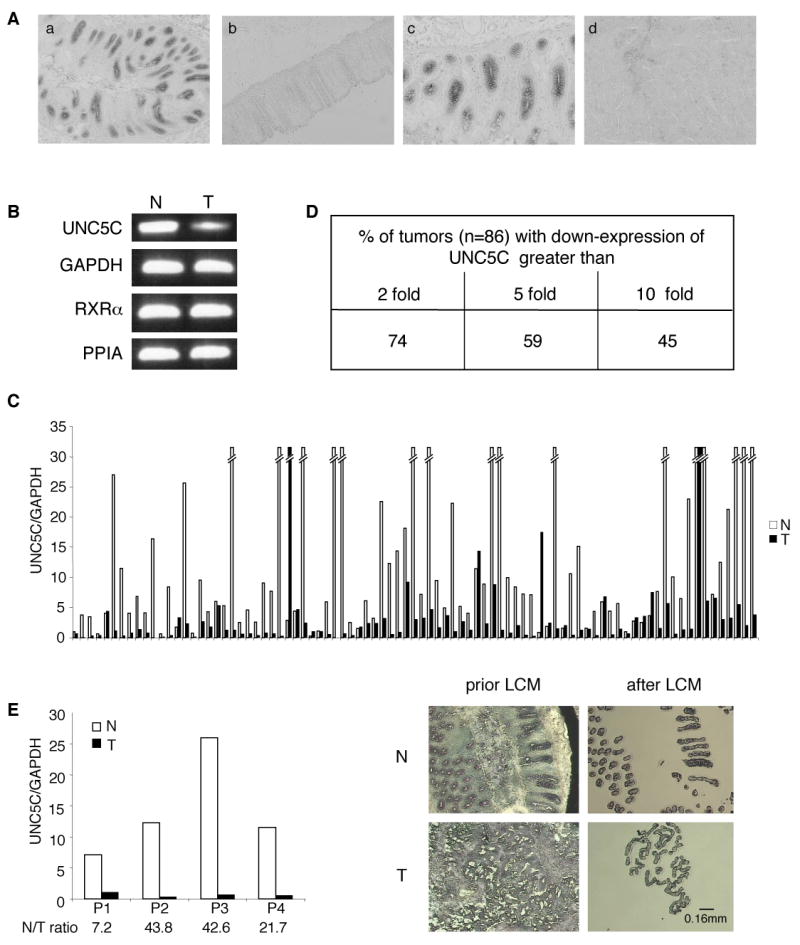
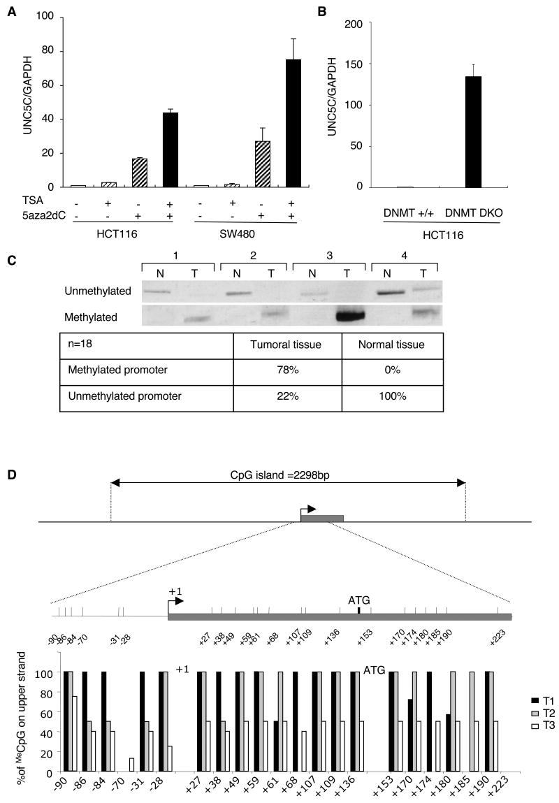
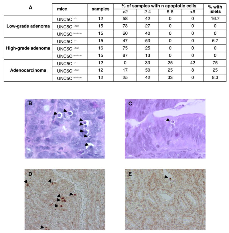
Methods: The level of UNC5C was analyzed in a panel of 86 primary sporadic colorectal carcinomas. Loss of heterozygosity in the UNC5C locus and epigenetic alterations in the UNC5C promoter were also analyzed. Intestinal tumor progression was monitored in mice bearing both UNC5C and APC1638N mutations, and apoptosis was measured in intestinal tumors developed in UNC5C/APC1638N mutant mice.

Results: We show here that UNC5C expression is down-regulated in a large fraction of human colorectal cancers, mainly through promoter methylation. Moreover, in mice, inactivation of UNC5C is associated with increased intestinal tumor progression and a decrease in tumor cell apoptosis.

Conclusions: The loss of UNC5C expression observed in human colorectal cancer is a selective advantage for tumor progression, in agreement with the dependence receptor hypothesis. Thus, the UNC5C dependence receptor is a tumor suppressor that regulates sporadic colorectal cancer.


Conflict of interest statement

No conflicts of interest exist

Figures

Figure 1
Figure 1. Loss of UNC5C expression in colorectal tumors
A, In situ hybridization performed in human colon on UNC5C. a: antisense probe on normal colon, b: sense probe, c: enlargement of a, d: antisense probe on adjacent tumor. Original magnifications: X50 (a,b,d), X100 (c). B,C,D, Quantitative real-time RT-PCR was performed using total RNA extracted from normal (N) and tumoral (T) tissues with specific human UNC5C primers and eleven couples of specific human genes primers showing the less variability in their expression between normal and colorectal tumoral tissues, as described in , (see Experimental procedures section). B, A PCR in the exponential phase of a representative pair tumor/normal tissue is shown. C, The expression levels in 86 colorectal tumors (T) and corresponding normal tissue (N) are given only as the ratio between UNC5C and GAPDH. Similar results were obtained with 10 other housekeeping genes described in Experimental procedures D, The percentage of patients showing a loss of UNC5C expression in tumor compared to normal tissue is indicated. E, Laser capture microdissection (LCM) was performed on 4 pairs of tumor/normal tissues and UNC5C expression was determined as in (B). Left: The expression levels in 4 colorectal tumors (T) and corresponding normal tissue (N) are given only as the ratio between UNC5C and GAPDH. Similar results were obtained with the other housekeeping RXRα, PPIA genes. Right: typical microscopic visualization of LCM on colon section from human normal (N) or tumoral biopsies (T). Sections are shown prior to LCM, and after LCM, as the captured material is confirmed under microscopic visualization prior to processing for RNA extraction.
Figure 2
Figure 2. Correlation between patient clinical features and UNC5C expression N/T ratio in a limited panel of patient
Correlation between UNC5C expression N/T ratio and TNM status classified in 4 stages from good to poor prognosis i.e. stage I (T1 or T2, N0M0), stage II (T3 or T4, N0M0), stage III (any T, N+M0) and stage IV (any T, any N, M+) . A Kruskall-Wallis test was used comparing the overall panel, p=0.001. A Mann-Whitney test was also used to compare stage I to stage IV (p=0.001), stage II to stage IV (p=0.004). The difference between stage III and stage IV (p=0.120) fails to be significant.
Figure 3
Figure 3. Epigenetic inactivation of UNC5C in human colorectal tumors
A, HCT116 and SW480 cells were treated with 5µM 5-Aza-2′-deoxycytidine (5aza2dc) for 48H and then with 0.3µM TSA for 24H. Quantitative real-time RT-PCR was performed with UNC5C primers relatively to GAPDH, RXRα and PPIA, as explained in Fig.1. The expression level is presented as a ratio between UNC5C and GAPDH. B, UNC5C expression was compared in HCT116 versus HCT116 DKO for DNMT1 and 3b by quantitative real-time RT-PCR, as described previously. C, Methylation-specific PCR (MSP) analysis of sodium-bisulfite-treated DNA from human colorectal biopsies of Normal tissues (N) and Tumoral tissues (T). Amplification using primers that hybridize specifically on sequences of the UNC5C promoter that are Unmethylated and/or Methylated (data shown only for 4 different biopsies, biopsy 4 showing both the methylated and unmethylated states of the UNC5C promoter in tumoral tissue, whereas in the three others, UNC5C promoter is completely methylated in tumoral tissue). Compiled results on 18 human colorectal biopsies are presented in the table. D, Change in MeCpG content in the region of the UNC5C promoter in three different tumors (T1,T2,T3). Upper: Representation of the DNA region surrounding the first exon (in grey) with transcriptional start site arrowed in black (Accession n°NM_003728). The locus includes a CpG island of 2298bp in length (%G+C=59; ObsCpG/ExpCpG=0.729; CG island searcher software). The region amplified by PCR primers is enlarged and the position of each CpG is indicated from the UNC5C transcription start site. Bottom: Ten PCR clones were sequenced from three different tumors (T1,T2,T3) and from the corresponding normal tissues (N1,N2,N3) (not shown as no methylation is detected) to determine the percentage of methylation at the CpG sites in the analysed region.
Figure 4
Figure 4. Increased intestinal tumor progression in UNC5C mutant mice
UNC5Crcm mice were backcrossed into an Apc1638N background and analysed for tumor development in 6 months old mice. A, Table showing the number (Nb) of Apc+/1638N UNC5Crcm/rcm, Apc+/1638N UNC5C+/rcm or Apc+/1638N UNC5C+/+ mice studied, the average number of tumors per mouse, the range, the number of tumor studied and the proportion of low-grade tumors and high-grade tumors including adenocarcinomas. Significance: χ2 test, p<0.001. B-G: representative lesions from Apc +/1638N UNC5Crcm/rcm mice: low grade adenoma (B,C), high grade adenoma (D,E) and adenocarcinoma (F, note the presence of muscularis (m) invasion indicated by arrows, G). C,E,G enlargement of respectively B,D,F, Original magnifications: B,D,F, X80; C,E,G, X250
Figure 5
Figure 5. Tumors from UNC5C mutant mice are less prone to apoptosis
A, Table showing the number of apoptotic cells per section of tumors (sample) issued from Apc+/1638N UNC5C+/+ mice, Apc+/1638N UNC5C+/rcm or Apc+/1638N UNC5Crcm/rcm. B,C, Representative microscopic images from adenocarcinoma from either Apc+/1638N UNC5C+/+ (B) or Apc+/1638N UNC5Crcm/rcm (C) mice. Note in adenocarcinomas from Apc+/1638N UNC5C+/+ mice, the presence of several (more than 3) apoptotic cells side by side that form islets of apoptotic cells. Apoptotic cells are shown by arrows. Original magnifications: X450. D,E, similar as in B, C except that TUNEL staining was performed as described in the Experimental procedures section.

Comment in

References

    1. Serafini T, Colamarino SA, Leonardo ED, Wang H, Beddington R, Skarnes WC, Tessier-Lavigne M. Netrin-1 is required for commissural axon guidance in the developing vertebrate nervous system. Cell. 1996;87:1001–14. - PubMed
    1. Keino-Masu K, Masu M, Hinck L, Leonardo ED, Chan SS, Culotti JG, Tessier-Lavigne M. Deleted in Colorectal Cancer (DCC) encodes a netrin receptor. Cell. 1996;87:175–85. - PubMed
    1. Forcet C, Stein E, Pays L, Corset V, Llambi F, Tessier-Lavigne M, Mehlen P. Netrin-1-mediated axon outgrowth requires deleted in colorectal cancer-dependent MAPK activation. Nature. 2002;417:443–7. - PubMed
    1. Ackerman SL, Kozak LP, Przyborski SA, Rund LA, Boyer BB, Knowles BB. The mouse rostral cerebellar malformation gene encodes an UNC-5-like protein. Nature. 1997;386:838–42. - PubMed
    1. Hong K, Hinck L, Nishiyama M, Poo MM, Tessier-Lavigne M, Stein E. A ligand-gated association between cytoplasmic domains of UNC5 and DCC family receptors converts netrin-induced growth cone attraction to repulsion. Cell. 1999;97:927–41. - PubMed

Publication types