. 2008 Jan;28(1):344-57.
doi: 10.1128/MCB.00617-07. Epub 2007 Oct 29.

A-raf and B-raf are dispensable for normal endochondral bone development, and parathyroid hormone-related peptide suppresses extracellular signal-regulated kinase activation in hypertrophic chondrocytes

Affiliations

A-raf and B-raf are dispensable for normal endochondral bone development, and parathyroid hormone-related peptide suppresses extracellular signal-regulated kinase activation in hypertrophic chondrocytes

Sylvain Provot et al. Mol Cell Biol. 2008 Jan.

Abstract

Parathyroid hormone-related peptide (PTHrP) and the parathyroid hormone-PTHrP receptor increase chondrocyte proliferation and delay chondrocyte maturation in endochondral bone development at least partly through cyclic AMP (cAMP)-dependent signaling pathways. Because data suggest that the ability of cAMP to stimulate cell proliferation involves the mitogen-activated protein kinase kinase kinase B-Raf, we hypothesized that B-Raf might mediate the proliferative action of PTHrP in chondrocytes. Though B-Raf is expressed in proliferative chondrocytes, its conditional removal from cartilage did not affect chondrocyte proliferation and maturation or PTHrP-induced chondrocyte proliferation and PTHrP-delayed maturation. Similar results were obtained by conditionally removing B-Raf from osteoblasts. Because A-raf and B-raf are expressed similarly in cartilage, we speculated that they may fulfill redundant functions in this tissue. Surprisingly, mice with chondrocytes deficient in both A-Raf and B-Raf exhibited normal endochondral bone development. Activated extracellular signal-regulated kinase (ERK) was detected primarily in hypertrophic chondrocytes, where C-raf is expressed, and the suppression of ERK activation in these cells by PTHrP or a MEK inhibitor coincided with a delay in chondrocyte maturation. Taken together, these results demonstrate that B-Raf and A-Raf are dispensable for endochondral bone development and they indicate that the main role of ERK in cartilage is to stimulate not cell proliferation, but rather chondrocyte maturation.


Figures
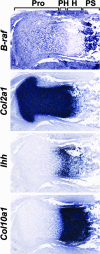
FIG. 1.
FIG. 1.
B-Raf is expressed in immature proliferative chondrocytes. Nonradioactive ISH analysis of E16.5 proximal tibia with the indicated chondrogenic markers. Note that B-raf mRNA is detected in the proliferative layer (Pro). Its expression declines in prehypertrophic (PH) chondrocytes, marked by Ihh, and is not detectable in mature hypertrophic (H) chondrocytes, marked by Col10a1, unlike Col2a1, which is expressed in the whole cartilaginous growth plate. Note that B-raf mRNA is also detected in bone (primary spongiosa [PS]).
FIG. 2.
FIG. 2.
Conditional removal of B-Raf from cartilage does not affect skeletogenesis. (A) Analysis of B-Raf expression by Western blot evaluation of primary chondrocytes isolated from newborn WT, CHET, and CKO littermates. Note that B-Raf protein expression is dramatically reduced in CKO chondrocytes compared to WT chondrocytes, whereas the expression of the housekeeping protein β-actin is unchanged. (B) Skeletal preparations (subjected to alizarin red and alcian blue staining) from newborn WT and CKO mice. (C) Blown-up image of skeletal preparations of forelimbs (FL) and hind limbs (HL) of WT and CKO newborn littermates. (D) Histological analysis (hematoxylin and eosin staining) of tibias of newborn WT, CHET, and CKO littermates. Note that the widths of each the proliferative (Pro) and hypertrophic (H) layers are comparable for all genotypes. (E) Nonradioactive ISH analysis of proximal tibias of E16.5 mice of the indicated genotypes, with the indicated chondrogenic markers. Note the similar intensities and distributions in WT, CHET, and CKO mice of Ihh and Col10a1, which mark prehypertrophic and hypertrophic chondrocytes, respectively. (F) BrdU assay of proximal tibias of E18.5 mice of the indicated genotypes. Note the similar distributions of black BrdU-positive cells in WT and CKO growth plates. The proliferation rates of chondrocytes (the numbers of BrdU-positive cells divided by the total numbers of cells counted, expressed as percentages) in E18.5 WT and CKO growth plates are given. The values indicated correspond to the average of two counts of cells from at least three animals of each genotype. Prehypertrophic and hypertrophic chondrocytes identified histologically were excluded from these counts.
FIG. 3.
FIG. 3.
B-Raf is not required for a PTHrP-induced increase in chondrocyte proliferation and delayed chondrocyte maturation. (A) ISH with the indicated chondrogenic markers and BrdU assay of metatarsal explants obtained from WT E15.5 hind limbs treated with PPR agonist [+PTH(1-34)] at 10−7 M or vehicle (control) for 24 h. Note that B-raf mRNA expression is not increased upon the activation of PTHrP signaling whereas chondrogenic maturation is repressed and proliferation is increased, as indicated by the repression of Ihh and Col10a1 mRNA expression and the increase of BrdU incorporation, respectively. (B) ISH with the indicated chondrogenic markers and BrdU assay of metatarsal explants obtained from B-Raf CKO E15.5 hind limbs treated with PPR agonist or vehicle (control) for 24 h. Note that chondrogenic maturation is repressed and proliferation is increased upon the activation of PTHrP signaling, in spite of the lack of B-Raf. (C) Histological analysis (hematoxylin and eosin staining) of tibias of E16.5 littermates of the indicated genotypes. Note the tremendous delay of endochondral bone development observed in mice homozygous (transgene/transgene [Tg/Tg]) for Col2-caPPR; the delay is characterized by the extension of the proliferative chondrocyte layer and the loss of the hypertrophic chondrocyte layer, with only a few maturing chondrocytes found on the side of the bone (asterisks). This ca-PPR-induced delay is not affected in the absence of B-Raf.
FIG. 4.
FIG. 4.
B-Raf conditional knockout in osteoblasts does not affect bone formation or the effects of PPR in bone. (A) Histological analysis of tibias of 2-week-old littermate mice expressing or not expressing B-Raf in bone. Note that mice with an Osterix-Cre-mediated conditional knockout of B-Raf in early osteoblasts (Osx-CKO) present amounts of trabecular and cortical bones (stained in pink) similar to those in control animals. (B) Histological analysis of tibias of 2-week-old littermate mice expressing or not expressing B-Raf in bone in the presence of caPPR in bone. The mid-diaphysis region of a control tibia is characterized by the absence of trabecular bone (no pink) and the presence of a homogenous compact cortical bone. The misexpression of Col1-caPPR in bone in mice hemizygous for the Col1-caPPR transgene (transgene/WT [Tg/+]) dramatically increases bone formation in the trabecular region and forms a porous cortical bone. The phenotype observed in mice expressing the Col1-caPPR transgene but the conditional knockout of B-Raf in osteoblasts (B-raf Col1-CKO) is identical to that observed in the presence of B-Raf (B-Raf Col1-CHET).
FIG. 5.
FIG. 5.
A-Raf is expressed in immature proliferative chondrocytes, and C-Raf is expressed in mature hypertrophic chondrocytes. (A) Radioactive ISH analysis of C-raf mRNA expression in proximal tibias of E16.5 WT and CKO animals. Note that C-raf mRNA is expressed almost exclusively in mature hypertrophic (H) chondrocytes in the growth plate, with only traces of expression in proliferative chondrocytes. Its expression is unchanged in CKO growth plates that lack B-Raf in chondrocytes. (B) Nonradioactive ISH analysis of A-Raf expression in proximal tibias of E18.5 WT and CKO animals. Note that A-raf mRNA expression is restricted to immature proliferative chondrocytes and, thus, A-Raf expression is identical to that of B-Raf. A-Raf expression is not significantly increased in CKO growth plates that lack B-Raf in chondrocytes. Pro, proliferative layer. (C) Western blot analysis of A-Raf protein expression in primary chondrocytes of newborn WT and CKO animals. Note that similar to the level of expression of the housekeeping protein β-actin, the level of A-Raf protein expression is unchanged in CKO chondrocytes that do not express B-Raf.
FIG. 6.
FIG. 6.
A-Raf-B-Raf double-KO animals present normal endochondral bone development. (A) Skeletal preparations from newborn WT and A-Raf-B-Raf double-KO mice (A-Raf KO-B-Raf CKO mice developed by using Col2-Cre to remove B-Raf from chondrocytes). (B) Detailed view of forelimbs (FL) and hind limbs (HL) of the mice corresponding to the samples presented in panel A. (C) Histological analysis of tibias of newborn WT and A-Raf-B-Raf double-KO littermates. Note the absence of histological differences between samples from the two genotypes. (D) Nonradioactive ISH analysis of proximal tibias of newborn mice of the indicated genotypes with the indicated chondrogenic markers. Note the similar intensities and distributions in WT and double-KO mice of Ihh and Col10a1, which mark prehypertrophic and hypertrophic chondrocytes, respectively. Note also that C-raf mRNA is not up-regulated in double-KO mice and is still restricted to hypertrophic chondrocytes.
FIG. 7.
FIG. 7.
ERK activation is independent of A-Raf and B-Raf, is restricted to mature hypertrophic chondrocytes, and is suppressed by PTHrP signals. (A) Immunohistochemistry analysis to detect phospho-ERK (activated ERK) on sections of proximal tibias of E17.5 WT mice, mice hemizygous for a transgene encoding a constitutively active version of PPR in cartilage (Col2-caPPR transgene/WT [Tg/+]), or mice homozygous for this transgene (Col2-caPPR Tg/Tg). The rectangles in blue and in red correspond to the regions of proliferative (Pro) and hypertrophic (H) chondrocytes, respectively, which are shown at higher magnification in the middle and lower panels, respectively. Note that in WT growth plates, phospho-ERK is detected essentially in mature hypertrophic chondrocytes, whereas ERK is activated in a dose-dependent manner in proliferative chondrocytes of transgenic animals. Note also that the signal appears weaker in hypertrophic chondrocytes of transgenic animals than in those of wild-type animals. In homozygous transgenic mice, only a few mature chondrocytes are present on one side of the tibia and these cells present only a weak phospho-ERK signal. (B) Immunohistochemistry analysis for the detection of phospho-ERK on sections of proximal tibias of E18.5 control mice or mice hemizygous (Tg/+) for the Col2-caPPR transgene and expressing B-Raf (B-raf WT) or not (B-raf CKO) in cartilage (Col2-Cre-mediated conditional knockout). Note that ERK is found activated in transgenic animals even in the absence of B-Raf in chondrocytes. The rectangles correspond to the regions of the growth plates shown at higher magnification in the lower panels. (C) Immunohistochemistry analysis for the detection of phospho-ERK on sections of proximal tibias of E17.5 control, PTHrP KO, and PTHrP-p57 double-KO mice. In the absence of PTHrP, chondrocyte maturation is accelerated, resulting in a shortening of the growth plate. The deletion of p57 partially inhibits this phenotype. Note that in the absence of PTHrP, the phospho-ERK signal is increased in hypertrophic chondrocytes but also in chondrocytes located at the articular surface and in the proliferative layer. This effect is abolished in PTHrP-p57 double-KO animals. (D) Immunohistochemistry analysis for the detection of phospho-ERK on sections of metatarsal explants obtained from WT E15.5 hind limbs treated with PPR agonist [+PTHrP(1-34)] or vehicle (control) for the indicated times. A higher-magnification image of the hypertrophic chondrocytes is shown for each condition. Note that PTHrP completely abolishes ERK activation after 1 h of treatment. This effect is transient, since some phospho-ERK signal is detected after 2 h of treatment. Explants treated for 24 h do not undergo chondrogenic maturation and thus do not present any phospho-ERK signal. (E) Immunohistochemistry analysis for the detection of phospho-ERK on sections of proximal tibias from newborn WT and A-Raf-B-Raf double-KO mice (A-Raf KO-B-Raf CKO mice developed by using Col2-Cre to remove B-Raf from chondrocytes). Note that the activation of ERK is not only restricted to hypertrophic chondrocytes but also independent of A-Raf and B-Raf.
FIG. 8.
FIG. 8.
Suppression of ERK activation delays hypertrophic chondrocyte maturation. (A) Metatarsals from E15.5 WT hind limbs were treated with the MEK1-MEK2 inhibitor U0126 or vehicle for the indicated time and photographed while in culture. The dark region visible at day 5 in the vehicle-treated metatarsal preparations corresponds to mineralized matrix (opaque to light) produced by mature chondrocytes. Note that U0126-treated metatarsals do not present any mineralized region, even after 6 days in culture. (B) E15.5 WT metatarsals treated for 2 or 6 days with U0126 or vehicle were processed for ISH with markers that cover different stages of chondrocyte maturation. The chondrogenic markers Ihh, Col10a1, and OP mark prehypertrophic, hypertrophic, and late-hypertrophic chondrocytes, respectively. Note that Ihh expression is affected only mildly after 6 days of treatment, whereas that of Col10a1 and OP is severely suppressed. Adjacent sections were subjected to immunohistochemistry analysis for phospho-histone H3 (P-H3; a mitotic marker) and phospho-ERK (P-ERK). Note that the numbers of mitotic cells in U0126- and vehicle-treated samples were comparable, although ERK activation was efficiently suppressed. A blown-up image of the regions of hypertrophic chondrocytes is shown for phospho-ERK signals.

Similar articles

Cited by

References

    1. Bastepe, M., L. S. Weinstein, N. Ogata, H. Kawaguchi, H. Juppner, H. M. Kronenberg, and U. I. Chung. 2004. Stimulatory G protein directly regulates hypertrophic differentiation of growth plate cartilage in vivo. Proc. Natl. Acad. Sci. USA 10114794-14799. - PMC - PubMed
    1. Benkhelifa, S., S. Provot, E. Nabais, A. Eychene, G. Calothy, and M. P. Felder-Schmittbuhl. 2001. Phosphorylation of MafA is essential for its transcriptional and biological properties. Mol. Cell. Biol. 214441-4452. - PMC - PubMed
    1. Calvi, L. M., N. A. Sims, J. L. Hunzelman, M. C. Knight, A. Giovannetti, J. M. Saxton, H. M. Kronenberg, R. Baron, and E. Schipani. 2001. Activated parathyroid hormone/parathyroid hormone-related protein receptor in osteoblastic cells differentially affects cortical and trabecular bone. J. Clin. Investig. 107277-286. - PMC - PubMed
    1. Chen, A. P., M. Ohno, K. P. Giese, R. Kuhn, R. L. Chen, and A. J. Silva. 2006. Forebrain-specific knockout of B-raf kinase leads to deficits in hippocampal long-term potentiation, learning, and memory. J. Neurosci. Res. 8328-38. - PubMed
    1. Chen, C., A. J. Koh, N. S. Datta, J. Zhang, E. T. Keller, G. Xiao, R. T. Franceschi, N. J. D'Silva, and L. K. McCauley. 2004. Impact of the mitogen-activated protein kinase pathway on parathyroid hormone-related protein actions in osteoblasts. J. Biol. Chem. 27929121-29129. - PubMed

Publication types

MeSH terms

Substances

LinkOut - more resources