. 2008 Jan;28(1):302-14.
doi: 10.1128/MCB.01044-07. Epub 2007 Oct 29.

Cancer-causing mutations in a novel transcription-dependent nuclear export motif of VHL abrogate oxygen-dependent degradation of hypoxia-inducible factor

Affiliations

Cancer-causing mutations in a novel transcription-dependent nuclear export motif of VHL abrogate oxygen-dependent degradation of hypoxia-inducible factor

Mireille Khacho et al. Mol Cell Biol. 2008 Jan.

Abstract

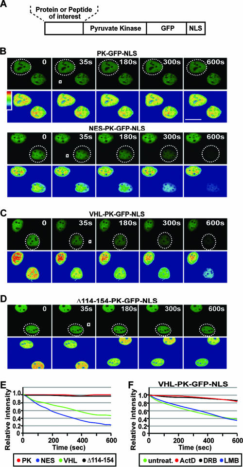
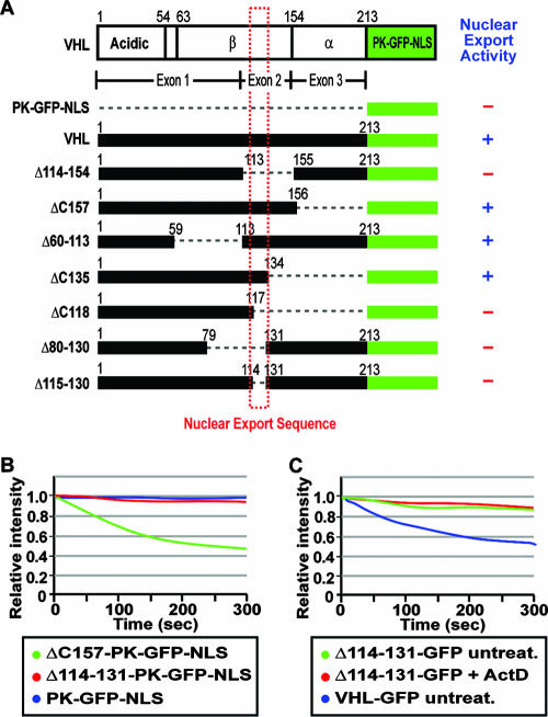
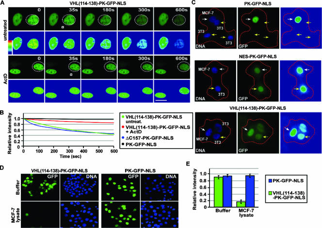
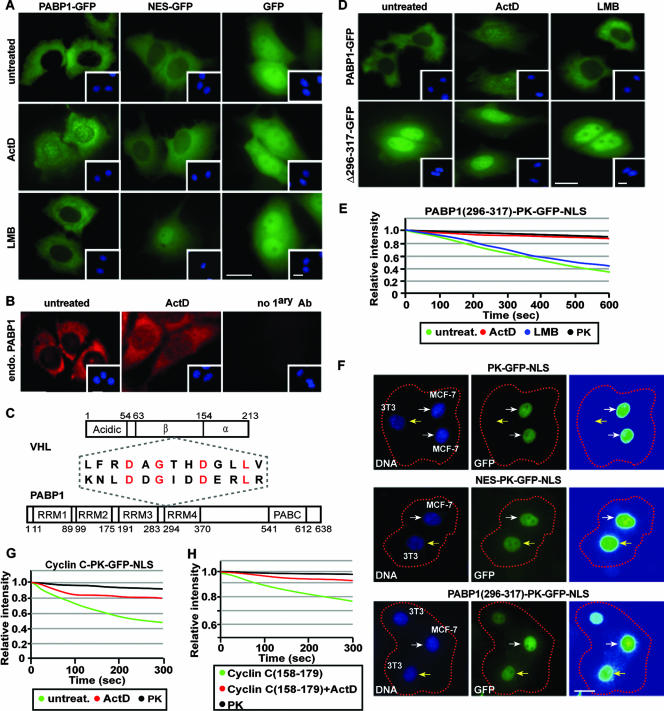
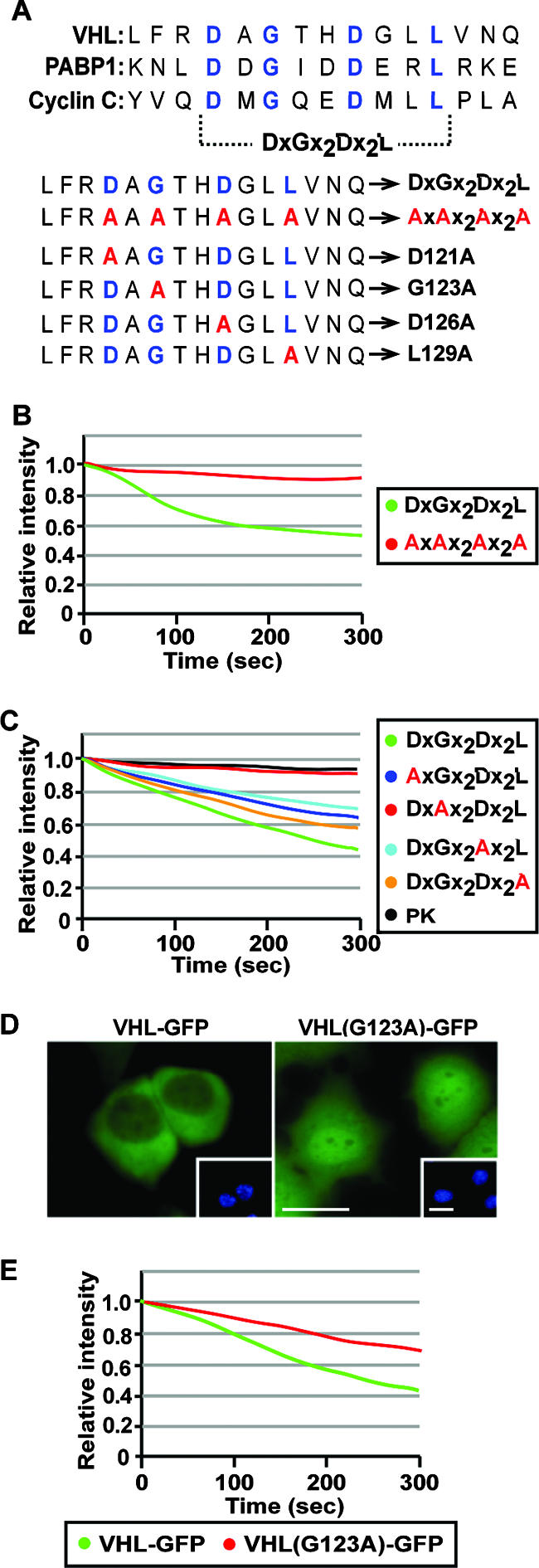
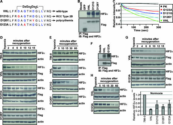
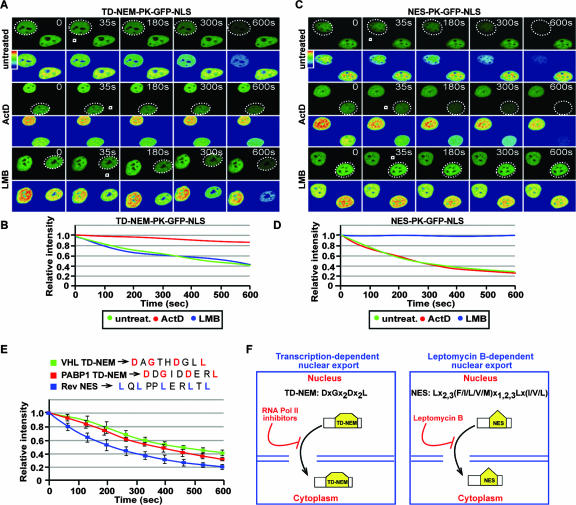
It is thought that degradation of nuclear proteins by the ubiquitylation system requires nuclear-cytoplasmic trafficking of E3 ubiquitin ligases. The von Hippel-Lindau (VHL) tumor suppressor protein is the substrate recognition component of a Cullin-2-containing E3 ubiquitin ligase that recruits hypoxia-inducible factor (HIF) for oxygen-dependent degradation. We demonstrated that VHL engages in nuclear-cytoplasmic trafficking that requires ongoing transcription to promote efficient HIF degradation. Here, we report the identification of a discreet motif, DXGX(2)DX(2)L, that directs transcription-dependent nuclear export of VHL and which is targeted by naturally occurring mutations associated with renal carcinoma and polycythemia in humans. The DXGX(2)DX(2)L motif is also found in other proteins, including poly(A)-binding protein 1, to direct its transcription-dependent nuclear export. We define DXGX(2)DX(2)L as TD-NEM (transcription-dependent nuclear export motif), since inhibition of transcription by actinomycin D or 5,6-dichlorobenzimidazole abrogates its nuclear export activity. Disease-causing mutations of key residues of TD-NEM restrain the ability of VHL to efficiently mediate oxygen-dependent degradation of HIF by altering its nuclear export dynamics without affecting interaction with its substrate. These results identify a novel nuclear export motif, further highlight the role of nuclear-cytoplasmic shuttling of E3 ligases in degradation of nuclear substrates, and provide evidence that disease-causing mutations can target subcellular trafficking.


Figures

FIG. 1.
FIG. 1.
Transcription-dependent nuclear export of VHL. (A) ActD alters the steady-state localization of VHL. MCF-7 cells transiently expressing VHL-GFP, NES-GFP, or GFP were either untreated or treated with LMB (10 μM), ActD (8 μM), or DRB (25 μg/ml). Insets are the corresponding Hoechst staining of the cells. Bars, 10 μm. (B and C) Cytoplasmic FLIP reveals that ActD decreases nuclear export of VHL. MCF-7 cells transiently expressing VHL-GFP were treated with ActD (2 μM) or LMB (10 μM) for 1 h. Cells were initially bleached in a large cytoplasmic region (dashed squares) to reduce cytoplasmic signal and then submitted to repetitive bleaching in a small cytoplasmic region (white squares). Cells were imaged between pulses, and the corresponding kinetics for loss of nuclear fluorescence were calculated and plotted on a graph (C) (see Materials and Methods). Bar, 10 μm. (D) Nuclear FLIP analysis was performed on cells treated as for panel B by repetitive bleaching in a small area in the nucleus. The loss of nuclear fluorescence was graphed. (E) ΔC157 exports from the nucleus in a transcription-dependent manner. ΔC157-GFP-transiently expressing cells were treated and analyzed as described for panel B. (F) Western blot analysis using anti-Flag and anti-GFP antibodies verified that the Flag- and GFP-tagged VHL, ΔC157, and GFP fusion proteins used for photobleaching experiments were not fragmented.
FIG. 2.
FIG. 2.
A transcription-dependent nuclear export sequence is encoded within the exon-2-encoded β-domain of VHL. (A) Schematic diagram of the PK-GFP-NLS fusion protein used for the live cell FLIP nuclear export assay, describing the region where protein or peptide sequences were fused. (B to E) MCF-7 cells transiently expressing the indicated constructs were submitted to cytoplasmic FLIP analysis, in which a small cytoplasmic region (white squares) within specific cells (a dashed circle outlines the cell nucleus) was repeatedly bleached. Kinetics for the loss of nuclear fluorescence from images obtained in panels B, C, and D were calculated and plotted on a graph (E). PK refers to the PK-GFP-NLS reporter construct, and NES, VHL, and Δ114-254 indicate the sequences fused to PK-GFP-NLS. Bar, 10 μm. (F) Cells were transiently transfected with VHL-PK-GFP-NLS and were either treated with ActD (2 μM), DRB (25 μg/ml), or LMB (10 μM) for 1 h or left untreated. Cytoplasmic FLIP was performed as described above to verify nuclear export activity.
FIG. 3.
FIG. 3.
Residues 114 to 131 are required for nuclear export of VHL. (A) Map of the nuclear export domain of VHL. The schematic diagram indicates deletion mutants of VHL that were submitted to cytoplasmic FLIP to assess the nuclear export activity. + and - indicate the ability or inability of the fusion protein to engage in nuclear export. (B) MCF-7 cells transiently transfected with PK-GFP-NLS, ΔC157-PK-GFP-NLS, or Δ114-131-PK-GFP-NLS were submitted to cytoplasmic FLIP, in which a small cytoplasmic region of a cell was bleached repetitively. The loss of nuclear GFP fluorescence was monitored over time and plotted on a graph. (C) Cells transiently expressing Δ114-131-GFP were initially bleached in a large cytoplasmic region followed by repetitive bleaching in a small cytoplasmic region after being treated for 1 h with 2 μM ActD or left untreated. Kinetics for the loss of nuclear fluorescence were calculated and plotted on a graph.
FIG. 4.
FIG. 4.
Residues 114 to 138 mediate transcription-dependent nuclear export of VHL. (A and B) MCF-7 cells were transiently transfected to express VHL(114-138)-PK-GFP-NLS. Cells were treated with a final concentration of 8 μM ActD or left untreated for 1 h before being subjected to photobleaching. Cytoplasmic FLIP was performed by repetitively bleaching a small cytoplasmic region (white squares) of a cell (the dotted circle outlines the cell nucleus). Cells were imaged between pulses, and the corresponding kinetics for the loss of nuclear fluorescence were calculated and are graphed (B). The nuclear export kinetics of ΔC157-PK-GFP-NLS revealed by a cytoplasmic FLIP analysis were plotted (B). Bar, 10 μm. (C) VHL(114-138) can mediate export in a polykaryon fusion assay. MCF-7 cells (donors) were fused with NIH 3T3 cells (acceptors) using PEG, and the transfer of nuclear fluorescence from donor to acceptor cells was monitored. Donor and acceptor cells were differentiated by a cell-specific Hoechst staining pattern. White arrows indicate donor cells, and yellow arrows indicate acceptor cells. Bar, 10 μm. (D and E) VHL(114-138) can mediate export in an in vitro nuclear export assay. MCF-7 cells transiently expressing the indicated constructs were permeabilized with digitonin, after which they were incubated with transport buffer containing ATP, GTP, and an ATP-regenerating system in the presence of buffer or MCF-7 cell lysate. Relative loss in nuclear fluorescence was calculated and plotted on a graph (E).
FIG. 5.
FIG. 5.
VHL and PABP1 share a common transcription-dependent nuclear export motif. (A) PABP1 exports by a transcription-dependent mechanism. MCF-7 cells transiently expressing PABP1-GFP, NES-GFP, or GFP alone were either untreated or treated with 8 μM ActD or 10 μM LMB for 3 h. Insets are the corresponding Hoechst staining of the cells. Bars, 10 μm. (B) Endogenous PABP1 is also sensitive to ActD treatment. MCF-7 cells were either left untreated or were treated with 8 μM ActD. Endogenous PABP1 was detected by immunofluorescence using an anti-PABP1 antibody. Insets show nuclei stained with Hoechst stain. A primary antibody exclusion (no 1ary Ab) control is also shown. Bar, 10 μm. (C) Schematic diagram depicting a region of alignment between the nuclear export sequence of VHL and PABP1. Conserved residues are indicated in red. (D) MCF-7 cells transiently expressing PABP1(Δ296-317)-GFP or PABP1-GFP were treated as described for panel A. Insets are the corresponding Hoechst staining of the cells. Bar, 10 μm. (E) Residues 296 to 317 of PABP1 encode a transcription-dependent nuclear export sequence. Transfected MCF-7 cells were treated with 2 μM ActD or 10 μM LMB for 1 h before being submitted to cytoplasmic FLIP. The corresponding loss of nuclear fluorescence was monitored, measured, and plotted on a graph. (F) PABP1(296-317) can mediate export in a polykaryon fusion assay. MCF-7 cells (donors) were fused with NIH 3T3 cells (acceptors) using PEG, and the transfer of nuclear fluorescence from donor to acceptor cells was monitored. Donor and acceptor cells were differentiated by a cell-specific Hoechst staining pattern. White arrows indicate donor cells, and yellow arrows indicate acceptor cells. Bar, 10 μm. (G) Full-length cyclin C can export the PK-GFP-NLS reporter in a transcription-dependent manner. Cells were treated with 2 μM ActD for 1 h or untreated, and the loss of nuclear fluorescence after cytoplasmic FLIP was plotted on a graph. (H) Residues 158 to 179 of cyclin C encode a transcription-dependent nuclear export motif. The indicated constructs were transiently expressed in MCF-7 cells and treated the same as for panel G. Cells were subjected to cytoplasmic FLIP, and the corresponding kinetics for loss of nuclear fluorescence were plotted on a graph.
FIG. 6.
FIG. 6.
Identification of key residues that mediate transcription-dependent nuclear export. (A) Sequence alignment depicting conserved residues (blue) in the transcription-dependent nuclear export motifs of VHL, PABP1, and cyclin C. Residues in red indicate alanine substitutions at key residues in the TD-NEM sequence of VHL. (B) Conserved residues in the DXGX2DX2L consensus sequence are essential for nuclear export. MCF-7 cells were transiently transfected with VHL(115-130)-PK-GFP-NLS (DxGx2Dx2L) or VHL(115-130AAAA)-PK-GFP-NLS (AxAx2Ax2A), where the four key residues were replaced with alanines. Cytoplasmic FLIP was performed, and the loss of nuclear fluorescence was plotted on a graph. (C) Single alanine substitutions of key residues within the DXGX2DX2L consensus sequence have a differential effect on nuclear export activity. MCF-7 cells transiently expressing the indicated PK-GFP-NLS-tagged constructs were subjected to cytoplasmic FLIP. The graph represents the loss of nuclear fluorescence. (D and E) A single amino acid substitution of G123A in full-length VHL affects steady-state localization and nuclear export activity. Steady-state localization of VHL(G123A)-GFP expressed in MCF-7 cells compared to wild-type VHL-GFP is shown (D). Insets are the corresponding Hoechst staining of the cells. Cells expressing VHL-GFP or VHL(G123A)-GFP were subjected to cytoplasmic FLIP, and the loss of nuclear fluorescence was monitored and graphed (E).
FIG. 7.
FIG. 7.
Cancer-causing mutations within TD-NEM of VHL abrogate nuclear export and oxygen-dependent degradation of HIF2α. (A) Sequence alignment depicting cancer-causing mutations (red) in the key TD-NEM residues of VHL that lead to RCC type 2B (D121G) and polycythemia (D126Y) in humans. G123A, which exhibits a defect in nuclear export as shown in Fig. 6, was also used to test its ability to mediate HIF degradation. (B) D121G and D126Y retain the ability to bind to HIF. Cells stably expressing Flag-tagged VHL-GFP, D121G-GFP, D126Y-GFP, and GFP were placed under hypoxic conditions and treated with 10 μM MG132 for 2 h before being harvested. Cell lysates were immunoprecipitated with anti-Flag beads and immunoblotted with anti-Flag and anti-HIF2α antibodies. (C) Cancer-causing mutants D121G and D126Y in TD-NEM decrease the nuclear export activity of VHL. Cells stably expressing VHL-GFP, D121G-GFP, D126Y-GFP, or G123A-GFP or transiently expressing PK-GFP-NLS were submitted to cytoplasmic FLIP as described previously. The loss of nuclear fluorescence was monitored and plotted on a graph. (D and E) Cells expressing D121G or D126Y exhibit a deficiency in HIF degradation. Stable cell lines of VHL-GFP, D121G-GFP, D126Y-GFP, and the VHL-defective cell line 786-0 were incubated for 20 h under hypoxic conditions before being reoxygenated by placing them in a normoxic environment, for the indicated time. Cells were lysed with 4% SDS and submitted to Western blot analysis using an anti-HIF2α antibody. Levels of VHL or its mutant counterparts were monitored using anti-Flag antibody, and actin was used to ensure equal loading of lysates. (F) G123A retains the ability to bind to HIF. Cells stably expressing the VHL point mutant, G123A, were treated the same as described for panel B. (G and H) Cells expressing G123A exhibit a deficiency in HIF degradation. G123A stably expressing cells were treated in the same manner as described for panels D and E. (I) D121G, D126Y, and G123A stable cells express higher normoxic HIF levels compared to wild-type VHL. Stable cell lines incubated under normoxic conditions were lysed with 4% SDS and submitted to Western blot analysis using anti-HIF2α and antiactin antibodies. HIF2α levels were normalized to actin and values, calculated relative to HIF2α levels in 786-0 cells, were plotted on a graph.
FIG. 8.
FIG. 8.
TD-NEM is a novel and efficient transcription-dependent nuclear export motif. (A to D) TD-NEM, contrary to the classical NES, mediates nuclear export in an ActD-sensitive but LMB-insensitive manner. Cells transiently expressing PK-GFP-NLS-tagged TD-NEM of VHL or NES of the Rev protein were either untreated or treated with ActD or LMB as previously described before being submitted to cytoplasmic FLIP. White squares indicate the bleached area, and the dotted circles outline the nucleus of the cell of interest. The loss of nuclear fluorescence was monitored, calculated, and plotted on a graph (B and D). (E) TD-NEM is an efficient nuclear export motif. Cells transiently expressing PK-GFP-NLS-tagged TD-NEM of VHL or PABP1, or the Rev NES, one of the strongest export signals, were submitted to cytoplasmic FLIP in order to compare the efficiency of nuclear export. (F) Model of nuclear export mediated by the TD-NEM consensus sequence compared to the classical NES (3). Nuclear export of TD-NEM is abrogated by RNA Pol II inhibitors, such as ActD and DRB, whereas NES activity is abrogated by LMB.

Similar articles

Cited by

References

    1. Afonina, E., R. Stauber, and G. N. Pavlakis. 1998. The human poly(A)-binding protein 1 shuttles between the nucleus and the cytoplasm. J. Biol. Chem. 27313015-13021. - PubMed
    1. Blondel, M., J. M. Galan, Y. Chi, C. Lafourcade, C. Longaretti, R. J. Deshaies, and M. Peter. 2000. Nuclear-specific degradation of Far1 is controlled by the localization of the F-box protein Cdc4. EMBO J. 196085-6097. - PMC - PubMed
    1. Bogerd, H. P., R. A. Fridell, R. E. Benson, J. Hua, and B. R. Cullen. 1996. Protein sequence requirements for function of the human T-cell leukemia virus type 1 Rex nuclear export signal delineated by a novel in vivo randomization-selection assay. Mol. Cell. Biol. 164207-4214. - PMC - PubMed
    1. Bonicalzi, M. E., I. Groulx, N. de Paulsen, and S. Lee. 2001. Role of exon 2-encoded beta-domain of the von Hippel-Lindau tumor suppressor protein. J. Biol. Chem. 2761407-1416. - PubMed
    1. Bruick, R. K., and S. L. McKnight. 2001. A conserved family of prolyl-4-hydroxylases that modify HIF. Science 2941337-1340. - PubMed

Publication types

MeSH terms

Substances