. 2007 Nov 26;204(12):2825-35.
doi: 10.1084/jem.20070994. Epub 2007 Oct 29.

Ets-1 is a negative regulator of Th17 differentiation

Affiliations

Ets-1 is a negative regulator of Th17 differentiation

Jacques Moisan et al. J Exp Med. .

Abstract

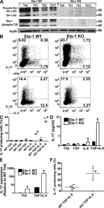
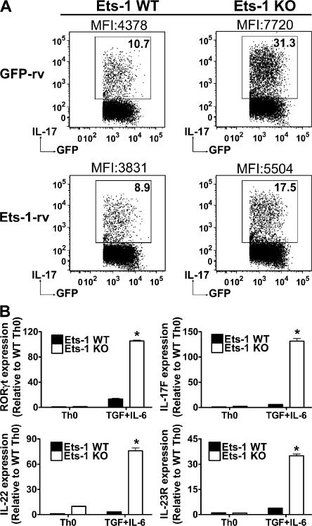
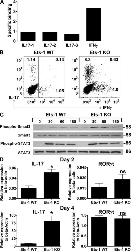
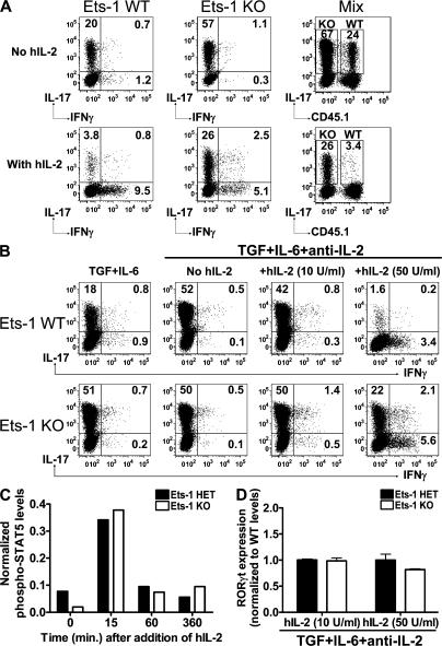
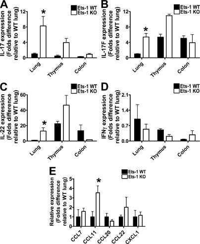
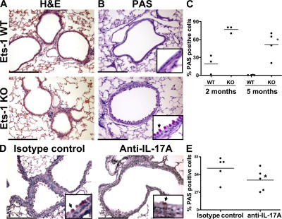
IL-17 is a proinflammatory cytokine that plays a role in the clearance of extracellular bacteria and contributes to the pathology of many autoimmune and allergic conditions. IL-17 is produced mainly by a newly characterized subset of T helper (Th) cells termed Th17. Although the role of Th17 cells in the pathology of autoimmune diseases is well established, the transcription factors regulating the differentiation of Th17 cells remain poorly characterized. We report that Ets-1-deficient Th cells differentiated more efficiently to Th17 cells than wild-type cells. This was attributed to both low IL-2 production and increased resistance to the inhibitory effect of IL-2 on Th17 differentiation. The resistance to IL-2 suppression was caused by a defect downstream of STAT5 phosphorylation, but was not caused by a difference in the level of RORgamma t. Furthermore, Ets-1-deficient mice contained an abnormally high level of IL-17 transcripts in their lungs and exhibited increased mucus production by airway epithelial cells in an IL-17-dependent manner. Based on these observations, we report that Ets-1 is a negative regulator of Th17 differentiation.


Figures

Figure 1.
Figure 1.
Ets-1 KO Th cells produce increased levels of IL-17. Total Th cells derived from WT or Ets-1 KO mice were cultured as described in the Materials and methods. (A) After 5 d in culture under Th0, Th1, Th2, or Th17 conditions, cells were restimulated with PMA/ionomycin for 10 min and lysed. Whole-cell extract was analyzed by Western blotting using antibodies against Ets-1 or phospho-T38 Ets-1. An antibody against Hsp90 was used as loading control. (B) Total Th cells from WT and Ets-1 KO mice were cultured under Th17 conditions for 5 d and restimulated with PMA/ionomycin. Cytokine production was assessed by ICS. The numbers represent the percentages of cells stained positive for the indicated cytokines. (C) Total Ets-1 KO and WT Th cells were differentiated for 5 d in the presence of WT irradiated splenocytes and indicated cytokines, and the production of IL-17 in response to PMA/ionomycin stimulation was analyzed by ICS. The percentages of IL-17–positive cells from five independent experiments are shown. (D) Some of the differentiated Th cells (at 1 million cells/ml) generated in C were restimulated with 0.1 μg/ml of plate-bound anti-CD3 for 24 h, and the level of IL-17 protein in the supernatant was measured by ELISA. (E) A fraction of the PMA/ionomycin-restimulated cells from C were analyzed for IL-17 expression by real-time PCR analysis. (F) Naive WT and Ets-1 KO Th cells were differentiated under Th17 conditions, and the production of IL-17 by the differentiated cells was analyzed by ICS after 5 d. Cumulative results of six independent experiments are shown. Data are presented as the mean ± the SD. *, P < 0.05.
Figure 2.
Figure 2.
Increased IL-17 production by Ets-1 KO Th cells is reversible, and is associated with increased expression of Th17 markers. (A) Total Th cells from WT and Ets-1 KO mice were infected 2 d after activation under Th17 skewing conditions with either empty virus (GFP-rv) or virus expressing full-length Ets-1 isoform (Ets-1-rv). The panels are gated on GFP-positive cells. The production of IL-17 by the transduced (GFP-positive) cells was examined by ICS. Mean fluorescence intensity (MFI) of the IL-17-positive cells is indicated. Results are representative of three independent experiments. (B) After 5 d in culture in the indicated conditions, differentiated WT and Ets-1 KO Th cells were restimulated for 4 h with PMA/ionomycin, and the transcript levels of IL-17F, IL-22, IL-23R, and RORγt were measured by real-time PCR. Data are presented as the mean ± the SD and are representative of three independent experiments. *, P < 0.05.
Figure 3.
Figure 3.
Ets-1 does not bind to the IL-17 promoter or interfere with early signaling events during Th17 differentiation. (A) CHIP was performed on WT Th17 cells using anti–Ets-1 antibody. Binding to conserved Ets sites in the IL-17 and IFN-γ genetic region was analyzed by real-time PCR. Relative binding was calculated as described in the Materials and methods. (B) Total WT and Ets-1 KO Th cells were plated on anti-CD3–coated plates and given anti-CD28, but in the absence of APC, and skewed to the Th17 lineage with IL-6 and TGFβ1 for 5 d. The production of indicated cytokines by the differentiated Th cells was measured by ICS. (C) Freshly isolated total WT and Ets-1 KO Th cells were stimulated with TGFβ1 and IL-6 for the indicated number of minutes. The levels of phospho-Smad3, total Smad2/3, phospho-STAT3, and total STAT3 were examined with Western blotting. (D) Naive Th cells were cultured under Th17 conditions, and RNA was harvested on day 2 or 4 and analyzed for expression of IL-17 and RORγt by real-time PCR. Data are representative of three independent experiments and are presented as the mean ± the SD. *, P < 0.05.
Figure 4.
Figure 4.
Increased Th17 differentiation in Ets-1 KO Th cells is dependent on IL-2. (A) Total Th cells from congenic CD45.1 C57BL/6 and CD45.2 Ets-1 KO mice were cultured separately or cocultured (Mix) at a 1:1 ratio under Th17 skewing conditions in the presence (added at 0 h) or absence of exogenous IL-2. The cells were stained for the CD45.1 isoform, and the production of indicated cytokines was analyzed by ICS after restimulation with PMA/ionomycin. (B) Total Th cells from WT and Ets-1 KO mice were cultured under Th17 conditions in the presence of anti–mouse IL-2 and various concentrations of hIL-2. Cytokine production was assessed by ICS. The numbers represent the percentages of cells stained positive for indicated cytokines. (C) Total Th cells from WT and Ets-1 KO mice were cultured for 3 d with TGFβ1, IL-6, anti–mouse IL-2, and hIL-2 (10 U/ml). Cells were subsequently rested for 2 h in the presence of anti–mouse IL-2 before being stimulated with 50 U/ml hIL-2 for the indicated amount of time. Cell lysates were harvested and subjected to Western blot analyses using antibody against phosphorylated or total STAT5. The blots were scanned and the levels of phosphorylated STAT5 were normalized to the total amount of STAT5. (D) After 5 d in culture in the indicated conditions, RNA from differentiated WT and Ets-1 KO Th17 cells was collected, and the transcript level of RORγt was measured by real-time PCR. Data are presented as the mean ± the SD and are from two independent experiments. *, P < 0.05.
Figure 5.
Figure 5.
In vivo evidence of increased Th17 differentiation in Ets-1 KO mice. Total RNA of indicated tissues from three WT and Ets-1 KO mice was extracted, and the transcript levels of IL-17 (A), IL-17F (B), IL-22 (C), and IFNγ (D) were quantified by real-time PCR. (E) Expression of various chemokines was analyzed in the lungs of WT and Ets-1 KO mice. Data are presented as the mean ± the SD. *, P < 0.05.
Figure 6.
Figure 6.
IL-17–dependent mucus overproduction by Ets-1 KO airway epithelial cells. Histological sections of inflated lungs from WT and Ets-1 KO mice were stained for hematoxylin and eosin (A) and PAS (B). Bars, 1 mm. The arrow in B points out PAS-positive epithelial cells. The percentages of PAS-positive epithelial cells observed in WT and KO mice (n = 3–4) at the indicated ages are shown in C. 2-mo-old Ets-1 KO mice were injected intraperitoneally with anti–IL-17 or control IgG every other day for 2 wk (6 injections in total) before killing. The histological sections of inflated lungs were stained with PAS (D). The percentages of PAS-positive epithelial cells are shown in F. Data are presented as the mean ± the SD. *, P < 0.05.

References

    1. Weaver, C.T., R.D. Hatton, P.R. Mangan, and L.E. Harrington. 2007. IL-17 family cytokines and the expanding diversity of effector t cell lineages. Annu. Rev. Immunol. 25:821–852. - PubMed
    1. Ziolkowska, M., A. Koc, G. Luszczykiewicz, K. Ksiezopolska-Pietrzak, E. Klimczak, H. Chwalinska-Sadowska, and W. Maslinski. 2000. High levels of IL-17 in rheumatoid arthritis patients: IL-15 triggers in vitro IL-17 production via cyclosporin A-sensitive mechanism. J. Immunol. 164:2832–2838. - PubMed
    1. Matusevicius, D., P. Kivisakk, B. He, N. Kostulas, V. Ozenci, S. Fredrikson, and H. Link. 1999. Interleukin-17 mRNA expression in blood and CSF mononuclear cells is augmented in multiple sclerosis. Mult. Scler. 5:101–104. - PubMed
    1. Molet, S., Q. Hamid, F. Davoine, E. Nutku, R. Taha, N. Page, R. Olivenstein, J. Elias, and J. Chakir. 2001. IL-17 is increased in asthmatic airways and induces human bronchial fibroblasts to produce cytokines. J. Allergy Clin. Immunol. 108:430–438. - PubMed
    1. Bettelli, E., M. Oukka, and V.K. Kuchroo. 2007. T(H)-17 cells in the circle of immunity and autoimmunity. Nat. Immunol. 8:345–350. - PubMed

Publication types

MeSH terms