. 2007 Nov 5;179(3):553-65.
doi: 10.1083/jcb.200703185. Epub 2007 Oct 29.

A molecular switch that controls cell spreading and retraction

Affiliations

A molecular switch that controls cell spreading and retraction

Panagiotis Flevaris et al. J Cell Biol. .

Abstract

Integrin-dependent cell spreading and retraction are required for cell adhesion, migration, and proliferation, and thus are important in thrombosis, wound repair, immunity, and cancer development. It remains unknown how integrin outside-in signaling induces and controls these two opposite processes. This study reveals that calpain cleavage of integrin beta(3) at Tyr(759) switches the functional outcome of integrin signaling from cell spreading to retraction. Expression of a calpain cleavage-resistant beta(3) mutant in Chinese hamster ovary cells causes defective clot retraction and RhoA-mediated retraction signaling but enhances cell spreading. Conversely, a calpain-cleaved form of beta(3) fails to mediate cell spreading, but inhibition of the RhoA signaling pathway corrects this defect. Importantly, the calpain-cleaved beta(3) fails to bind c-Src, which is required for integrin-induced cell spreading, and this requirement of beta(3)-associated c-Src results from its inhibition of RhoA-dependent contractile signals. Thus, calpain cleavage of beta(3) at Tyr(759) relieves c-Src-mediated RhoA inhibition, activating the RhoA pathway that confines cell spreading and causes cell retraction.


Figures

Figure 1.
Figure 1.
A calpain cleavage–resistant mutation of β3: R760E. (A) WT β3 cytoplasmic domain sequence and the R760E mutation. (B) Cells expressing WT and R760E mutant αIIbβ3 were solubilized, treated with or without purified μ-calpain, and immunoblotted with anti-β3 C-terminal antibody Ab762, calpain cleavage–indicator antibody Ab759, and the anti-β3 extracellular domain antibody mAb 15. Note that although R760E showed lower reactivity with Ab762 because of mutation in the epitope (TYR760GT), calpain caused minimal loss of Ab762 reactivity or gain of Ab759 reactivity, which indicates calpain resistance. (C) Analysis of stable αIIbβ3 expression in cells expressing WT and R760E mutant integrins, using the monoclonal anti-αIIbβ3 antibody D57. (D) WT and R760E cells spreading on fibrinogen were permeabilized and stained with mAb 15 (green) and purified Ab759 (red). Ab759 staining of R760E cells was reduced compared with WT β3. Bar, 10 μm.
Figure 2.
Figure 2.
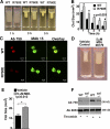
Effects of cleavage-resistant β3 mutation, R760E, and calpain inhibition on clot retraction and β3 cleavage. (A) Fibronectin-depleted plasma mixed with WT or R760E cells was induced to coagulate with 2 U/ml thrombin at 37°C for the indicated times, and then photographed. (B) Quantification of clot size at various time points in A. Error bars are the SD from three separate experiments. (C) Clots containing WT or R760E cells were fixed at 1 h, cryosectioned, stained with purified Ab759 (red) or mAb 15 (green), and observed by confocal microscopy. Note the reduced Ab759 staining of R760E cells. Bars, 10 μm. (D) Clot retraction by WT cells preincubated with either 5 μM of calpain inhibitor MDL 28170 or vehicle control. (E) Quantification of clot sizes in D (mean ± SD). (F) Washed platelets from WT and μ-calpain knockout mice were incubated in an aggregometer with or without 0.1 U/ml thrombin for 7 min. β3 cleavage at Y759, which was detected using cleavage-specific antibody Ab759, was enhanced in the WT compared with the calpain knockout. Ab8053 recognizes β3 extracellular domain and serves as a loading control.
Figure 3.
Figure 3.
Effects of cleavage-resistant and -mimicking β3 mutations and ROCK inhibitors on cell spreading and clot retraction. (A) Indicated cell lines were incubated on fibrinogen-coated chamber slides at 37°C and photographed at various times. Images at 20 min are shown. (B) Sizes of spread cells were quantified as percentage of original sizes using Image J software. Histogram shows the mean ± SEM. (C and D) 5 μM each of ROCK inhibitors Y-27632 and H-1152 inhibited clot retraction in platelets (D) and in CHO cells expressing WT β3 (C). (E) Spreading of WT cells and cells expressing a calpain cleavage–mimicking β3 mutant (Δ759) on fibrinogen for 1 h. Defective spreading of Δ759 cells was corrected by 2 μM each of ROCK inhibitors H-1152 and Y-27632. (F) Quantification of cell spreading from cells in E, using image J software. Histogram shows the mean ± SEM. (G) 5 μM each of ROCK inhibitors H-1152 and Y-27632 enhance spreading in WT cells but not in cleavage-resistant R760E mutants at 20 min. (H) Quantification of cell spreading from cells in G, using image J software. Histogram shows the mean ± SEM.
Figure 4.
Figure 4.
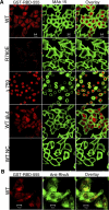
Calpain cleavage of β3 stimulates RhoA activation. (A) WT, R760E, and Δ759 cells spreading on fibrinogen for 1 h were fixed and allowed to react with Alexa Fluor 555–labeled rhotekin (GST-RBD-555 [red]) to indicate RhoA activation. To estimate nonspecific binding, fluorescent rhotekin binding was also performed in the presence of unlabeled rhotekin (NC) or 20 mM glutathione (glut). MAb 15 (green) serves as a marker for cell margins and integrin β3 expression. (B) Activated RhoA, indicated by GST-RBD-555 (red), colocalizes with anti-RhoA (green). Bars, 10 μm.
Figure 5.
Figure 5.
Inhibition of GST-RBD binding to active RhoA by cell-permeable C3 transferase. Cells expressing WT αIIbβ3 were preincubated with or without 5 μg/ml of cell-permeable C3 transferase and were allowed to spread on fibrinogen-coated surfaces for 1 h. Cells were stained with GST-RBD-555 (red) to detect active RhoA and with mAb 15 (green) to mark integrin surface expression. Bars, 10 μM.
Figure 6.
Figure 6.
Coimmunoprecipitation of c-Src with WT β3 and β3 mutants. (A) β3 immunoprecipitates from CHO cells expressing WT, R760E mutant, and Δ759 mutant integrins were probed on Western blots with antibodies against β3 (mAb 15) or c-Src. c-Src binding to the integrin cytoplasmic domain is abolished in the calpain cleavage–mimicking mutant Δ759 but not in the calpain cleavage–resistant mutant R760E. (B) Lysates were also immunoblotted with antibody to c-Src.
Figure 7.
Figure 7.
The effect of calpain cleavage of β3 on c-Src–dependent inhibition of RhoA-mediated cell retraction. (A) WT cells incubated with or without 10 μM PP2 or PP2 plus 4 μM of ROCK inhibitors H-1152 or Y-27632 were allowed to spread on fibrinogen for 1 h. Inhibition of cell spreading by PP2 is reversed by ROCK inhibitors. Bar, 10 μm. (B) Spreading of WT cells transfected with GFP alone, a dominant-negative c-Src (DN c-Src) plus GFP, N19-RhoA plus GFP, or N19-RhoA and dominant-negative c-Src plus GFP. Note that N19-RhoA reversed the inhibitory effect of dominant-negative c-Src. (C) R760E cells are allowed to adhere to fibrinogen in the presence of PP2 or vehicle control DMSO and are stained with GST-RBD-555 (red) and mAb 15 (green). Defective RhoA activation in R760E cells is corrected by PP2. Bars, 10 μM. (D) Quantitation of cell sizes in A using Image J (mean ± SEM from four random fields). (E) Quantitation of cell sizes in B using Image J (mean ± SEM from three experiments). (F) Stain of GST-RBD-555 in cells from C was quantified using LSM 5 software (mean ± SEM from three experiments). (G) 10 μM of SFK inhibitor PP2 accelerates clot retraction in platelets.
Figure 8.
Figure 8.
The effect of c-Src activation on the integrin-cleavage–dependent inhibition of cell spreading. (A) WT, R760E, and Δ759 cells were either in suspension or allowed to spread on fibrinogen for 30 min, and then solubilized and analyzed by Western blot with antibodies specific for c-Src or for Src phosphorylated at Y416 as an indicator of Src activation. Note that c-Src activation is similar in all three cell lines. (B) Δ759 cells were transfected with either GFP or GFP plus a constitutively active mutant of c-Src, E378G. Nonadherent transfected cells were solubilized and immunoblotted with antibodies against total c-Src, Y416-phosphorylated c-Src, and β3 (Ab8053, to verify equal loading). (C) Δ759 cells were transfected with GFP alone or GFP plus a constitutively active mutant of c-Src, E378G. The transfected cells were allowed to spread on fibrinogen-coated surfaces. Note that active c-Src cannot reverse the spreading defect in Δ759 cells. (D) Quantitation of cell sizes in C using Image J software (mean ± SEM from three experiments).
Figure 9.
Figure 9.
A new calpain-dependent switch that controls cell spreading and retraction. This schematic illustrates the calpain-dependent switch that controls the direction of integrin signaling. In this model, ligand binding to integrins induces not only Rac- and cdc42-dependent cell spreading signals, but also phosphorylation of the cytoplasmic domain of β3 and elevation of intracellular calcium levels that activate calpain. Tyrosine phosphorylation at Y759 of β3 protects the c-Src binding site in the C-terminal domain of β3 from calpain cleavage. The β3-associated c-Src facilitates cell spreading by inhibiting RhoA-dependent cell retraction signals. In spread cells, Y759 of β3 becomes dephosphorylated, allowing β3 cleavage by calpain. Removal of the c-Src binding site in β3 by calpain relieves the local inhibitory effect of c-Src on the RhoA-ROCK signaling pathway, activates RhoA-dependent cell retraction, and thus switches the outcome of local integrin signaling from mediating cell spreading to retraction. However, the possible involvement of additional pathways is not totally excluded.

Similar articles

Cited by

References

    1. Amano, M., M. Ito, K. Kimura, Y. Fukata, K. Chihara, T. Nakano, Y. Matsuura, and K. Kaibuchi. 1996. Phosphorylation and activation of myosin by Rho-associated kinase (Rho-kinase). J. Biol. Chem. 271:20246–20249. - PubMed
    1. Arias-Salgado, E.G., S. Lizano, S. Sarkar, J.S. Brugge, M.H. Ginsberg, and S.J. Shattil. 2003. Src kinase activation by direct interaction with the integrin beta cytoplasmic domain. Proc. Natl. Acad. Sci. USA. 100:13298–13302. - PMC - PubMed
    1. Arias-Salgado, E.G., F. Haj, C. Dubois, B. Moran, A. Kasirer-Friede, B.C. Furie, B. Furie, B.G. Neel, and S.J. Shattil. 2005. a. PTP-1B is an essential positive regulator of platelet integrin signaling. J. Cell Biol. 170:837–845. - PMC - PubMed
    1. Arias-Salgado, E.G., S. Lizano, S.J. Shattil, and M.H. Ginsberg. 2005. b. Specification of the direction of adhesive signaling by the integrin beta cytoplasmic domain. J. Biol. Chem. 280:29699–29707. - PubMed
    1. Arthur, W.T., and K. Burridge. 2001. RhoA inactivation by p190RhoGAP regulates cell spreading and migration by promoting membrane protrusion and polarity. Mol. Biol. Cell. 12:2711–2720. - PMC - PubMed

Publication types