

Review
doi: 10.1083/jcb.200708001.
Epub 2007 Oct 29.
Skin and bones: the bacterial cytoskeleton, cell wall, and cell morphogenesis
Affiliations
- PMID: 17967949
- PMCID: PMC2064785
- DOI: 10.1083/jcb.200708001
Item in Clipboard
Review
Skin and bones: the bacterial cytoskeleton, cell wall, and cell morphogenesis
J Cell Biol.
.
Abstract
The bacterial world is full of varying cell shapes and sizes, and individual species perpetuate a defined morphology generation after generation. We review recent findings and ideas about how bacteria use the cytoskeleton and other strategies to regulate cell growth in time and space to produce different shapes and sizes.
Figures
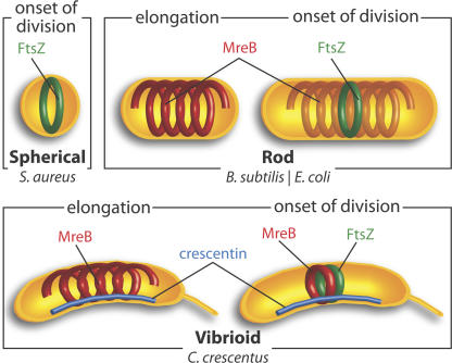
The bacterial cytoskeleton. The only cytoskeletal element present in spherical bacteria such as S. aureus (top left) is the tubulin-like cell division protein FtsZ (green), which localizes in a ring at the onset of cell division, recruits other cell division proteins, and defines the division plane. Most rod-shaped bacteria (top right) also contain one or more actin-like MreB homologues (red), which exhibit helix-like localization patterns and are essential for cell width control. At the onset of cell division, the FtsZ ring forms and defines the division plane. C. crescentus, a vibrioid bacterium (bottom), contains a third cytoskeletal element, the intermediate filament-like crescentin (blue), which is required for cell curvature and localizes at the inner curvature of cells. At the onset of division in C. crescentus, MreB exhibits FtsZ-dependent relocalization from a helix-like to a ring-like pattern at the FtsZ ring location.
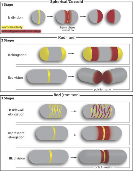
Cell wall growth patterns. Yellow and red colors show where new cell wall synthesis is occurring and where cell wall material has been inserted within a particular stage, respectively. (top) Spherical (coccoid) bacteria synthesize a new cell wall at division only, when a division septum bisects the cell (left) and is remodeled to form two new hemispheres (middle). Each daughter cell has one new hemisphere produced during division (right). (middle) A few known rod-shaped bacteria (e.g., Corynebacterium) lack MreB and grow from their poles to elongate between rounds of division; thus, they have two stages of growth: elongation and division. During elongation, active wall synthesis at the cell poles leads to lengthening of the cell cylinder. During division, the division septum snaps in two to form two new poles. (bottom) Most rod-shaped bacteria have at least one MreB homologue and can have two elongation stages. The first is characterized by patchy or helical cell wall insertion along the sidewall of the cell cylinder between inert poles. Subsequently, before the onset of division, preseptal elongation occurs in an FtsZ ring–dependent fashion, during which wall synthesis is localized adjacent to the FtsZ ring. Finally, cell division occurs.
References
-
- Aaron, M., G. Charbon, H. Lam, H. Schwarz, W. Vollmer, and C. Jacobs-Wagner. 2007. The tubulin homologue FtsZ contributes to cell elongation by guiding cell wall precursor synthesis in Caulobacter crescentus. Mol. Microbiol. 64:938–952. - PubMed
-
- Ausmees, N., J.R. Kuhn, and C. Jacobs-Wagner. 2003. The bacterial cytoskeleton: an intermediate filament-like function in cell shape. Cell. 115:705–713. - PubMed
