. 2007 Nov 13;116(20):2260-8.
doi: 10.1161/CIRCULATIONAHA.107.703330. Epub 2007 Oct 29.

Mutation in glycerol-3-phosphate dehydrogenase 1 like gene (GPD1-L) decreases cardiac Na+ current and causes inherited arrhythmias

Affiliations

Mutation in glycerol-3-phosphate dehydrogenase 1 like gene (GPD1-L) decreases cardiac Na+ current and causes inherited arrhythmias

Barry London et al. Circulation. .

Abstract

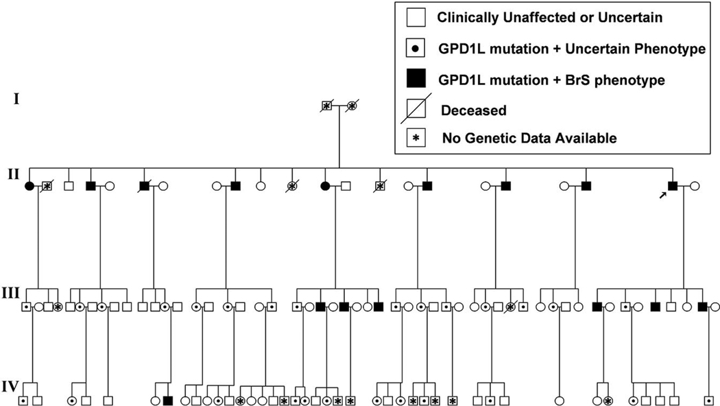
Background: Brugada syndrome is a rare, autosomal-dominant, male-predominant form of idiopathic ventricular fibrillation characterized by a right bundle-branch block and ST elevation in the right precordial leads of the surface ECG. Mutations in the cardiac Na+ channel SCN5A on chromosome 3p21 cause approximately 20% of the cases of Brugada syndrome; most mutations decrease inward Na+ current, some by preventing trafficking of the channels to the surface membrane. We previously used positional cloning to identify a new locus on chromosome 3p24 in a large family with Brugada syndrome and excluded SCN5A as a candidate gene.

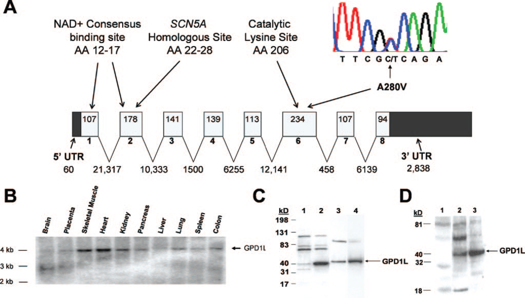
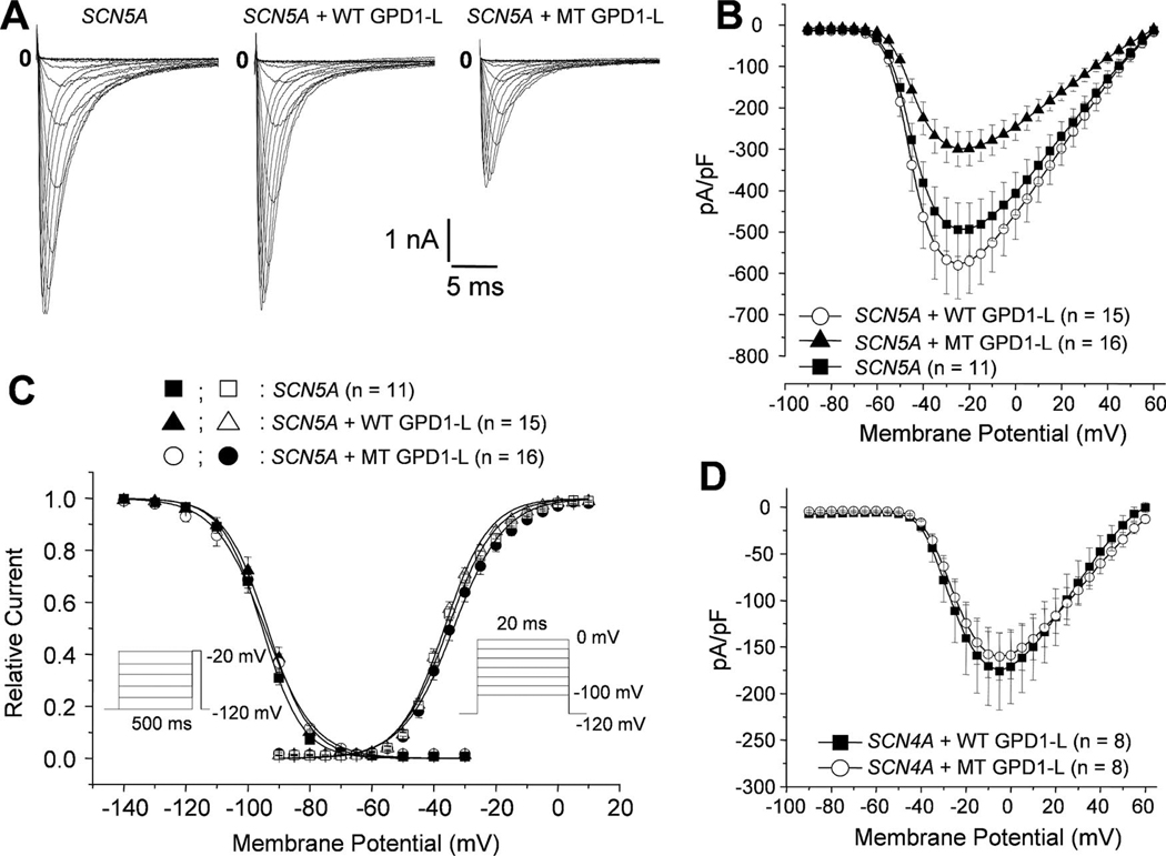
Methods and results: We used direct sequencing to identify a mutation (A280V) in a conserved amino acid of the glycerol-3-phosphate dehydrogenase 1-like (GPD1-L) gene. The mutation was present in all affected individuals and absent in >500 control subjects. GPD1-L RNA and protein are abundant in the heart. Compared with wild-type GPD1-L, coexpression of A280V GPD1-L with SCN5A in HEK cells reduced inward Na+ currents by approximately 50% (P<0.005). Wild-type GPD1-L localized near the cell surface to a greater extent than A280V GPD1-L. Coexpression of A280V GPD1-L with SCN5A reduced SCN5A cell surface expression by 31+/-5% (P=0.01).

Conclusions: GPD1-L is a novel gene that may affect trafficking of the cardiac Na+ channel to the cell surface. A GPD1-L mutation decreases SCN5A surface membrane expression, reduces inward Na+ current, and causes Brugada syndrome.


Figures

Figure 1
Figure 1
Pedigree of the FN family. The arrow indicates the proband; squares indicates men; circles, women. Children of genotypically unaffected individuals are not shown. BrS indicates Brugada syndrome.
Figure 2
Figure 2
A mutation in GPD1-L causes Brugada syndrome. A, Gene structure of GPD1-L. Exons 1 through 8 are indicated by boxes and introns by lines; sizes (bp) are shown inside or below, respectively. Inset, Point mutation (C→T) in exon 6 at amino acid (AA) 280 (A280V) identified in the FN family by DNA sequencing. An area of close homology between amino acids 22 through 28 of GPD1-L and amino acids 830 through 836 in the DIS4-S5 intracellular loop of SCN5A and the area homologous to the putative catalytic site of GPD are noted. B, Expression of GPD1-L RNA. A human multitissue Northern blot (Ambion) was probed with radiolabeled GPD1-L cDNA. C, GPD1-L protein expression by Western blot using a rabbit polyclonal antibody: lane 1, mock-transfected HEK cells; lane 2, HEK cells transfected with human WT GPD1-L; lane 3, total protein from mouse heart; and lane 4, crude membrane fraction from mouse heart. The mouse GPD1-L protein runs slightly larger than human GPD1-L. D, Western blot of GPD1-L in human heart: lane 1, control using preimmune serum; lane 2, total protein from human left ventricle; and lane 3, positive control from COS-7 cells transfected with human WT GPD1-L. Protein (10 µg) was loaded for the HEK/COS-7 cell immunoblots; 20 µg protein was loaded for heart total protein and membrane fraction immunoblots.
Figure 3
Figure 3
The A280V GPD1-L mutation decreases INa. A, Representative currents from an HEK cell line transfected with SCN5A (left) or cotransfected with SCN5A and WT (middle) or A280V GPD1-L (right). Tracings are for 20-ms depolarizations every 5 seconds from a holding potential of −120 to 50 mV in 10-mV steps. The capacitance for each cell is as follows: SCN5A, 8.34 pF; SCN5A+WT GPD1-L, 9.25 pF; and SCN5A+MT GPD1-L, 8.80 pF. B, Current-voltage relationship for peak INa. Mean cell capacitance for each group is as follows: SCN5A, 7.88±0.47 pF (n=11); SCN5A+WT GPD1-L, 8.61±0.53 pF (n=15); and SCN5A+MT GPD1-L, 8.22±0.40 pF (n=16). C, Steady-state activation and inactivation curves. Inset, Protocols. D, Current-voltage relationship for cells cotransfected with SCN4A and WT vs A280V GPD1-L. MT indicates mock transfected.
Figure 4
Figure 4
The A280V mutation reduces localization of GPD1-L adjacent to the cell surface. Confocal fluorescence microscopy of COS-7 cells (A and B) and HEK 293 cells (C and D) transiently transfected with WT GPD1-L–GFP (A and C) or A280V GPD1-L–GFP (B and D). Arrows indicate regions consistent with membrane staining.
Figure 5
Figure 5
The GPD1-L mutation decreases SCN5A membrane expression. A, SCN5A immunofluorescence imaged on a confocal microscope of an HEK cell cotransfected with SCN5A and either WT GPD1-L–GFP (left) or A280V GPD1-L–GFP (right). B, Quantification of SCN5A expression near the cell surface in cells cotransfected with SCN5A and WT vs A280V-GPD1-L–GFP (n=16 each). Total cell surface area designated as membrane was not different between the groups (WT, 16.8%; A280V, 16.9%; P=NS). No difference existed in total SCN5A protein expression by Western blot (data not shown). C, Surface expression assayed by biotinylation in HEK 293 cells constitutively expressing SCN5A that were mock infected or infected with AAV-WT GPD1-L (WT GPD1L) vs AAV-A280V GPD1-L (MT GPD1-L). Total protein (30 µg) from each group was loaded onto the immunoblot. In this experiment, there was a 45% decrease in cell surface expression of SCN5A, normalized to total protein, in cells infected with AAV-A280V vs WT GPD1-L.

References

    1. Antzelevitch C, Brugada P, Borggrefe M, Brugada J, Brugada R, Corrado D, Gussak I, LeMarec H, Nademanee K, Perez Riera AR, Shimizu W, Schulze-Bahr E, Tan H, Wilde A. Brugada syndrome: report of the Second Consensus Conference: endorsed by the Heart Rhythm Society and the European Heart Rhythm Association. Circulation. 2005;111:659–670. - PubMed
    1. Brugada J, Brugada P. Further characterization of the syndrome of right bundle branch block, ST segment elevation, and sudden cardiac death. J Cardiovasc Electrophysiol. 1997;8:325–331. - PubMed
    1. Brugada P, Brugada J. Right bundle branch block, persistent ST segment elevation and sudden cardiac death: a distinct clinical and electrocardiographic syndrome: a multicenter report. J Am Coll Cardiol. 1992;20:1391–1396. - PubMed
    1. Grant AO. Electrophysiological basis and genetics of Brugada syndrome. J Cardiovasc Electrophysiol. 2005;16:S3–S7. - PubMed
    1. Chen Q, Kirsch GE, Zhang D, Brugada R, Brugada J, Brugada P, Potenza D, Moya A, Borggrefe M, Breithardt G, Ortiz-Lopez R, Wang Z, Antzelevitch C, O’Brien RE, Schulze-Bahr E, Keating MT, Towbin JA, Wang Q. Genetic basis and molecular mechanism for idiopathic ventricular fibrillation. Nature. 1998;392:293–296. - PubMed

Publication types

MeSH terms