

Protease-activated receptor-1 contributes to cardiac remodeling and hypertrophy
- PMID: 17967980
- PMCID: PMC2848478
- DOI: 10.1161/CIRCULATIONAHA.107.692764
Protease-activated receptor-1 contributes to cardiac remodeling and hypertrophy
Abstract
Background: Protease-activated receptor-1 (PAR-1) is the high-affinity receptor for the coagulation protease thrombin. It is expressed by a variety of cell types in the heart, including cardiomyocytes and cardiac fibroblasts. We have shown that tissue factor (TF) and thrombin contribute to infarct size after cardiac ischemia-reperfusion (I/R) injury. Moreover, in vitro studies have shown that PAR-1 signaling induces hypertrophy of cardiomyocytes and proliferation of cardiac fibroblasts. The purpose of the present study was to investigate the role of PAR-1 in infarction, cardiac remodeling, and hypertrophy after I/R injury. In addition, we analyzed the effect of overexpression of PAR-1 on cardiomyocytes.
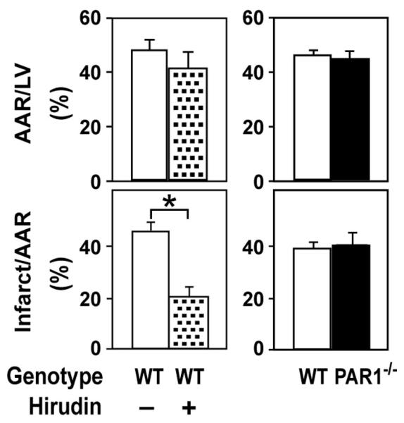
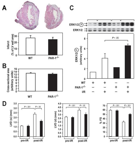
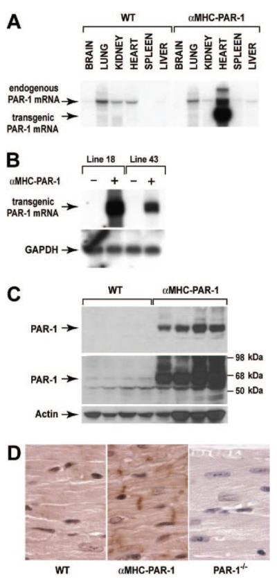
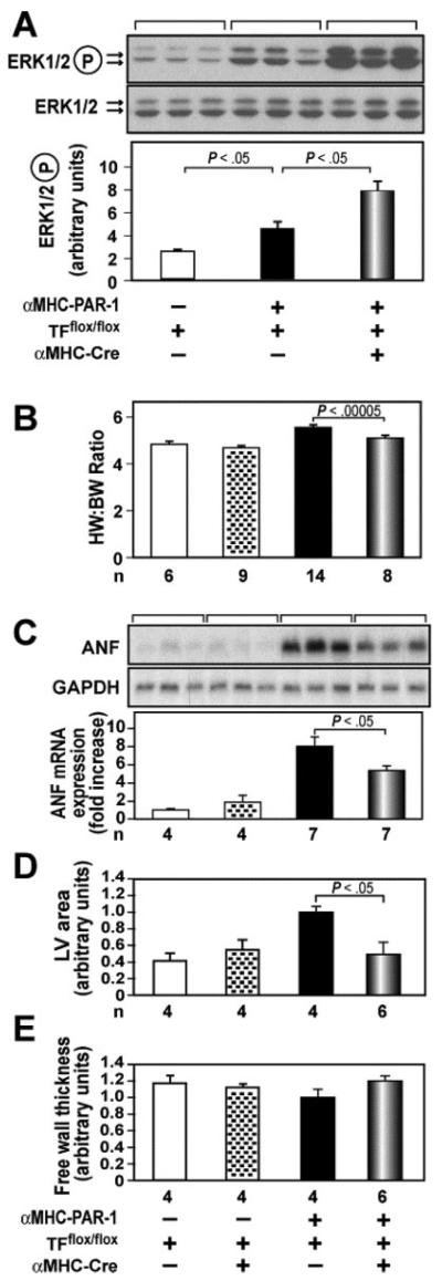
Methods and results: We found that PAR-1 deficiency reduced dilation of the left ventricle and reduced impairment of left ventricular function 2 weeks after I/R injury. Activation of ERK1/2 was increased in injured PAR-1(-/-) mice compared with wild-type mice; however, PAR-1 deficiency did not affect infarct size. Cardiomyocyte-specific overexpression of PAR-1 in mice induced eccentric hypertrophy (increased left ventricular dimension and normal left ventricular wall thickness) and dilated cardiomyopathy. Deletion of the TF gene in cardiomyocytes reduced the eccentric hypertrophy in mice overexpressing PAR-1.
Conclusions: Our results demonstrate that PAR-1 contributes to cardiac remodeling and hypertrophy. Moreover, overexpression of PAR-1 on cardiomyocytes induced eccentric hypertrophy. Inhibition of PAR-1 after myocardial infarction may represent a novel therapy to reduce hypertrophy and heart failure in humans.
Figures


Comment in
-
Letter by Strande regarding article "Protease-activated receptor-1 contributes to cardiac remodeling and hypertrophy".Circulation. 2008 Jun 17;117(24):e495; author reply e496. doi: 10.1161/CIRCULATIONAHA.107.758219. Circulation. 2008. PMID: 18559709 No abstract available.
Similar articles
-
Protease activated receptor-2 contributes to heart failure.PLoS One. 2013 Nov 27;8(11):e81733. doi: 10.1371/journal.pone.0081733. eCollection 2013. PLoS One. 2013. PMID: 24312345 Free PMC article.
-
Letter by Strande regarding article "Protease-activated receptor-1 contributes to cardiac remodeling and hypertrophy".Circulation. 2008 Jun 17;117(24):e495; author reply e496. doi: 10.1161/CIRCULATIONAHA.107.758219. Circulation. 2008. PMID: 18559709 No abstract available.
-
Tissue factor cytoplasmic domain exacerbates post-infarct left ventricular remodeling via orchestrating cardiac inflammation and angiogenesis.Theranostics. 2021 Sep 3;11(19):9243-9261. doi: 10.7150/thno.63354. eCollection 2021. Theranostics. 2021. PMID: 34646369 Free PMC article.
-
Protease-activated receptors and myocardial infarction.IUBMB Life. 2011 Jun;63(6):383-9. doi: 10.1002/iub.441. Epub 2011 Mar 24. IUBMB Life. 2011. PMID: 21438116 Free PMC article. Review.
-
The role of the tissue factor-thrombin pathway in cardiac ischemia-reperfusion injury.Semin Vasc Med. 2003 May;3(2):193-8. doi: 10.1055/s-2003-40677. Semin Vasc Med. 2003. PMID: 15199482 Review.
Cited by
-
Straight to the heart: Pleiotropic antiarrhythmic actions of oral anticoagulants.Pharmacol Res. 2019 Jul;145:104257. doi: 10.1016/j.phrs.2019.104257. Epub 2019 May 2. Pharmacol Res. 2019. PMID: 31054953 Free PMC article. Review.
-
Protease activated receptor-2 contributes to heart failure.PLoS One. 2013 Nov 27;8(11):e81733. doi: 10.1371/journal.pone.0081733. eCollection 2013. PLoS One. 2013. PMID: 24312345 Free PMC article.
-
Direct thrombin inhibition with dabigatran attenuates pressure overload-induced cardiac fibrosis and dysfunction in mice.Thromb Res. 2017 Nov;159:58-64. doi: 10.1016/j.thromres.2017.09.016. Epub 2017 Sep 21. Thromb Res. 2017. PMID: 28982031 Free PMC article.
-
Direct interrogation of context-dependent GPCR activity with a universal biosensor platform.Cell. 2024 Mar 14;187(6):1527-1546.e25. doi: 10.1016/j.cell.2024.01.028. Epub 2024 Feb 26. Cell. 2024. PMID: 38412860 Free PMC article.
-
Serum protease-activated receptor (PAR-1) levels as a potential biomarker for diagnosis of inflammation in type 2 diabetic patients.Inflammopharmacology. 2022 Oct;30(5):1843-1851. doi: 10.1007/s10787-022-01049-0. Epub 2022 Aug 16. Inflammopharmacology. 2022. PMID: 35974263
References
-
- Jessup M, Brozena S. Heart failure. N Engl J Med. 2003;348:2007–2018. - PubMed
-
- Brown RD, Ambler SK, Mitchell MD, Long CS. The cardiac fibroblast: therapeutic target in myocardial remodeling and failure. Annu Rev Pharmacol Toxicol. 2005;45:657–687. - PubMed
-
- Blaxall BC, Spang R, Rockman HA, Koch WJ. Differential myocardial gene expression in the development and rescue of murine heart failure. Physiol Genomics. 2003;15:105–114. - PubMed
-
- Li DY, Tao L, Liu H, Christopher TA, Lopez BL, Ma XL. Role of ERK1/2 in the anti-apoptotic and cardioprotective effects of nitric oxide after myocardial ischemia and reperfusion. Apoptosis. 2007;11:923–930. - PubMed
-
- Lips DJ, Bueno OF, Wilkins BJ, Purcell NH, Kaiser RA, Lorenz JN, Voisin L, Saba-El-Leil MK, Meloche S, Pouyssegur J, Pages G, De Windt LJ, Doevendans PA, Molkentin JD. MEK1-ERK2 signaling pathway protects myocardium from ischemic injury in vivo. Circulation. 2007;109:1938–1941. - PubMed
Publication types
MeSH terms
Substances
Grants and funding
LinkOut - more resources
Full Text Sources
Medical
Molecular Biology Databases
Miscellaneous
