. 2008 Apr;17(2):265-79.
doi: 10.1007/s11248-007-9152-5. Epub 2007 Oct 30.

Generation and characterization of transgenic zebrafish lines using different ubiquitous promoters

Affiliations

Generation and characterization of transgenic zebrafish lines using different ubiquitous promoters

Christopher T Burket et al. Transgenic Res. 2008 Apr.

Abstract

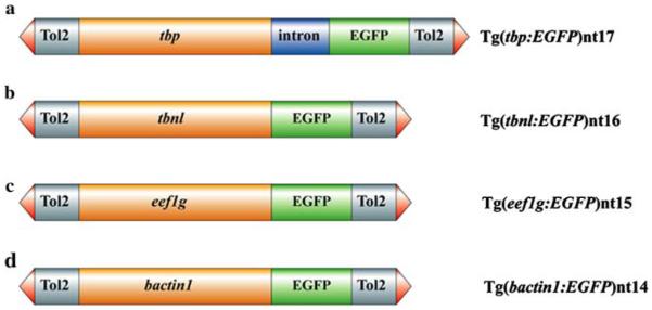
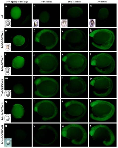
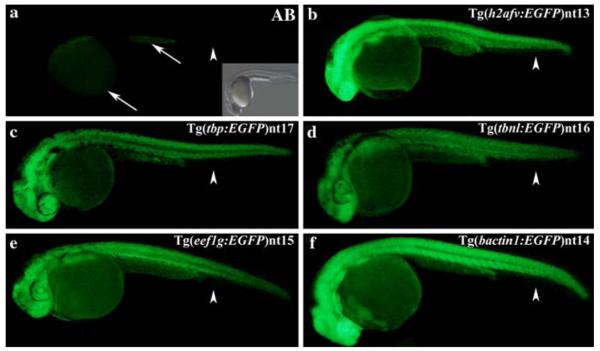
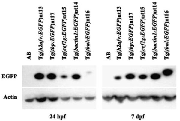
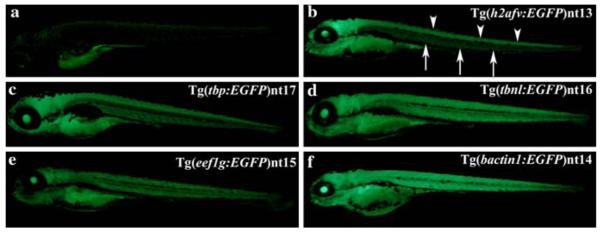
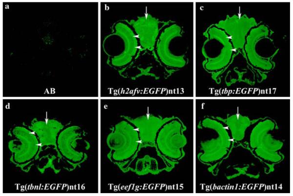
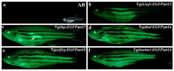
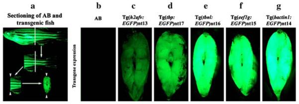
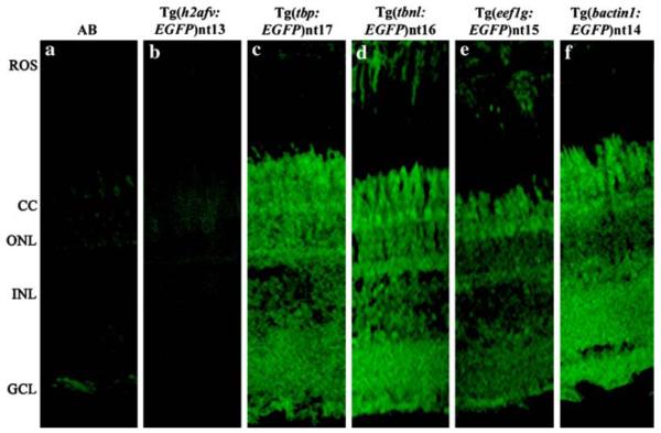
Two commonly used promoters to ubiquitously express transgenes in zebrafish are the Xenopus laevis elongation factor 1 alpha promoter (XlEef1a1) and the zebrafish histone variant H2A.F/Z (h2afv) promoter. Recently, transgenes utilizing these promoters were shown to be silenced in certain adult tissues, particularly the central nervous system. To overcome this limitation, we cloned the promoters of four zebrafish genes that likely are transcribed ubiquitously throughout development and into the adult. These four genes are the TATA box binding protein gene, the taube nuss-like gene, the eukaryotic elongation factor 1-gamma gene, and the beta-actin-1 gene. We PCR amplified approximately 2.5 kb upstream of the putative translational start site of each gene and cloned each into a Tol2 expression vector that contains the EGFP reporter transgene. We used these four Tol2 vectors to independently generate stable transgenic fish lines for analysis of transgene expression during development and in the adult. We demonstrated that all four promoters drive a very broad pattern of EGFP expression throughout development and the adult. Using the retina as a well-characterized component of the CNS, all four promoters appeared to drive EGFP expression in all neuronal and non-neuronal cells of the adult retina. In contrast, the h2afv promoter failed to express EGFP in the adult retina. When we examined EGFP expression in the various cells of the blood cell lineage, we observed that all four promoters exhibited a more heterogenous expression pattern than either the XlEef1a1 or h2afv promoters. While these four ubiquitous promoters did not express EGFP in all the adult blood cells, they did express EGFP throughout the CNS and in broader expression patterns in the adult than either the XlEef1a1 or h2afv promoters. For these reasons, these four promoters will be valuable tools for expressing transgenes in adult zebrafish.


Figures

Fig. 1
Fig. 1
Structure of the tbp:EGFP, tbnl:EGFP, eef1g:EGFP and bactin1:EGFP transgenes. Each promoter is 2.5 kb (orange boxes). The Tol2 sequences that flank each transgene include the terminal inverted repeats (red arrowheads) and approximately 500 bp (grey boxes). The EGFP transgene is 900 bp in length (green boxes). The tbp:EGFP transgene contains the β-globin intron (a), which is 644 bp and represented as the blue box
Fig. 2
Fig. 2
Developmental time course of EGFP expression in transgenic embryos. AB embryos were collected at the same time points from either 50% epiboly through the 30+ somite stage as the transgenic lines. EGFP fluorescence was not detected in any of the AB embryos (a–d). EGFP expression was present from the bud stage through the 30+ somite stage in the Tg(h2afv:EGFP)nt13 (e–h), Tg(tbp:EGFP)nt17 (i–l), and Tg(eef1g:EGFP)nt15 lines (q–t). EGFP expression was detected in the Tg(tbnl:EGFP)nt16 and Tg(bactin1:EGFP)nt14 lines from the 10–14 somite through the 30+ somite stages (Panels m–p and u–x, respectively) Embryos that are difficult to see in the fluorescent field have an inset of a bright field image
Fig. 3
Fig. 3
EGFP expression between 24 and 36 hpf in the transgenic embryos. The AB embryos possessed only a very low level of autofluorescence in the yolk sac and anal yolk extension at 24–36 hpf (Panel a, arrows). Furthermore, no fluorescence was detected in the fin fold (Panel a, arrowhead). A bright field image showing the AB embryo is inset. The EGFP expression was ubiquitous in the Tg(h2afv:EGFP)nt13, Tg(tbp:EGFP)nt17, Tg(tbnl:EGFP)nt16, Tg(eef1g:EGFP) nt15, and Tg(bactin1:EGFP)nt14 lines (Panels b–f), except for the absence of EGFP expression in the fin folds (arrowheads)
Fig. 4
Fig. 4
Immunoblot of EGFP expression at 24 hpf and 7 dpf. Total protein extracts from AB and five transgenic lines were isolated at 24 hpf and 7 dpf, electrophoresed through 4–12% SDS-PAGE, and transferred to PVDF membrane. Duplicate membranes were incubated with either anti-GFP or anti-actin antibodies, which served as a loading control. The immunoblots revealed a decrease in EGFP expression from 24hfp to 7 dpf in the Tg(h2afv:EGFP)nt13 line and increased expression in the Tg(eef1g:EGFP)nt15 and the Tg(tbnl:EGFP)nt16 lines. The Tg(tbnl:EGFP)nt16 line also revealed an EGFP protein that was slightly larger than the EGFP in the other transgenic lines. This increased molecular weight is due to an AUG codon that is in frame and upstream of the EGFP open reading frame, which yields a fusion protein
Fig. 5
Fig. 5
Ubiquitous transgene expression at 7 dpf. Similar to 24 hpf, a low level of autofluorescence was detected at 7 dpf in the AB fry (Panel a). The Tg(h2afv:EGFP)nt13 transgenic fish expressed EGFP in the brain and spinal cord (Panel b, arrowheads), while the body had a much lower level of EGFP expression (arrows). In comparison, the other four transgenic lines exhibited broader expression throughout the body, brain and spinal cord (Panels c–f). EGFP continued to be absent from the fin fold in all transgenic lines
Fig. 6
Fig. 6
Ubiquitous transgene expression throughout the head 7 dpf. At 7 dpf, EGFP expression was immunolocalized throughout the brain, retina, and other tissues of the head in all five transgenic lines (Panels b–f, arrows and arrowheads, respectively). The lens in each transgenic line appears to have a distinct EGFP expression pattern that was not further analyzed in this paper. However, EGFP appears to be expressed in the lens of Tg(tbp:EGFP)nt17, Tg(tbnl:EGFP)nt16, Tg(eef1g:EGFP)nt15, and Tg(bactin1:EGFP)nt14 lines (Panels c–f), but not in the Tg(h2afv:EGFP)nt13 transgenic fish (Panel b). As expected, AB fish stained with the same antibody did not show any substantial EGFP staining in the head (Panel a)
Fig. 7
Fig. 7
EGFP expression persists in a ubiquitous pattern in the adult transgenic fish, with the exception of the adult fins. EGFP expression is readily visible from the jaw to the girdle of the tail fin in the Tg(tbp:EGFP)nt17, Tg(tbnl:EGFP)nt16, Tg(eef1g:EGFP)nt15, and Tg(bactin1:EGFP)nt14 lines (Panels c–f). In contrast, the Tg(h2afv:EGFP)nt13 transgenic fish (Panel b), exhibited dramatically reduced EGFP expression in the anterior and posterior regions. In several fish, the female reproductive organs expressed elevated levels of EGFP (Panel c, arrow). There was no fluorescence detected in the AB fish (Panel a). A brightfield image of the adult AB fish is shown (Panel a, inset)
Fig. 8
Fig. 8
Visualizing distribution of EGFP expression in the transgenic fish body. EGFP expression was analyzed in tissue posterior to the anus/urogenital pore and anterior to the dorsal fin. The white lines represent where transverse cuts were made in each fish (Panel a). Arrowheads indicate the tissues that were visualized for EGFP expression in the transgenic lines. The tbp, tbnl, eef1g and bactin1 promoters directed EGFP expression ubiquitously in the body muscle (Panels d–g). While EGFP expression was seen in the body muscle from Tg(h2afv:EGFP)nt13 transgenic zebrafish, the reduced expression in some regions is obvious (Panel c). There was no EGFP expression detected in comparably treated AB fish taken at the same settings (Panel b)
Fig. 9
Fig. 9
EGFP is expressed in the retina from the new transgenes. EGFP was immunolocalized in the retinal sections of wild-type fish (Panel a) and the five transgenic lines (Panels b-f). Only a low level of autofluorescence was observed in the AB and Tg(h2afv:EGFP)nt13 lines (Panels a and b, respectively), which confirmed the silencing of the h2afv:EGFP transgene (Thummel et al. 2006b). In contrast, EGFP was immunolocalized throughout the retina in the Tg(tbp:EGFP) nt17, Tg(tbnl:EGFP)nt16, Tg(eef1g:EGFP)nt15, and Tg(bactin1:EGFP)nt14 lines (Panels c-f). ROS, rod outer segments; CC, cone cell layer; ONL, outer nuclear layer; INL, inner nuclear layer; GCL, ganglion cell layer

Similar articles

Cited by

References

    1. Amsterdam A, Lin S, Hopkins N. The Aequorea victoria green fluorescent protein can be used as a reporter in live zebrafish embryos. Dev Biol. 1995;171(1):123–129. - PubMed
    1. Amsterdam A, Lin S, Moss LG, Hopkins N. Requirements for green fluorescent protein detection in transgenic zebrafish embryos. Gene. 1996;173:99–103. - PubMed
    1. Bai S, Thummel R, Godwin AR, Nagase H, Itoh Y, Li L, Evans R, McDermott J, Seiki M, Sarras MP., Jr Matrix metalloproteinase expression and function during fin regeneration in zebrafish: analysis of MT1-MMP, MMP2 and TIMP2. Matrix Biol. 2005;24(4):247–260. - PubMed
    1. Bretaud S, Li Q, Lockwood BL, Kobayashi K, Lin E, Guo S. A choice behavior for morphine reveals experience-dependent drug preference and underlying neural substrates in developing larval zebrafish. J Neurosci. 2007;146(3):1109–1116. - PubMed
    1. Brinster RL, Allen JM, Behringer RR, Gelinas RE, Palmiter RD. Introns increase transcriptional efficiency in transgenic mice. Proc Natl Acad Sci USA. 1988;85(3):836–840. - PMC - PubMed

Publication types

MeSH terms